Intraluminal methods of ablating nerve tissue

a nerve tissue and intraluminal technology, applied in the field of system and method of treating interior tissue regions of the body, can solve the problems of reducing requiring lifelong dependence, and affecting the ability of the patient to tolerate the pain of swallowing, so as to prevent or delay the opening of the sphincter, reduce the compliance of the tissue, and heat the sphincter tissue.

Inactive Publication Date: 2013-05-23
RECOR MEDICAL INC
View PDF8 Cites 51 Cited by
  • Summary
  • Abstract
  • Description
  • Claims
  • Application Information

AI Technical Summary

Benefits of technology

[0017]The present invention seeks to heat sphincter tissues using ultrasound energy. The preferred method is to use ultrasound energy to heat tissue and thus create necrotic regions (lesions) in the tissue. The lesions tighten the tissue by shrinking it (through dessication, protein denaturation, and disruption of collagen bonds), and / or bulking it (with new collagen formation). The lesions also prevent or delay opening of the sphincter by reducing the compliance of the tissue in either or both the radial and longitudinal directions as the sphincter is forced to expand and shorten when the internal pressure increases. The lesions also interrupt nerve pathways responsible for sphincter relaxations. In general, during the heating process, the invention employs means to minimize heat damage to the mucosal layer of the sphincter. However, in the case of Barrett's Esophagus, selective heating of the intestinal metaplasia on the luminal surface of the esophagus is preferred. Ultrasound may also be used (continuously or in pulsed mode) to create shock waves that cause mechanical disruption through cavitation that create the desired tissue effects. While this invention relates broadly to many tissue sphincters in the body, the focus of the disclosure will be on the treatment of a dysfunctional lower esophageal sphincter (LES) responsible for GERD.
[0018]The key advantage of an ultrasound ablation system over others is that a uniform annulus of tissue can be heated simultaneously. Alternatively, the transducers can be designed so that only user-defined precise regions of the circumference are heated. Ultrasound also penetrates tissue deeper than RF or simple thermal conduction, and therefore can be delivered with a more uniform temperature profile. Thus lesions can be created at deeper locations than could be safely achieved with RF needles puncturing the tissue. Similarly, the deeper heating and uniform temperature profile also allow for an improved ability to create a cooling gradient at the surface. Relatively low power can be delivered over relatively long durations to maximize tissue penetration but minimize surface heating. If only surface heating is desired, as in the case of Barrett's Esophagus, the acoustic energy can be focused at or just before the tissue surface. Another means to selectively heat the tissue surface is to place a material against the tissue, between the tissue and the transducer, that selectively absorbs acoustic energy and preferentially heats at the tissue interface. A device using ultrasound for ablation may also be configured to allow diagnostic imaging of the tissue to determine the proper location for therapy and to monitor the lesion formation process.
[0022]Preferred ultrasonic transducers may be energized to produce unfocused acoustic energy from the transducer surface in the range from 10 W / cm2 to 100 W / cm2, usually from 30 W / cm2 to 70 W / cm2. The transducer will usually be energized at a duty cycle in the range from 10% to 100%, more usually from 70% to 100%. Focused ultrasound may have much higher energy densities, but will typically use shorter exposure times and / or duty cycles. In the case of heating the tissue, the transducer will usually be energized under conditions which cause a temperature rise in the tissue to a tissue temperature in the range from 55° C. to 95° C., usually from 60° C. to 80° C. In such instances, it will usually be desirable to cool the luminal surface, which is a mucosal surface in the case of the esophagus which may treated by the present invention, in order to reduce the risk of injury.

Problems solved by technology

Complications of the disease can progress to difficulty and pain in swallowing, stricture, perforation and bleeding, anemia, and weight loss.
Most patients respond to drug therapy, but it is palliative in that it does not cure the underlying cause of sphincter dysfunction, and thus requires lifelong dependence.
As with other highly invasive procedures, antireflux surgery is associated with the risk of complications such as bleeding and perforation.
In addition, a significant proportion of individuals undergoing laparascopic fundoplication report difficulty swallowing (dysphagia), inability to vomit or belch, and abdominal distention.
The result is shrinkage of the tissue and interruption of vagal afferent pathways some believe to play a role in the transient relaxations of the LES.
The above endoscopic techniques all require the penetration of the esophageal wall with a needle-like device, which entails the additional risks of perforation or bleeding at the puncture sites.
Use of the plication technique requires many operational steps and over time sutures have been reported to come loose and / or the tissue folds have diminished or disappeared.
Lesion size is also limited by the needle size.
Limitations of the design require additional steps of rotating the device to achieve additional lesions.
Dysfunction of the anal sphincter leads to fecal incontinence, the loss of voluntary control of the sphincter to retain stool in the rectum.
However, multiple sessions are often necessary, and patients need to be highly motivated.
Surgical interventions suffer from the same disadvantages discussed above with respect to GERD.
Potential device complications and use limitations are similar to those described for GERD.
Dysfunction of the urinary sphincter leads to urinary incontinence, the loss of voluntary control of the sphincter to retain urine in the bladder.
U.S. Pat. No. 6,073,052 describes a method of sphincter treatment using a microwave antennae and specific time and temperature ranges, and U.S. Pat. No. 6,321,121 a method of GERD treatment using a non-specific energy source, with limited enabling specifications.
Such collagen formation may in some cases, at least, result from cavitation or other injury-producing application of the vibrational energy.

Method used


Image

  • Intraluminal methods of ablating nerve tissue
  • Intraluminal methods of ablating nerve tissue
  • Intraluminal methods of ablating nerve tissue

Examples


Embodiment Construction

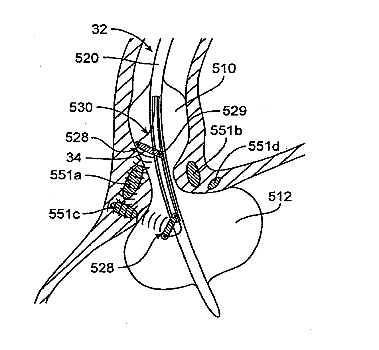
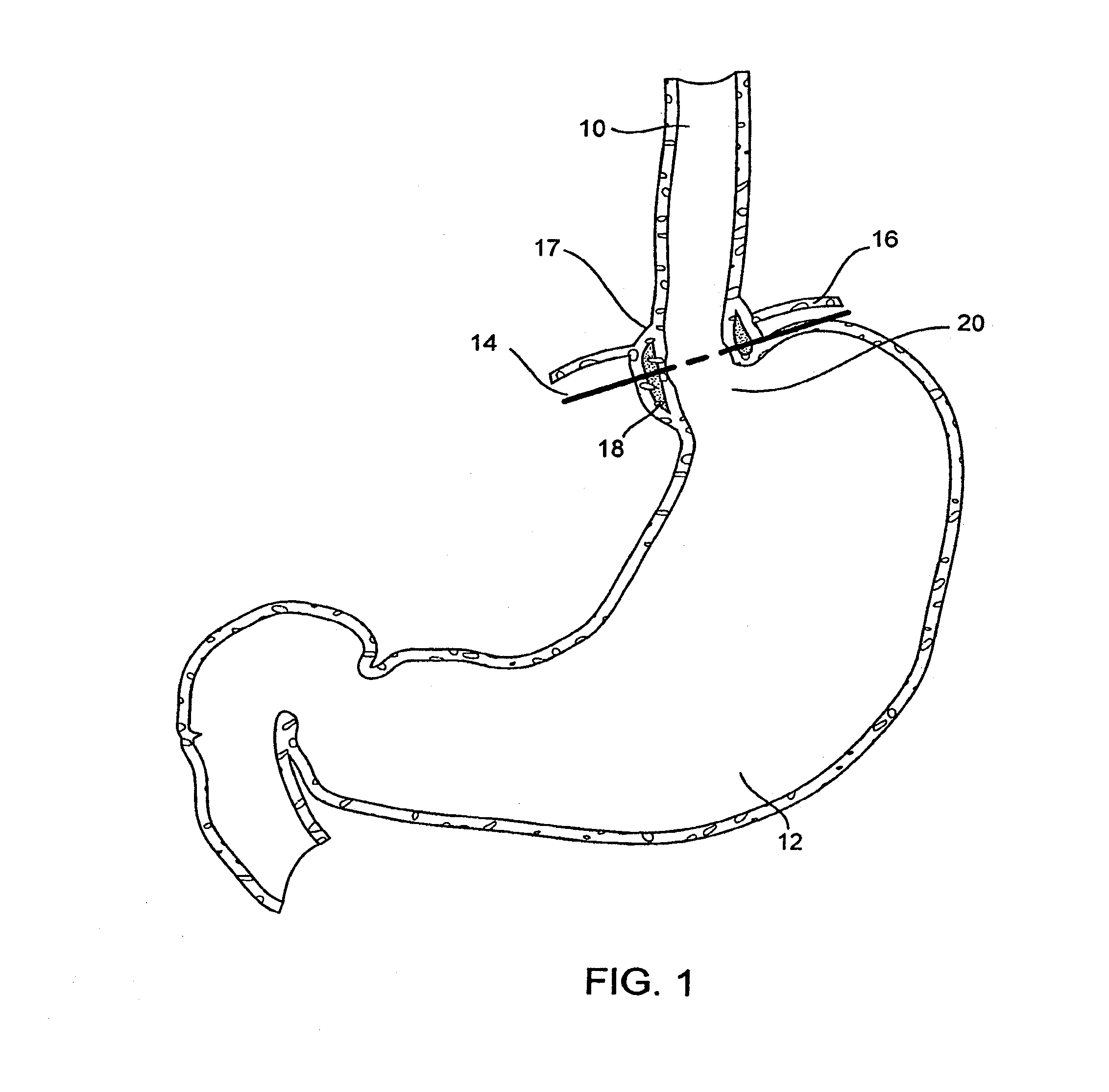
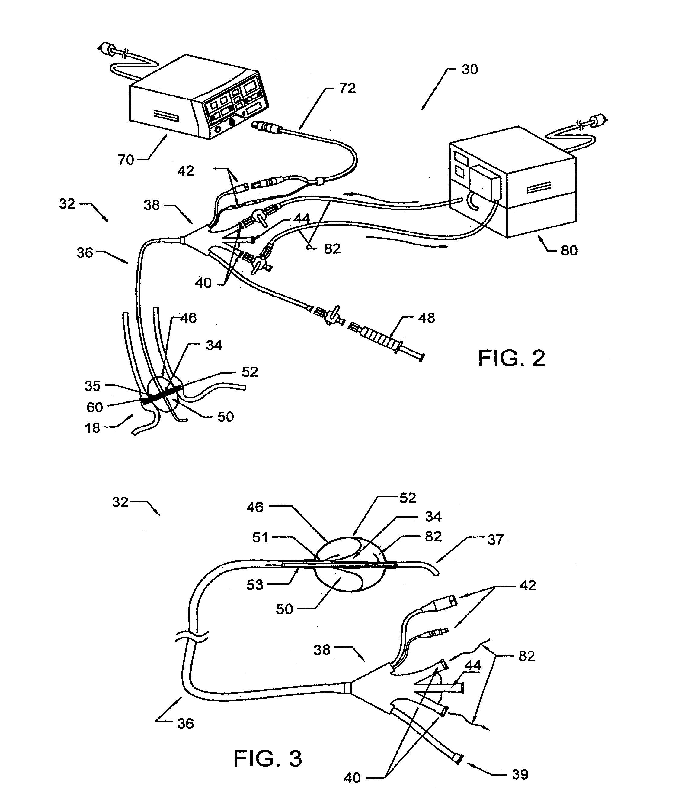
[0096]This Specification discloses various catheter-based systems and methods for treating dysfunction of sphincters and adjoining tissue regions in the body. The systems and methods are particularly well suited for treating these dysfunctions in the upper gastrointestinal tract, e.g., in the lower esophageal sphincter (LES) and adjacent cardia of the stomach. For this reason, the systems and methods will be described in this context.

[0097]Still, it should be appreciated that the disclosed systems and methods are applicable for use in treating other dysfunctions elsewhere in the body, which are not necessarily sphincter-related. For example, the various aspects of the invention have application in procedures requiring treatment of hemorrhoids, or incontinence, or restoring compliance to or otherwise tightening interior tissue or muscle regions. The systems and methods that embody features of the invention are also adaptable for use with systems and surgical techniques that are not n...


PUM


Abstract

Methods and apparatus for treating gastroesophageal reflex and other luminal conditions provide for delivering acoustic energy to a body lumen to remodel tissue surrounding the body lumen. In the case of treating GERD, a catheter carrying an ultrasonic or other vibrational transducer is introduced to the lower esophageal sphincter, and acoustic energy is delivered to the sphincter in order to tighten or bulk the sphincter such that reflex is reduced.

Description

CROSS-REFERENCE TO RELATED APPLICATIONS[0001]This application is a continuation application of U.S. patent application Ser. No. 13 / 478,825, filed May 23, 2012, which is a continuation application of U.S. patent application Ser. No. 10 / 611,838, filed on Jun. 30, 2003 and now abandoned, which claims priority benefit of U.S. Provisional Application No. 60 / 393,339, filed Jul. 1, 2002, and U.S. Provisional Application No. 60 / 419,317, filed Oct. 16, 2002, the entireties of all of which are hereby incorporated by reference herein.BACKGROUND[0002]1. Field[0003]In a general sense, the invention is directed to systems and methods for treating interior tissue regions of the body. More specifically, the invention is directed to systems and methods for treating dysfunction in body sphincters and adjoining tissue, e.g., in and around the lower esophageal sphincter and cardia of the stomach.[0004]2. Description of the Background Art[0005]The gastrointestinal (GI) tract extends from the mouth to th...

Claims


Application Information

Patent Timeline
Patent Type & Authority Applications(United States)
IPC IPC(8): A61N7/02A61B17/00A61N7/00
CPCA61B2017/0046A61N2007/0091A61N2007/0078A61N2007/0095A61B18/1492A61B18/18A61B18/1815A61B18/20A61N7/022A61B17/2202A61N2007/003A61N2007/0043A61N2007/0065A61N2007/0082A61N7/02A61B8/4281
Inventor SCHAER, ALAN
Owner RECOR MEDICAL INC
Why Eureka
Try Eureka