

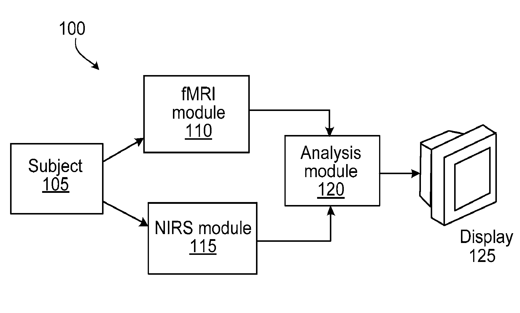
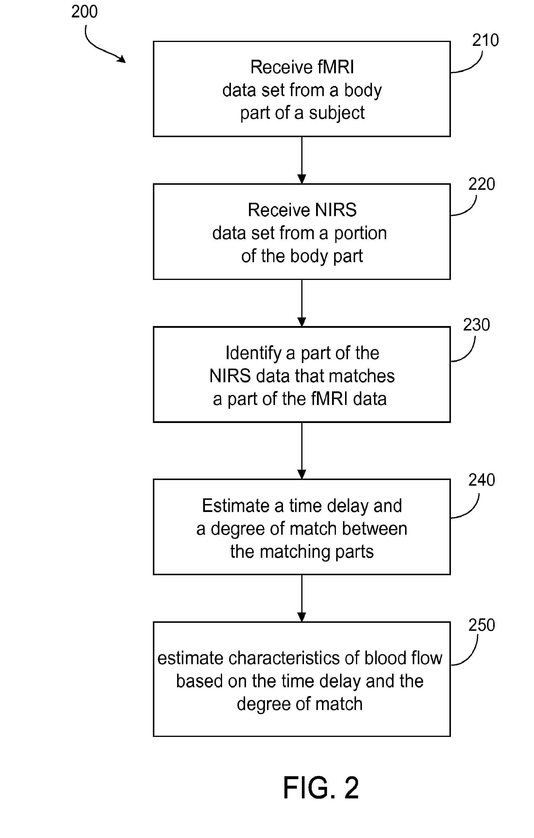
Multi-modal imaging of blood flow
a multi-modal imaging and blood flow technology, applied in mammography, medical science, diagnostics, etc., can solve the problems of low sensitivity, low resolution, and certain risks
- Summary
- Abstract
- Description
- Claims
- Application Information
AI Technical Summary
Benefits of technology
Problems solved by technology
Method used
Image

Examples
example 1
[0080]In this example, the high temporal resolution of NIRS (about 80 ms) was combined with the high spatial resolution of fMRI (about 3.5×3.5×4 mm) to investigate the physiological origin of LFOs detected by both modalities in human brain during a resting state of a subject. The goals of the resting state study were: 1) to examine the temporal and spatial characteristics of LFOs as measured with NIRS; 2) to determine the spatial extent of NIRS LFO in brain; and 3) to investigate temporal changes in the spatial patterns of LFO signal (identified by NIRS) throughout the brain in the BOLD fMRI data.
[0081]Both NIRS and BOLD fMRI signals are sensitive to blood-oxygenation-related changes in the brain and both are able to accurately sample the low frequency band (<0.1 Hz), in general, there exists a strong temporal correlation between measurements of low-frequency physiological processes obtained using both techniques simultaneously. Conversely, these two imaging modalities have differen...
example 2
Spectral Separation of NIRS Signals
[0112]In this example, a temporal trace of NIRS was first spectrally separated into four bands using a zero delay Fourier domain bandpass filter in Matlab. The four bands were defined as very low frequency oscillation (VLF), VLF0.8 Hz. The temporal signal from each band was then resampled according to the TR of the fMRI acquisition. FIG. 12 shows the process for one subject. FIGS. 12a, 12c, 12f, 12i and 12l show the power spectra for (a) original data, (b) VLF, (c) LFO, (d) Resp and (e) Card, respectively. FIGS. 12b, 12d, 12g, 12j and 12m show the corresponding time traces, respectively. FIGS. 12e, 12h, 12k and 12n show the enlarged sections of the corresponding time traces, respectively. The resampled points (separated by 1.5 s) in the enlarged sections are shown as closed circles. Regressor interpolation at progressive time delays can then be applied on each spectrally separated band to assess the spatiotemporal evolutions of VLF, LFO, respiratio...
example 3
Noise Removal from BOLD Data
[0113]The following examples illustrate how some of the factors described herein affect the efficacy of noise removal from BOLD data. In the following examples, different types of NIRS data (deoxy-hemoglobin, Δ[HbR] and total hemoglobin Δ[tHb])were used alone and in combination with one another, the effect of using a plurality of frequency bands were compared, and data from different NIRS detection paths were compared. The probe placement for the experiment was substantially same as that shown in FIG. 5.
[0114]Choice of NIRS Type
[0115]As shown in FIGS. 13A and 13B, two independently determined NIRS quantities −Δ[HbR] and Δ[tHb] were used to illustrate the physiological variation in the BOLD data, and were found to be effective in different areas. For example, the Δ[tHb] regressors worked effectively near large vessels and in gray matter. Because the amount of physiological variance is essentially additive, both types of NIRS signals were used. When regress...
PUM
 Abstract
Description
Claims
Application Information