Biological substance detection method

a detection method and biological substance technology, applied in the direction of fluorescence/phosphorescence, material analysis, instruments, etc., can solve the problems of easy influence of staining and difficult simultaneous observation

Active Publication Date: 2013-06-20
KONICA MINOLTA INC +1
View PDF4 Cites 22 Cited by
  • Summary
  • Abstract
  • Description
  • Claims
  • Application Information

AI Technical Summary

Benefits of technology

[0033]According to the present invention, in simultaneous staining for morphological and immunological observation, the method can discriminate...

Problems solved by technology

However, the staining with an enzyme label such as DAB staining develops a color similar to the color in HE staining to preclude the distinction between the staining by an enzyme label and the HE staining, which makes the simultaneous observation difficul...

Method used


Image

  • Biological substance detection method

Examples


examples

[0087]Hereinafter, the present, invention will be described in detail by Examples, but is not limited thereto.

[0088][Preparation of Samples]

[0089](Sample 1: Fluorescent Nanoparticles)

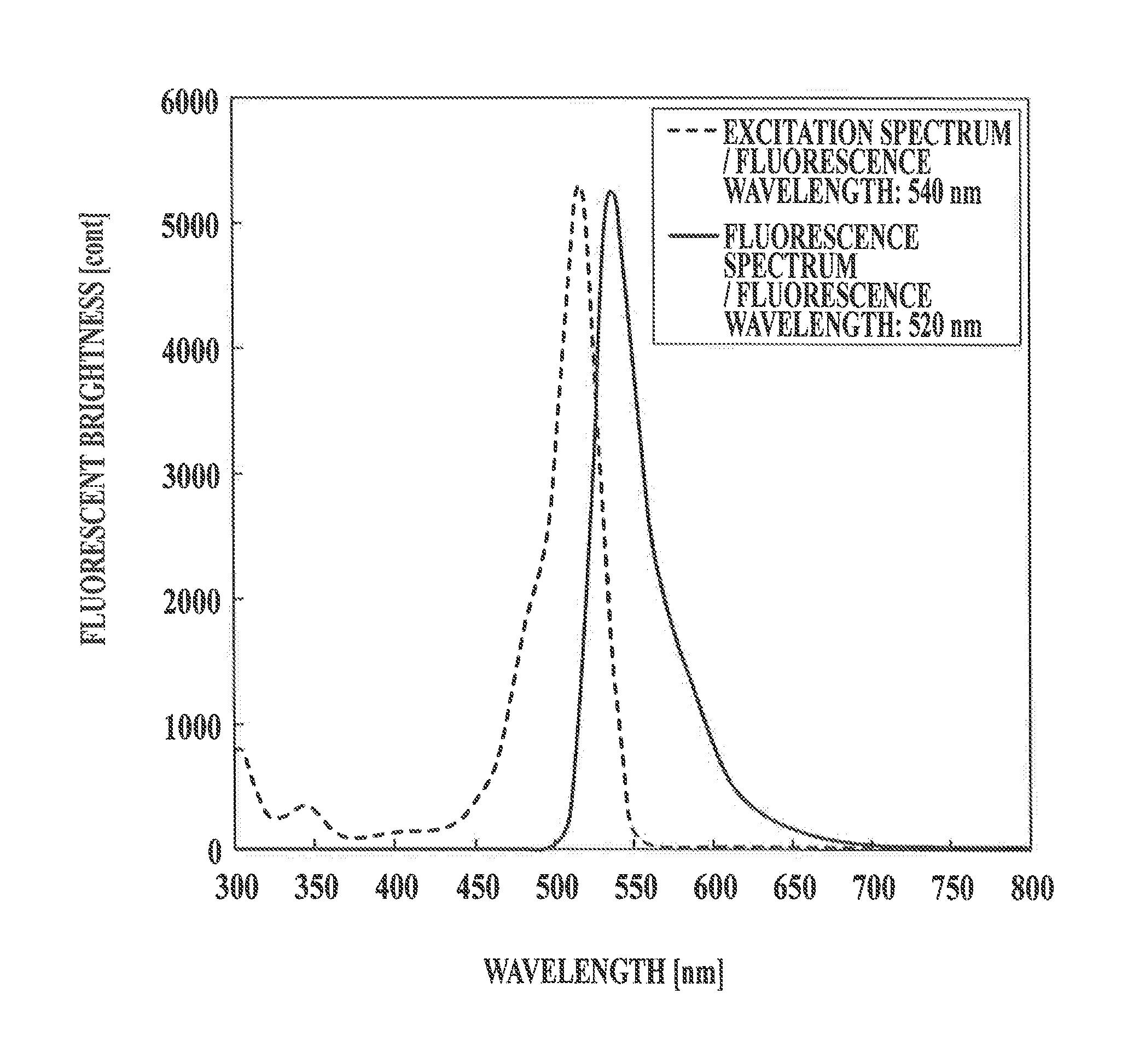
[0090]CdSe / ZnS fluorescent nanoparticles (Qdot655, Invitrogen Corporation) modified with PEG having amino groups at the ends were prepared as fluorescent nanoparticles for antibody binding.

[0091]Meanwhile, anti-human ER antibodies were subjected to reduction treatment with 1M dithiothreitol (DTT), and excess DTT was removed by a gel filtration column to obtain a solution of the reduced antibodies capable of bonding so silica particles.

[0092]The fluorescent nanoparticles for antibody binding and the reduced antibodies were mixed in PBS containing 2 mM EDTA, followed by reaction for 1 hour. The reaction was stopped by adding 10 mM mercaptoetnanol. The obtained solution was concentrated with a centrifugal filter, and subsequently, the unreached antibodies and the like were removed by a gel filtration colum...


PUM


Abstract

A biological substance detection method for detecting a biological substance specifically in a pathological specimen, comprising a step of immunologically staining the pathological specimen using a fluorescent label, a step of staining the pathological specimen with a staining reagent for morphology observation purposes (eosin) to observe the morphology of the pathological specimen, a step of irradiating the stained pathological specimen, with excited light to cause the emission of a fluorescent and detecting the biological substance in the pathological specimen. In the step of immunologically staining the pathological specimen, a special fluorescent particle for which the excitation wavelength appears in a region that is different from the excitation wavelength region of eosin is used as the fluorescent label.

Description

TECHNICAL FIELD[0001]The present invention relates to a biological substance detection method, and specifically relates to tissue staining for multiply staining a tissue with fluorescent labels.BACKGROUND ART[0002]As one of medical diagnoses, a pathological diagnosis is performed. A pathologist diagnoses a disease using a tissue section taken from a human body and notifies a clinician of the necessity or unnecessity of a therapy or operation. On the basis of conditions of a patient and pathological diagnosis, a physician determines a drug treatment plan, and a surgeon determines whether or not an operation should be performed.[0003]In pathological diagnosis, it is widely performed to prepare a tissue sample by slicing a tissue specimen obtained through resection of an organ or needle biopsy into a thickness of about several micrometers and observe an enlarged image of the tissue sample with an optical microscope for obtaining various findings. In many oases, the sample is prepared b...

Claims


Application Information

Patent Timeline
IPC IPC(8): G01N21/64
CPCG01N21/6428G01N33/582G01N2021/6439G01N21/6458G01N21/6486G01N33/4833G01N2021/6441
Inventor TAKANASHI, KENSAKUGOUDA, HIDEKIOKADA, HISATAKENAKANO, YASUSHIGONDA, KOHSUKETAKEDA, MOTOHIROOHUCHI, NORIAKI
Owner KONICA MINOLTA INC
Why Eureka
Try Eureka