Method for neuroepithelial cells differentiation from pluripotent stem cells and medium using same

a technology of neuroepithelial cells and stem cells, applied in the direction of embryonic cells, cell culture active agents, artificial cell constructs, etc., can solve the problems of poor efficiency of neural differentiation, contaminated non-neural cells, and large cost, and achieve the effect of shortening time cost and high purity

Inactive Publication Date: 2013-06-20
NATIONAL CHUNG HSING UNIVERSITY
View PDF2 Cites 4 Cited by
  • Summary
  • Abstract
  • Description
  • Claims
  • Application Information

AI Technical Summary

Benefits of technology

[0015]Upon the method, it could efficiently shorten the time cost for acquiring the neuroepithelial cells from pluripotent stem cells with higher purity. The acquired neuroepi

Problems solved by technology

Furthermore, there are some non-neural cells contaminated with neuroepithelial cells during the adhesion process within the culture system.
Although the aforesaid methods to generate the neuroepithelial cells from stem cells, the disadvantages such as poor efficiency of neural differentiation, expansive cost, risk resulted from virus-mediated genetic manipulations and non-neural cells

Method used


Image

  • Method for neuroepithelial cells differentiation from pluripotent stem cells and medium using same
  • Method for neuroepithelial cells differentiation from pluripotent stem cells and medium using same
  • Method for neuroepithelial cells differentiation from pluripotent stem cells and medium using same

Examples


example 1

Generation of Embryoid Body from Embryonic Stem Cells

[0045]The human embryonic stem cells, TW1 cells, are cultured at 37° C. and 5% CO2, first. Following the pre-culture, the aggregated clones of embryonic stem cells are selected for the further suspension culture within DMEM-F12 containing 20% knock-out replacement serum (KSR, Invitrogen, USA) at 37° C. and 5% CO2 for two days to generate the embryoid bodies.

example 2

Induction of the Neural Differentiation of Pluripotent Stem Cells into Neuroepithelial Cells

[0046]The embryoid bodies generated in example 1 are collected in the 15 mL centrifuge tube and placed at room temperature for descending the embryoid bodies and discarding the supernatant. Prepare 500 mL of first neural induction medium which contains the basic constitutions listed in table 1 and additive drugs including 0.5 μM BIO, 10 μM SB431542 and 10 ng / ml FGF2.

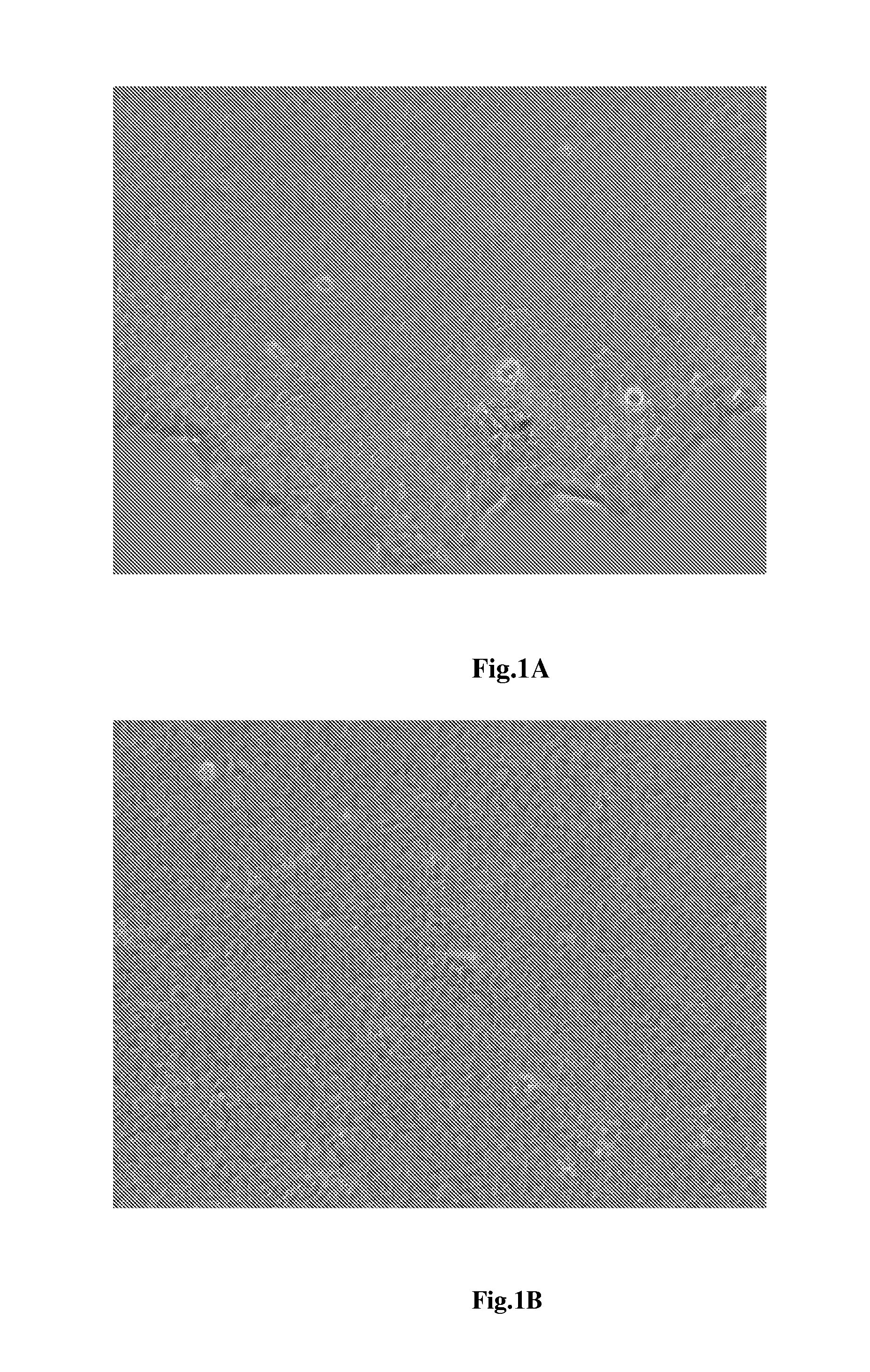
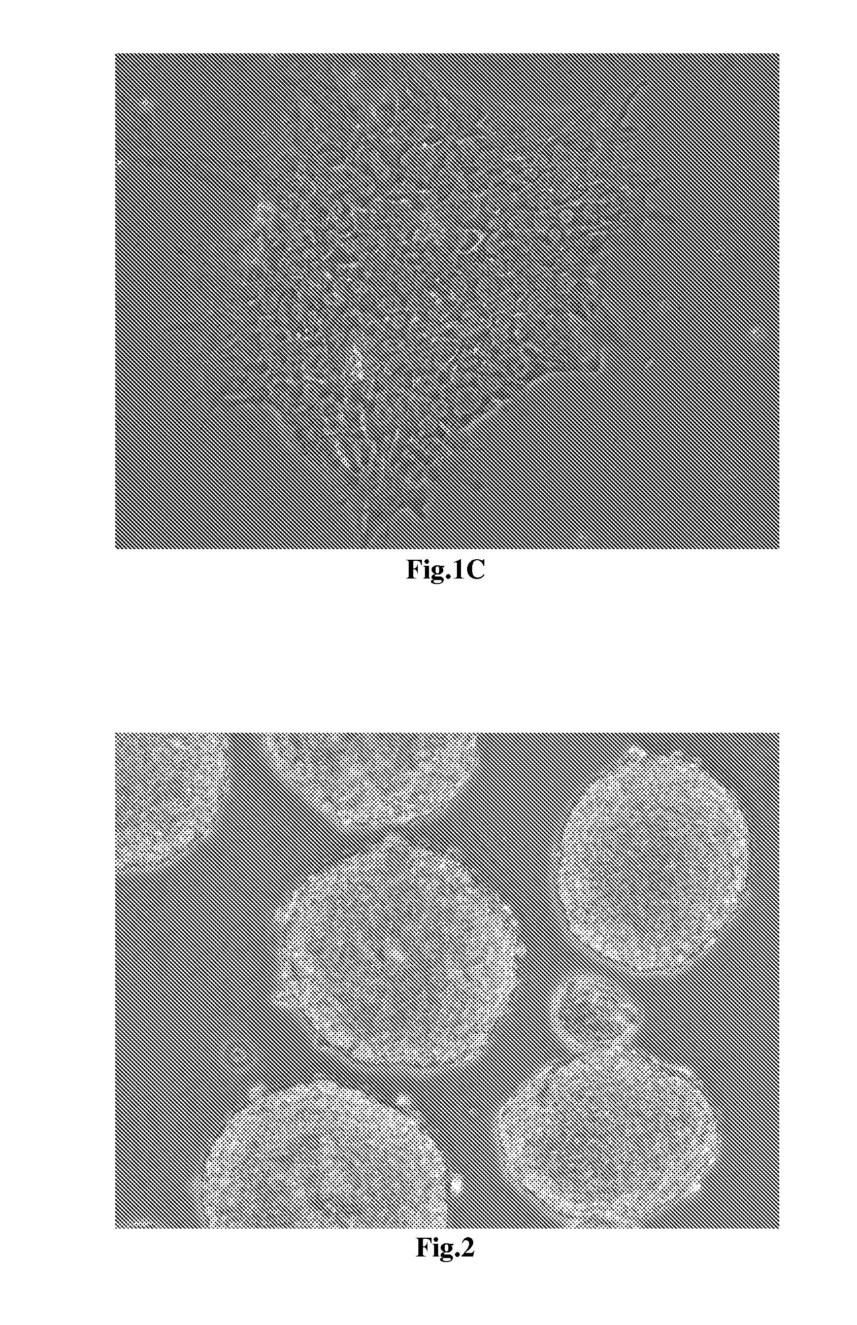
[0047]Notably, the announcement has to be emphasized here is that the working concentration of these additive drugs in the first neural induction medium are not restricted on what we indicated. The working concentration of BIO is between 0.05 μM to 0.5 μM; the working concentration of SB431542 is between 1 μM to 100 μM; and the working concentration of FGF2 is between 1 ng / ml to 100 ng / ml. The collected cells are further cultured with the first neural induction medium for two days to generate neuroepithelial cells as shown in FIG....

example 3

Further Induction of Neural Differentiation into Neuroepithelial Cells

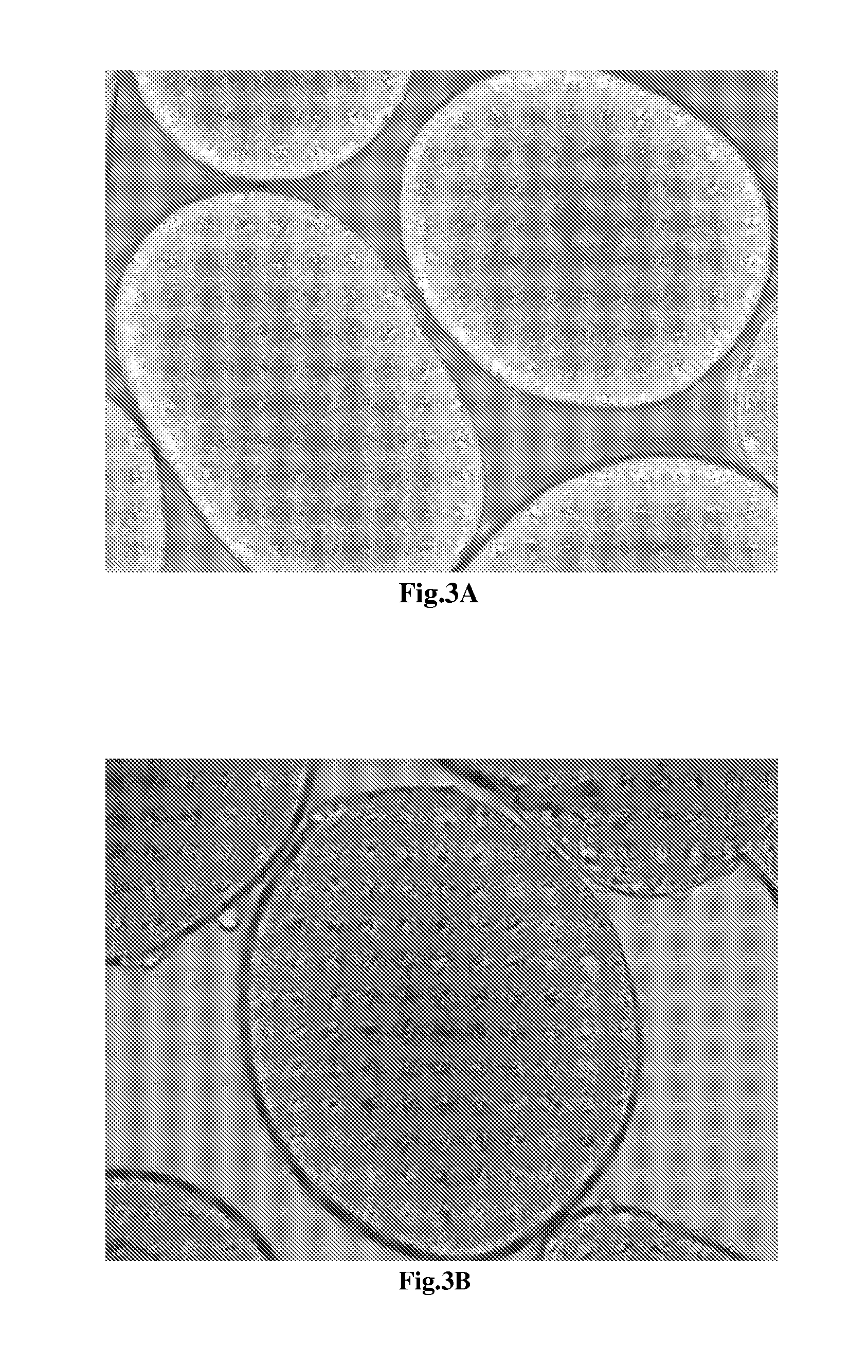
[0048]The neuroepithelial cells gained in example 2 are switched from first neural induction medium to secondary neural induction medium which is added with 10 ng / ml FGF2 for maintaining the further differentiation into neuroepithelial cells. Herein, the constitutions of secondary neural induction medium are shown in table 2.

[0049]The cells finishing the neural differentiation are shown in FIG. 3 and FIG. 4 with 100×, 200×, and 400× magnifications under microscope. In FIG. 3A, the cultured cells reveal the homogenous morphology and form the globular structure units through tightly tubular aggregation at the edge. The FIG. 3B reveals the globular structure unit which contain the tightly tubular aggregation at the edge and rosette formations resembling the early neural tube in the center. The FIG. 3C shows that the cultured cells in the center of the globular unit form rosette clump resembling the neural tube-like s...


PUM


Abstract

The present invention discloses a neural induction medium comprising Wnt-signal agonist, TGFβ-signal inhibitor and FGF-signal agonist for inducing neural differentiation. The neural induction medium used in a culture system is capable for inducing the neural differentiation of stem cells into neuroepithelial cells which are useful for the clinical applications. Therein, the neuroepithelial cells can further differentiate into mature neurons for the practical applications including regeneration medicine and drug discovery for neural disorders.

Description

FIELD OF THE INVENTION[0001]The present invention relates to a method for neuroepithelial cells differentiation and a medium using same. Specifically, the present invention discloses a method for neuroepithelial cells differentiation from pluripotent stem cells and medium using same.BACKGROUND OF THE INVENTION[0002]Stem cells are the undifferentiated cells which exhibit the capacities for self-renewing and differentiating into more than two kinds of mature somatic cells. They are classified into totipotent stem cells, pluripotent stem cells, multipotent stem cell and bipotent stem cell according to their differentiation capacity. Furthermore, they cloud be also classified into embryonic stem cells, somatic stem cells and induced pluripotent stem cells (iPSCs) upon their origins. Herein, human embryonic stem cells are derived from the inner cell mass of the pre-implantation blastocyst and reveal the pluripotency for differentiating into all adult somatic cells. In addition, iPSCs are...

Claims


Application Information

Patent Timeline
IPC IPC(8): C12N5/0797C12N5/0735
CPCC12N5/0623C12N2506/02C12N2501/415C12N2501/115
Inventor SU, HONG-LINCHEN, SHENG-MEI
Owner NATIONAL CHUNG HSING UNIVERSITY
Why Eureka
Try Eureka