Assessment of tissue or lesion depth using temporally resolved light scattering spectroscopy

a technology of light scattering and tissue depth, which is applied in the field of optical interrogation configuration, can solve the problems of limiting the ability to characterize the depth, contributing to the observed “noise” in the data, and all signals arriving earlier than about 45 ps contain no useful information, etc., and achieves the cost of implementing this invention, fast response time, and rapid reduction of costs.

Active Publication Date: 2013-09-26
LAWRENCE LIVERMORE NAT SECURITY LLC
View PDF8 Cites 71 Cited by
  • Summary
  • Abstract
  • Description
  • Claims
  • Application Information

AI Technical Summary

Benefits of technology

[0019]Recent advances in laser and light sources have provided multiple methods to produce short pulses with broad spectrum via generation of a super-continuum by photonic fibers or other nonlinear materials. Also, compact ultrashort laser sources operating at different w

Problems solved by technology

Consequently, as the depth of the lesion increases, the signal detected continuously decreases, limiting the ability to characterize the depth of lesions deeper than about 5-8 mm and also contributing to the observed “noise” in the data.
Thus, if the

Method used


Image

  • Assessment of tissue or lesion depth using temporally resolved light scattering spectroscopy
  • Assessment of tissue or lesion depth using temporally resolved light scattering spectroscopy
  • Assessment of tissue or lesion depth using temporally resolved light scattering spectroscopy

Examples


Embodiment Construction

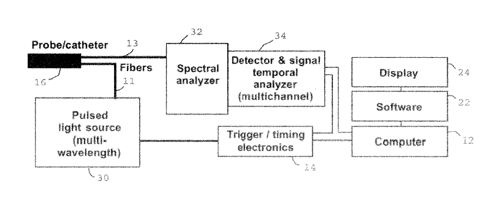
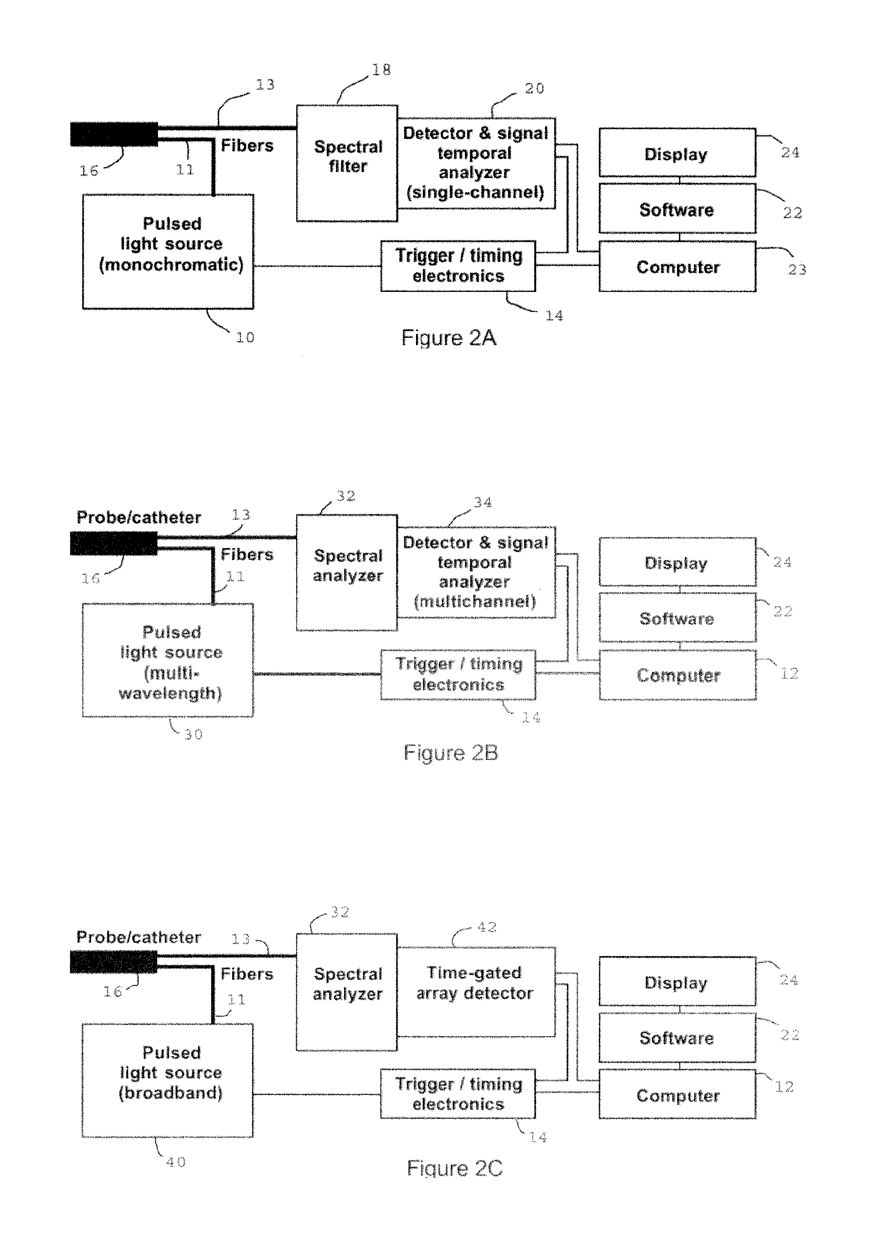
[0032]The present invention teaches a different method to assess the lesion spatial parameters in real time with higher precision, especially of deeper ablation lesions. The present method can use the same catheter designs as in the parent case, incorporating optical elements in the tip of the catheter (most commonly in the form of one or more emitting and one or more receiving optical fibers). A schematic of the ablation catheter incorporating optical fibers in an arrangement that is suitable for the current invention is shown in FIGS. 2(a) and 2(b) of the parent case. The present method can also use the ejection of light into the tissue as in the parent case, e.g.; however, while the parent application teaches a method that uses the spectral profile of the receive light in order to assess the lesion depth, the present invention uses the temporal profile of the received light pulse in order to assess the lesion depth.

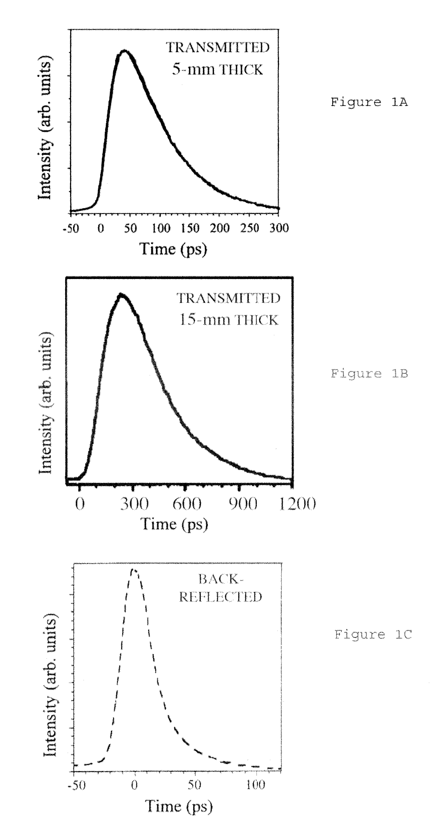
[0033]FIGS. 1A-1C show experimental measurements that represent t...


PUM


Abstract

A method is described to enhance the ability to evaluate the depth of a tissue component or a lesion having optical properties different from a surrounding tissue using time resolved optical methods. This invention may be particularly suitable for the evaluation of lesion depth during RF ablation (irreversible tissue modification/damage) using specially designed devises (catheters) that deliver heat in a localized region for therapeutic reasons. The technique allows for increased ability to evaluate the depth of the ablated lesion or detect the presence of other processes such as micro-bubble formation and coagulation with higher sensitivity compared to that offered by steady state spectroscopy. The method can be used for in-vivo, real-time monitoring during tissue ablation or other procedures where information on the depth of a lesion or tissue is needed. Exemplary uses are found in tissue ablation, tissue thermal damage, lesion and tissue depth assessment in medical applications.

Description

CROSS-REFERENCE TO RELATED APPLICATIONS[0001]This application is a continuation-in-part of U.S. application Ser. No. 11 / 414,009 titled “Fiber Optic Evaluation of Tissue Modification,” filed Apr. 27, 2006, incorporated herein by reference, which is a continuation-in-part of U.S. application Ser. No. 11 / 281,853, titled “Fiber-Optic Evaluation of Cardiac Tissue Ablation,” filed Nov. 17, 2005, incorporated herein by reference, which claims priority to U.S. Provisional Application No. 60 / 629,166, titled “Fiber-optic Evaluation of Cardiac Tissue Ablation and Optical Spectroscopy,” filed on Nov. 17, 2004, incorporated herein by reference.STATEMENT REGARDING FEDERALLY SPONSORED RESEARCH OR DEVELOPMENT[0002]The United States Government has rights in this invention pursuant to Contract No. DE-AC52-07NA27344 between the U.S. Department of Energy and Lawrence Livermore National Security, LLC, for the operation of Lawrence Livermore National Laboratory.BACKGROUND OF THE INVENTION[0003]1. Field o...

Claims


Application Information

Patent Timeline
IPC IPC(8): A61B5/00
CPCA61B5/0084A61B5/0086A61B18/22A61B2017/00061A61B18/082A61B5/0075A61B18/24A61B2017/00066A61B2018/00351A61B2018/00357A61B2018/0212A61B18/1492A61B2090/062
Inventor DEMOS, STAVROS G.
Owner LAWRENCE LIVERMORE NAT SECURITY LLC
Why Eureka
Try Eureka