Hybrid slot-scanning grating-based differential phase contrast imaging system for medical radiographic imaging

a technology of differential phase contrast and imaging system, which is applied in the field of hybrid slotscanning grating-based differential phase contrast imaging system for medical radiographic imaging, can solve the problems of poor contrast compared with bone images, incompatibility with a typical clinical environment, and cost of synchrotron radiation sour

Inactive Publication Date: 2013-10-03
CARESTREAM HEALTH INC
View PDF7 Cites 44 Cited by
  • Summary
  • Abstract
  • Description
  • Claims
  • Application Information

AI Technical Summary

Benefits of technology

[0017]In accordance with one embodiment, the invention can provide a method that can include providing a beam shaping assembly comprising a beam limiting apparatus and a source grating G0, providing an x-ray grating interferometer comprising a phase grating G1, and an analyzer grating G2, and offsetting a pitch of the analyzer grating G2 relative to a pitch of an interference pattern produced by the phase grating G1 at a prescribed distance from the phase grating G1.

Problems solved by technology

However, for soft tissues including vessels, cartilages, lungs, and breast tissues with little absorption, this provides poor contrast compared with bone images.
However, there are various practical problems associated with all three techniques such as efficiency and limited field of view.
A synchrotron radiation source is costly and incompatible with a typical clinical environment.
Both techniques are also limited by the accepted beam divergence of only a very small angle (a few mrad) due to the use of crystal optics.
The free-space propagation technique is limited in efficiency since it requires high spatial coherence, which can only be obtained from an x-ray source with a very small focal spot.
Although some of the techniques yield excellent results for specific applications, none is very widely used and none has so far found application in medical diagnostics.
The grating-based PCI method with a standard x-ray tube is limited by the loss of visibility of the interference fringes at the detector due to the broad spectrum of the x-ray tube.
Given the limitations of the current grating fabrication techniques (e.g., silicon wafer size, structure height, and grating uniformity), the manufacturing cost of such large gratings will be extremely high.
For a grating-based PCI system having a divergent cone beam (or fan beam) geometry and a large FOV, the phase contrast image quality is generally inferior in the edge regions of the detector.
As a result, these gratings would cause a shadowing effect of the phase grating and the scan effect of the analyzer grating at larger angles, degrading the image quality.
In all x-ray imaging systems, scattered radiation from the object has been shown to degrade the image quality in terms of subject contrast and contrast-to-noise ratio significantly.
However, intrinsic to the anti-scatter grid method is the attenuation of a significant fraction of the primary x-rays.

Method used


Image

  • Hybrid slot-scanning grating-based differential phase contrast imaging system for medical radiographic imaging
  • Hybrid slot-scanning grating-based differential phase contrast imaging system for medical radiographic imaging
  • Hybrid slot-scanning grating-based differential phase contrast imaging system for medical radiographic imaging

Examples


Embodiment Construction

[0036]The following is a detailed description of exemplary embodiments according to the application, reference being made to the drawings in which the same reference numerals identify the same elements of structure in each of the several figures.

[0037]To be useful for clinical imaging, the phase contrast imaging systems must meet various requirements including: (i) use of a standard broadband x-ray source; (ii) a large field of view (FOV) of many centimeters (e.g., 24 cm×30 cm for a typical mammography system); (iii) a reasonably compact design comparable to current radiographic imaging systems (e.g., the source-to-detector distance is about 65 cm for a typical mammography system); and / or (iv) a reasonable exposure time and dose (e.g., the mean exposure for a typical mammography system is about 5 mR).

[0038]1. System Configuration

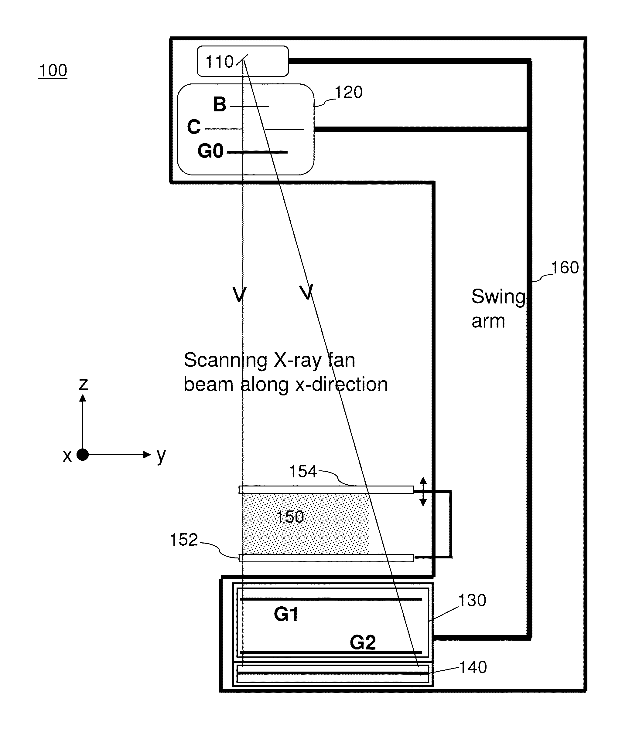
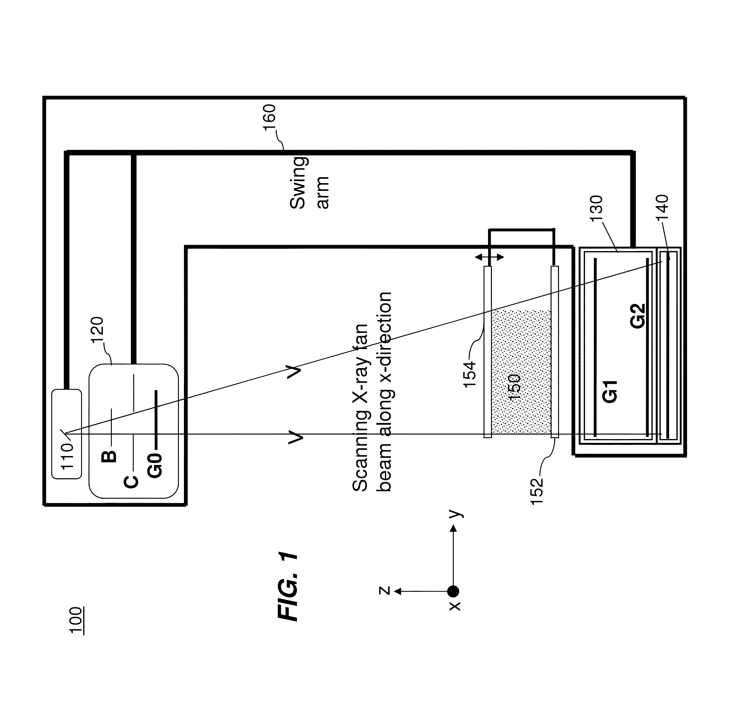
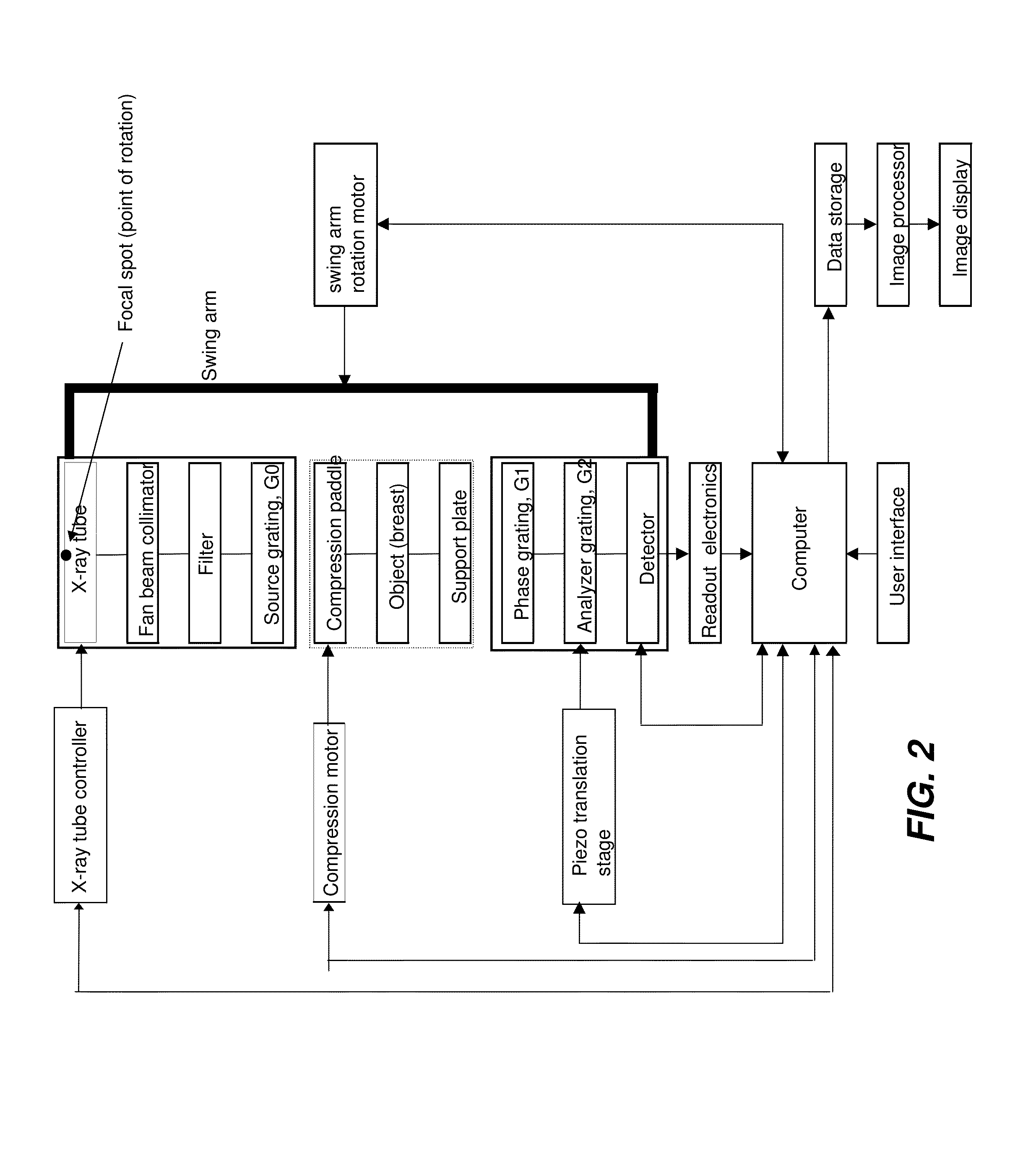
[0039]FIG. 1 is a diagram that shows an exemplary embodiment of a slot-scanning phase-contrast imaging system in accordance with the application. As shown i...


PUM


Abstract

Embodiments of methods and apparatus are disclosed for obtaining a phase-contrast digital mammography system and methods for same that can include an x-ray source for radiographic imaging; a beam shaping assembly including a filter or a tunable monochromator, a collimator, a source grating, an x-ray grating interferometer including a phase grating, and an analyzer grating; and an x-ray detector; where the source grating, the phase grating, and the analyzer grating are aligned in such a way that the grating bars of these gratings are parallel to each other.

Description

CROSS REFERENCE TO RELATED APPLICATIONS[0001]This is application claims priority to U.S. provisional patent application 61 / 617,948, filed Mar. 30, 2012, entitled HYBRID SLOT-SCANNING GRATING-BASED DIFFERENTIAL PHASE CONTRAST IMAGING SYSTEM FOR MAMMOGRAPHY, which is hereby incorporated by reference in its entirety.FIELD OF THE INVENTION[0002]The application generally relates to digital x-ray imaging methods / system, and more specifically, to methods and / or systems for acquiring multiple image information of an object (e.g., medical radiographic imaging) using a grating-based differential phase contrast imaging technique with a slot-scanning configuration.BACKGROUND OF THE INVENTION[0003]Conventional medical x-ray imaging devices are based on the attenuation through photoelectric absorption of the x-rays penetrating the object to be imaged. However, for soft tissues including vessels, cartilages, lungs, and breast tissues with little absorption, this provides poor contrast compared wit...

Claims


Application Information

Patent Timeline
Patent Type & Authority Applications(United States)
IPC IPC(8): A61B6/00
CPCA61B6/502A61B6/484A61B6/4035A61B6/508G21K2207/005A61B6/06
Inventor YIP, KWOK L.WOJCIK, TIMOTHY J.SHAFER, MARK E.JADRICH, BRADLEY S.BATURIN, PAVLO
Owner CARESTREAM HEALTH INC
Why Eureka
Try Eureka