

Cancer imaging with therapy: theranostics
a technology of cancer imaging and theranostics, applied in the field of genetic constructs, can solve the problems of unacceptably high background noise, unsatisfactory inability to provide sufficient specific localization of imaging agents, etc., to achieve high level of precise imaging, low background noise, and low background noise.
- Summary
- Abstract
- Description
- Claims
- Application Information
AI Technical Summary
Benefits of technology
Problems solved by technology
Method used
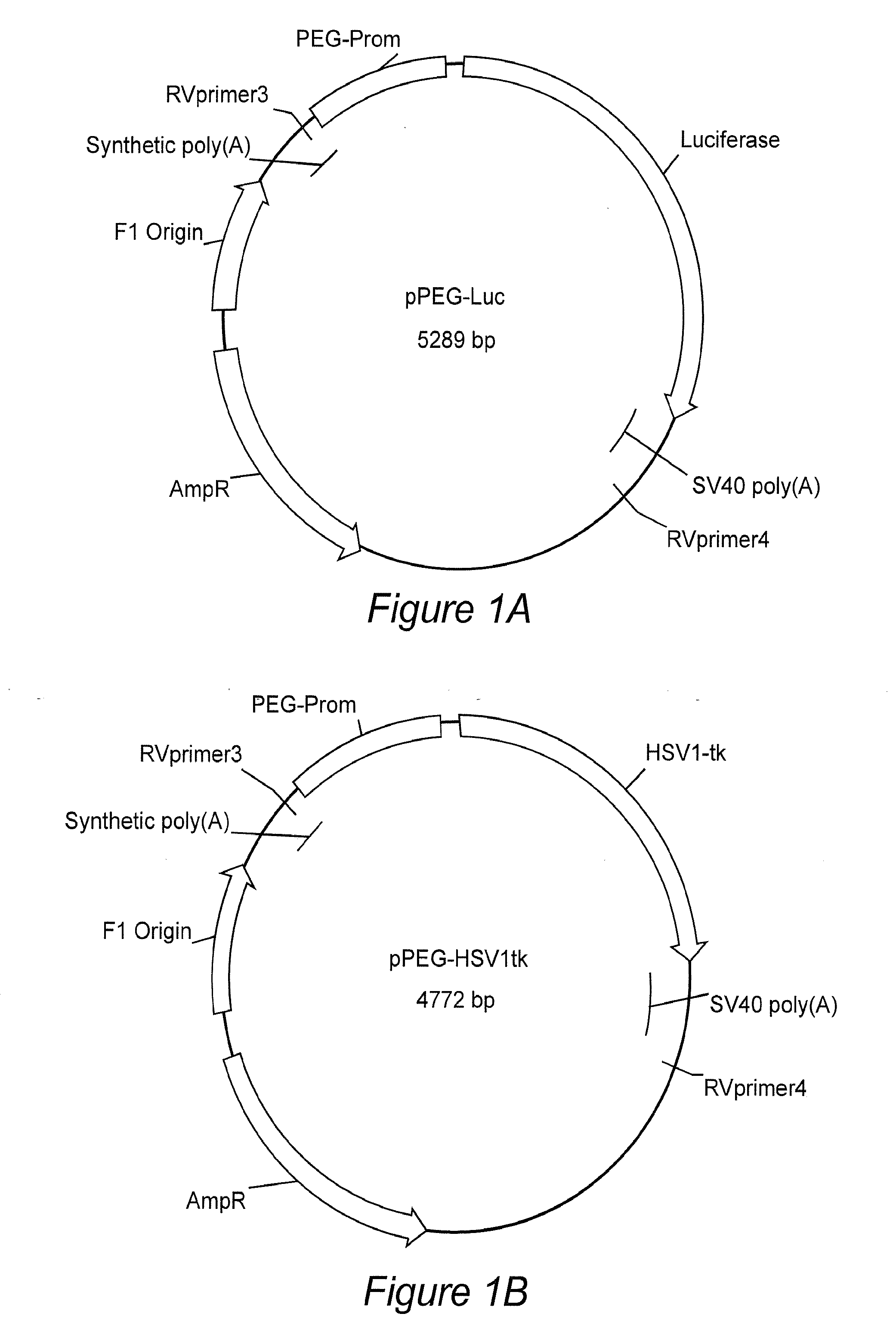
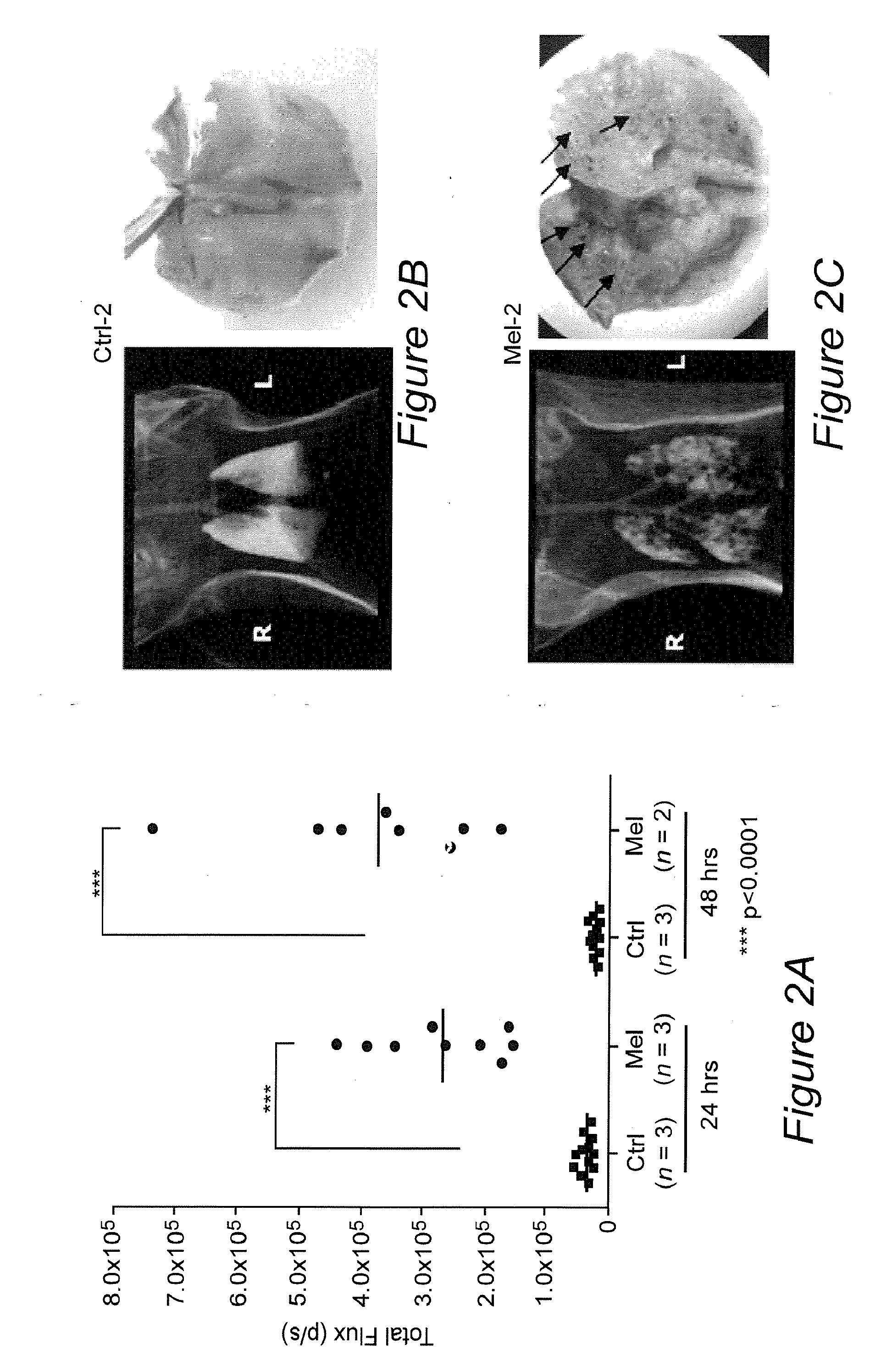
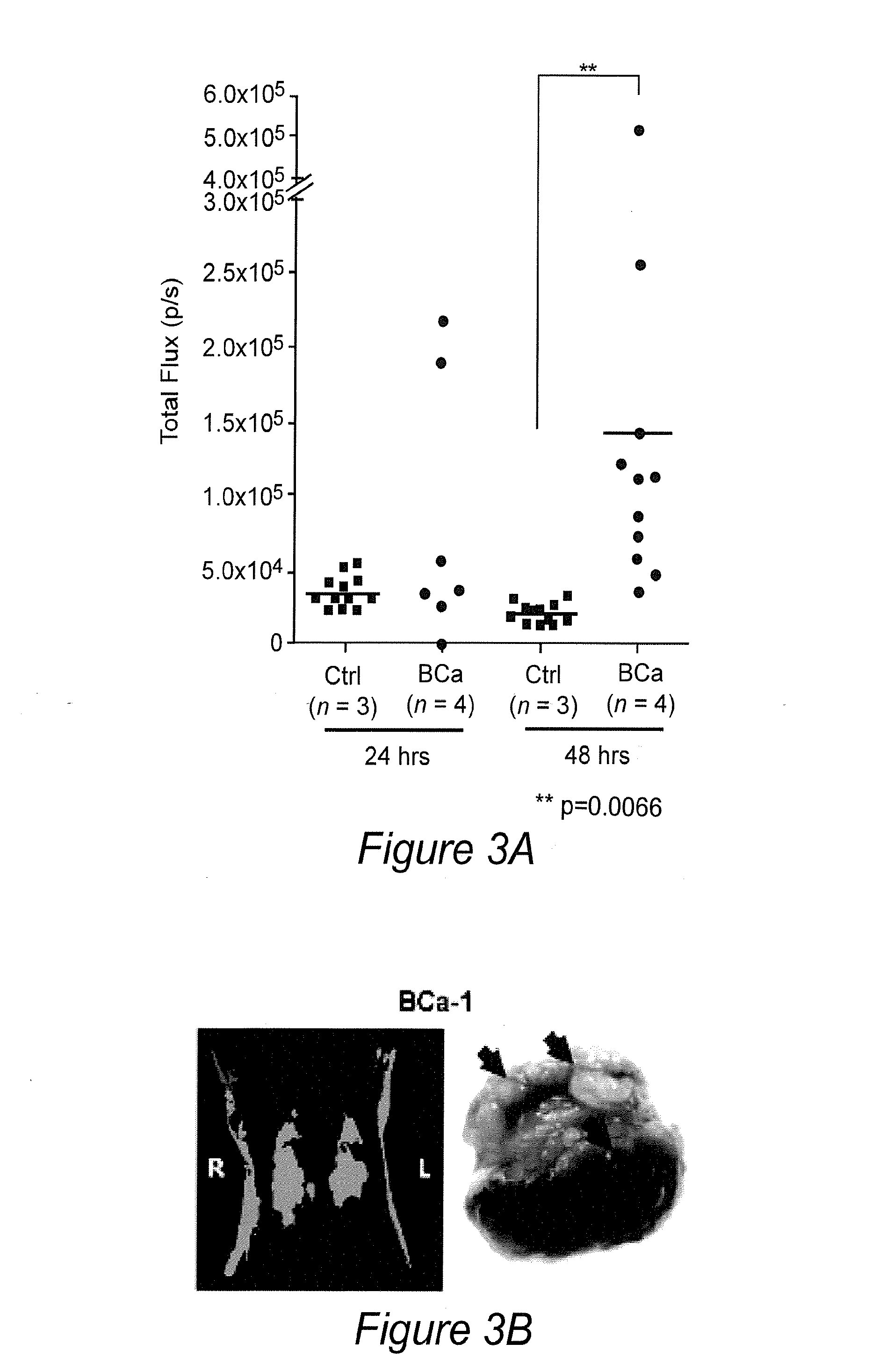
Image

Examples
example 1
REFERENCES FOR EXAMPLE 1
[0088]1. Blasberg, R. G. & Tjuvajev, J. G. Molecular-genetic imaging: current and future perspectives. J Clin Invest 111, 1620-1629 (2003).[0089]2. Zhang, Y., et al. ABCG2 / BCRP expression modulates D-Luciferin based bioluminescence imaging. Cancer Res 67, 9389-9397 (2007).[0090]3. Uhrbom, L., Nerio, E. & Holland, E. C. Dissecting tumor maintenance requirements using bioluminescence imaging of cell proliferation in a mouse glioma model. Nat Med 10, 1257-1260 (2004).[0091]4. Kishimoto, H., et al. In vivo imaging of lymph node metastasis with telomerase-specific replication-selective adenovirus. Nat Med 12, 1213-1219 (2006).[0092]5. Padmanabhan, P., et al. Visualization of telomerase reverse transcriptase (hTERT) promoter activity using a trimodality fusion reporter construct. J Nucl Med 47, 270-277 (2006).[0093]6. Freytag, S. O., et al. Phase I trial of replication-competent adenovirus-mediated suicide gene therapy combined with IMRT for prostate cancer. Mol Th...
example 2
REFERENCES FOR EXAMPLE 2
[0130]1. Su Z Z, Sarkar D, Emdad L, Duigou G J, Young C S H, Ware J, Randolph A, Valerie K, and Fisher P B. Targeting gene expression selectively in cancer cells by using the progression-elevated gene-3 promoter. Proc Natl Acad Sci USA 2005; 102(4):1059-1064.[0131]2. Su Z, Shi Y, Fisher P B. Cooperation between AP1 and PEA3 sites within the progression elevated gene-3 (PEG-3) promoter regulate basal and differential expression of PEG-3 during progression of the oncogenic phenotype in transformed rat embryo cells. Oncogene 2000; 19(30):3411-21.[0132]3. Su Z Z, Shi Y, Fisher P B. Subtraction hybridization identifies a transformation progression-associated gene PEG-3 with sequence homology to a growth arrest and DNA damage-inducible gene. Proc Natl Acad Sci USA 1997; 94(17):9125-30.[0133]4. Su Z Z, Goldstein N I, Jiang H, Wang M N, Duigou G J, Young C S, Fisher P B. PEG-3, a nontransforming cancer progression gene, is a positive regulator of cancer aggressivenes...
PUM
| Property | Measurement | Unit |
|---|---|---|
| quantitative real time PCR | aaaaa | aaaaa |
| total volume | aaaaa | aaaaa |
| total volume | aaaaa | aaaaa |
Abstract
Description
Claims
Application Information