Cancer imaging with therapy: theranostics

a technology of cancer imaging and theranostics, applied in the field of genetic constructs, can solve the problems of unacceptably high background noise, unsatisfactory specific localization of imaging agents, and none of these techniques have provided sufficient specific localization of imaging agents, and achieves a high level of precise delivery of anti-tumor agents, low background noise, and low cost.

Inactive Publication Date: 2020-12-10
VIRGINIA COMMONWEALTH UNIV +1
View PDF1 Cites 0 Cited by
  • Summary
  • Abstract
  • Description
  • Claims
  • Application Information

AI Technical Summary

Benefits of technology

[0012]The treatment aspect of the invention provides a high level of precise delivery of anti-tumor agents to cancer cells, even when delivery is made systemically, since the anti-tumor agents associated with the methods are only expressed within cancer cells. This advantageously results in few or no side effects for patients being treated by the method.
[0013]Similarly, the imaging aspect of the invention provides a high level of precise imaging of cancer cells and tumors with little or no background signal. Importantly, since there is little or no background “noise”, the imaging techniques of the invention enable the facile detection of metastatic cancer, even metastatic cancer that is not detectable with other methods due to e.g., the very small size of a newly developing tumor, or the diffuse pattern of cancer cells which do not actually form, a tumor. As is well known in the art, early detection of tumors can significantly improve the outcome of tumor treatment. Similarly, detection of cancerous tissues before formation of a tumor will provide significant benefits.
[0014]The combined imaging and treatment methods are advantageous over the prior art in many ways. A combined approach to imaging and therapy is more efficient and requires fewer procedures, and hence less effort, on the part of the patient and the cancer specialist. Since activity is confined to cancer cells, side effects are reduced. In addition, the combined imaging and treatment method provides the ability to accurately monitor the effects of prior treatment concomitantly with providing treatment and this provides a cancer treatment specialist with an invaluable and accurate window on the progress of therapy, permitting therapeutic parameters to be fine-tuned in close conjunction with treatment.

Problems solved by technology

But many efforts at tumor-specific imaging are fraught by nonspecific localization of the putstive targeted agents, eliciting unacceptably high background noise.
Unfortunately, to date, none of these techniques has provided sufficient specific localization of imaging agents, and unacceptably high background noise is still prevalent.
However, they are also still hampered by nonspecific delivery of and-tumor agents to normal cells, resulting in horrendous side effects for patients.
This lack of specificity also results in lower efficacy of treatments due to the want of a capability to deliver active agents in a focused manner where they are most needed, i.e., to cancer cells alone.
In addition, there is currently no efficacious way to image and / or treat cancerous cells and tissues which are caused by spontaneous metastasis.

Method used


Image

  • Cancer imaging with therapy: theranostics
  • Cancer imaging with therapy: theranostics
  • Cancer imaging with therapy: theranostics

Examples


example 1

References for Example 1

[0101]1. Blasberg, R. G. & Tjuvajev, J. G. Molecular-genetic imaging current and future perspectives. J Clin Invest 111, 1620-1629 (2003).

[0102]2. Zhang, Y., et al. ABCG2 / BCRP expression modulates D-Lucilerin based bioluminescence imaging. Cancer Res 67, 9389-9397 (2007).

[0103]3. Uhrbom, L., Nerio, E. & Holland, E. C. Dissecting tumor maintenance requirements using bioluminescence imaging of cell proliferation in a mouse glioma model. Nat Med 10, 1257-1260 (2004).

[0104]4. Kishimow, H., et al. In vivo imaging of lymph node metastasis with telomerase-specific replication-selective adenovirus. Nat Med 12, 1213-1219 (2006).

[0105]5. Padmanahhan, P., et al. Visualization of telomerase reverse transcriptase (hTERT) promoter activity using a trimodality fusion reporter construct. J Nucl Med 47, 270-277 (2006).

[0106]6. Freytag, S. O., et al. Phase I trial of replication-competent adenovirus-mediated suicide gene therapy combined with IMRT for prostate cancer. Mol Ther...

example 2

Overview:

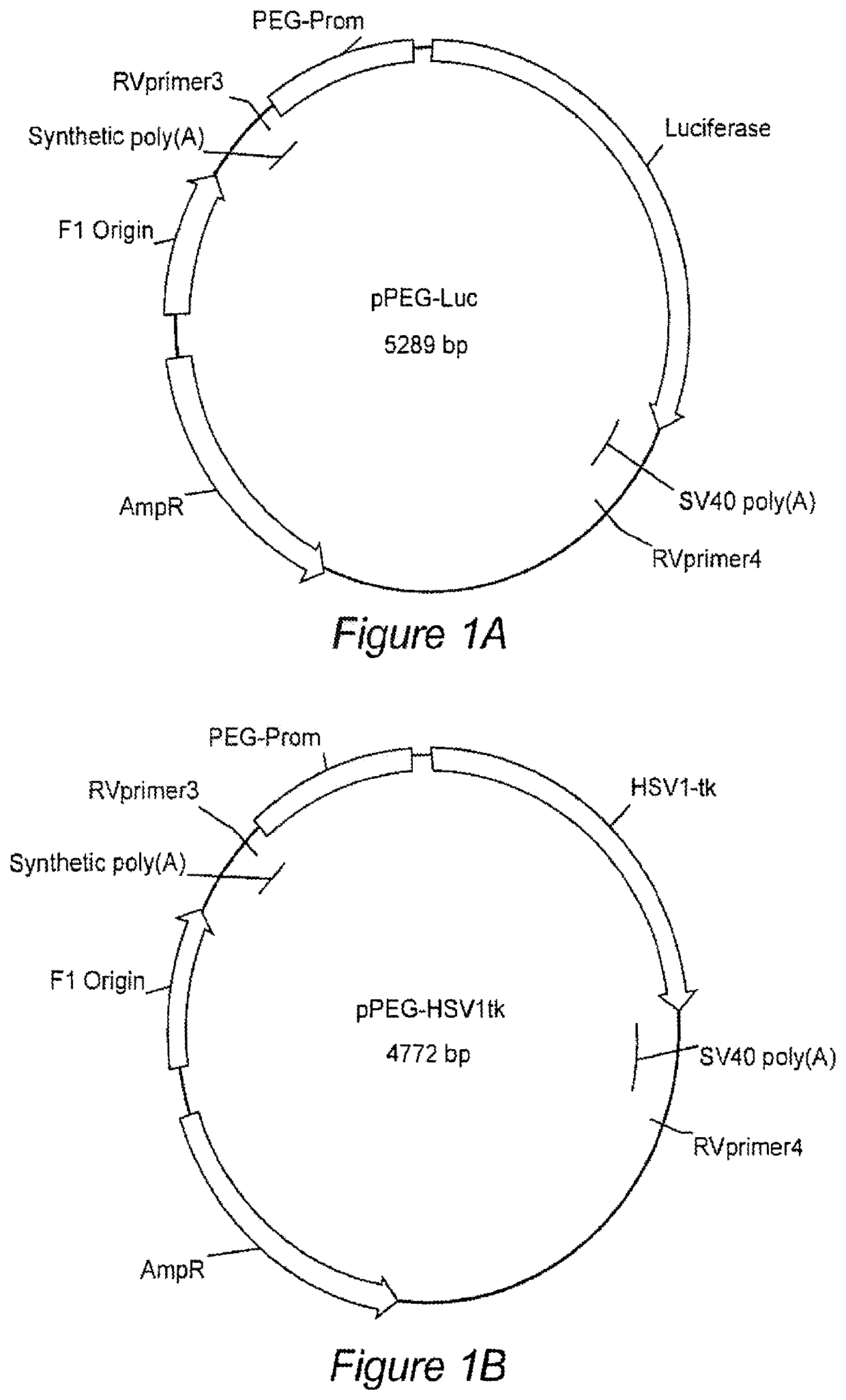
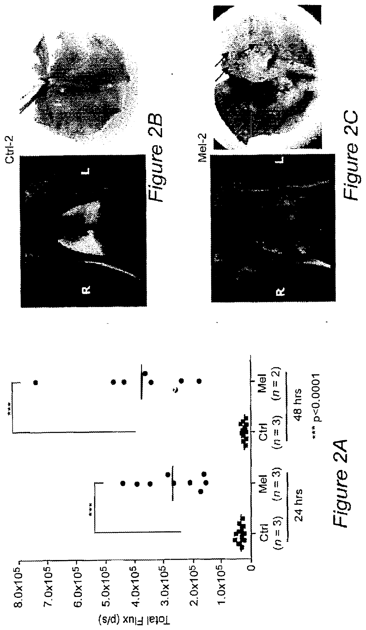
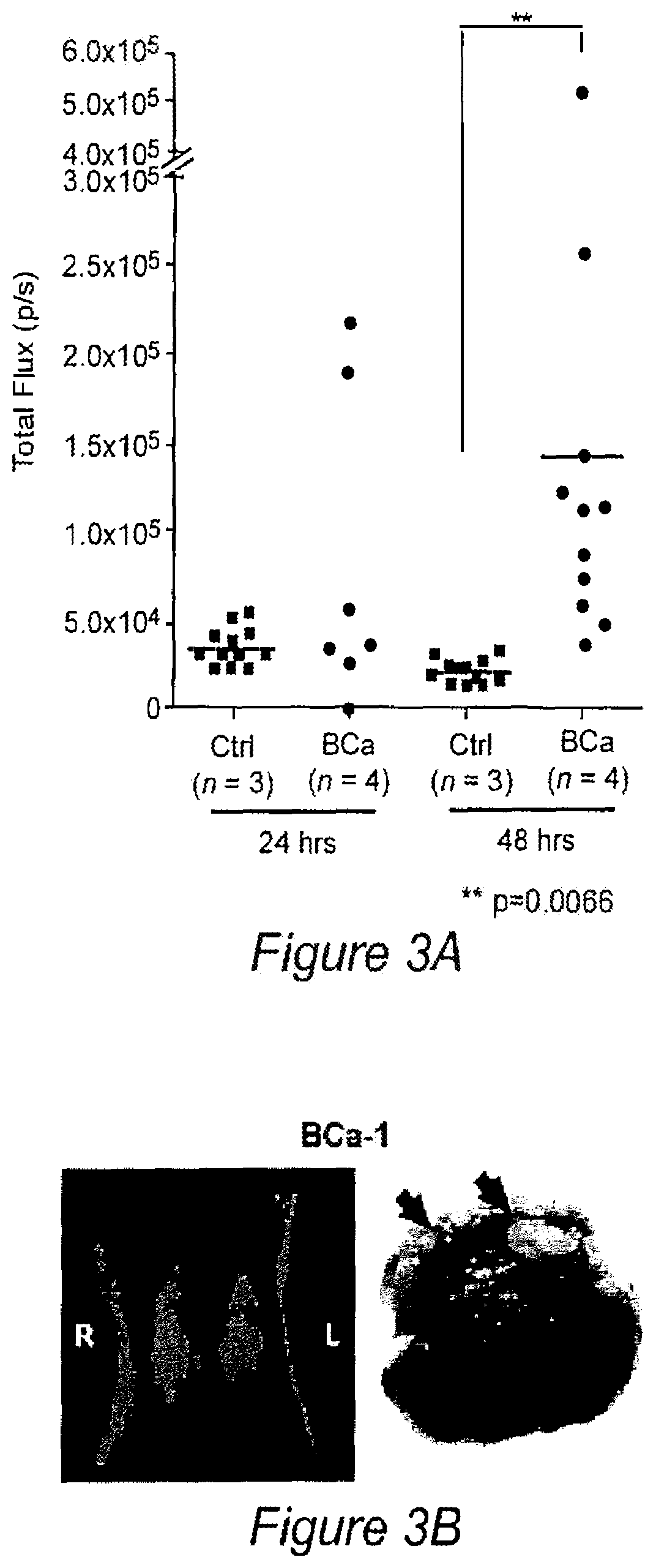
[0139]Targeted imaging of cancer remains a significant but elusive goal. Such imaging could provide early diagnosis, aid in treatment planning and profoundly benefit therapeutic monitoring. We identified the minimal promoter region of progression elevated gene-3 (PEG-Prom)1,2 derived from a rodent PEG-3 gene through subtraction hybridization', whose expression directly correlates with malignant transformation and tumor progression in rodent tumors3,4, as well as in human tumors, including cancer cell lines derived from tumors in the brain, prostate, breast, melanoma, and pancreas5-9. Based on these findings, we hypothesized and subsequently confirmed that systemic delivery of the PEG-Prom linked to and regulating an imaging construct would enable tumor-specific expression of reporter genes, not only within a primary tumor, but also in associated metastases in a manner broadly applicable to tumors of different tissue origin or subtype10. PEG-Prom is responsive directly to el...

example 3

References for Example 3

[0171]1. Bhang H-eC, Gabrielson K L, Laterra J, Fisher P B, Pomper M G. Tumor-specific imaging through progression elevated gone-3 promoter-driven gene expression. Nature medicine. 2011;17:123-1302,

[0172]2. Kuho H. Gardner T A, Wada Y, Koeneman K S, Gotoh A, Yang L, et al. Phase I dose escalation clinical trial of adenovirus vector carrying osteocalcin promoter-driven herpes simplex virus thymidine kinase in localized and metastatic hormone-refractory prostate cancer. Human gene therapy. 2003;14:227-41,

[0173]3. Su Z Z, Sarkar D, Emdad L, Duigou G J, Young C S, Ware j, et al. Targeting gene expression selectively in cancer cells by using the progression-elevated gene-3 promoter. Proceedings of the National Academy of Sciences of the United States of America. 2003;102:1059-64.

[0174]4. Lee S G, Su Z Z, Emdad L. Sarkar D. Fisher P B. Astrocyte elevated gene-1 (AEG-1) is a target gene of oncogenic Ha-ras requiring phosphatidylinositol 3-kinase and c-Myc. Proceedin...


PUM

PropertyMeasurementUnit
quantitative real time PCRaaaaaaaaaa
volumeaaaaaaaaaa
volumeaaaaaaaaaa

Abstract

Genetic constructs comprising reporter genes operably linked to cancer specific or cancer selective promoters (such as the progression elevated gene-3 (PEG-3) promoter and astrocyte elevated gene 1 (AEG-1) promoter) are provided, as are methods for their use in cancer imaging, cancer treatment, and combined imaging and treatment protocols, e.g. for imaging and / or treating spontaneous metastasis. Transgenic animals in which a reporter gene is linked to a cancer specific or cancer selective promoter, and which may be further genetically engineered, bred or selected to have a predisposition to develop cancer, are also provided.

Description

CROSS REFERENCE TO RELATED APPLICATIONS[0001]The present application is a continuation of U.S. patent application Ser. No. 14 / 182,690, filed Feb. 18, 2014, which is a continuation-in-part of U.S. patent application Ser. No. 13 / 881,777, filed Jun. 13, 2013, which claims the benefit of and priority to U.S. Provisional Patent Application No. 61 / 766,473, filed Feb. 19, 2013, which are herein incorporated by reference in their entireties for all purposes. U.S. patent application Ser. No. 13 / 881,777 is a U.S. National Stage Application under 35 U.S.C. § 371 of International Application No. PCT / US2011 / 058249, filed Oct. 28, 2011, which claims priority to and the benefit of U.S. Provisional Patent Application No. 61 / 407,714, filed Oct. 28, 2010, which are herein incorporated by reference in their entireties for all purposes.BACKGROUND OF THE INVENTIONSequence Listing[0002]The instant application contains a Sequence Listing which has been filed electronically in ASCII format and is hereby in...

Claims


Application Information

Patent Timeline
Patent Type & Authority Applications(United States)
IPC IPC(8): A61K51/04A61K49/00A01K67/027
CPCC12N2830/60A61K49/0045C12N2800/107A01K2267/0331A01K67/0275A61K49/0054A01K2227/105A01K2217/15A01K2267/0393A01K2217/052A61K51/0491A01K2217/206C12N2830/85
Inventor POMPER, MARTIN GILBERTBHANG, HYO-EUNFISHER, PAUL
Owner VIRGINIA COMMONWEALTH UNIV
Why Eureka
Try Eureka