Bispecific Anti-VEGF/Anti-ANG-2 Antibodies and their use in the Treatment of Ocular Vascular Diseases

Active Publication Date: 2014-01-16
ROCHE GLYCART AG
View PDF3 Cites 46 Cited by
  • Summary
  • Abstract
  • Description
  • Claims
  • Application Information

AI Technical Summary

Benefits of technology

[0044]The antibodies according to the invention have highly valuable properties due to their specific modifications in the Fc part/constant region causing a benefit for a patient suffering from ocular vascular diseases. They show high stability in the intravitreal environment and slow diffusion from the eye (compared to smaller antibody fragments without a constant heavy chain region), where the actual disease is located and treated (so treatment schedule can potentially be improved compared to non-IgG like antibodies like e.g. Fab and (Fab)2 fragments). Surprisingly compared to unmodified IgG antibodies the half-life in the eye after intravitreal application of the antibodies with the mutations I253A, H310A, and H435A in the constant region (with no more FcRn binding) was similar (only slightly reduced) (Tables 17a and 18a and FIGS. 7D and 7E), whereas the diffusion from the eye into the blood serum was similar (Table 15 and FIG. 7B). This highly valuable as it is desired for the treatment of ocular vascular diseases related to ANG2 and/or VEGF it to eliminate VEGF and Ang2 from the eye (e. via the transportation into the blood serum as anti-ANG2/ANG2 antibody complex or anti-VEGF/VEGF antibody complex). The antibodies according to the invention are cleared on the other hand quite rapidly from serum when compared to unmodifie

Problems solved by technology

They are the leading causes of visual loss in industrialized nations.
Since the retina consists of well-defined layers of neuronal, glial, and vascular elements, relatively small disturbances such as those seen in vascular proliferation or edema can lead to significant loss of visual function.
Ischemic retinopathies are characterized by loss or dysfunction of the retinal vasculature which results in a reduction of blood flow and hypoxia.
The retina responds to hypoxia by generating signals to grow new blood vessels, but these new vessels are usually fragile and disorganized.
It is the growth of these abnormal new vessels that creates most of the threat to vision since they can leak, hemorrhage or lead to scarring that may end in retinal

Method used


Image

  • Bispecific Anti-VEGF/Anti-ANG-2 Antibodies and their use in the Treatment of Ocular Vascular Diseases
  • Bispecific Anti-VEGF/Anti-ANG-2 Antibodies and their use in the Treatment of Ocular Vascular Diseases
  • Bispecific Anti-VEGF/Anti-ANG-2 Antibodies and their use in the Treatment of Ocular Vascular Diseases

Examples


example 1

Expression and Purification

Transient Transfections in HEK293-F System

[0273]The bispecific antibodies were generated by transient transfection with the respective plasmids (e.g. encoding the heavy and modified heavy chain, as well as the corresponding light and modified light chain) using the HEK293-F system (Invitrogen) according to the manufacturer's instruction. Briefly, HEK293-F cells (Invitrogen) growing in suspension either in a shake flask or in a stirred fermenter in serum-free FreeStyle™ 293 expression medium (Invitrogen) were transfected with a mix of the four expression plasmids and 293Fectin™ or fectin (Invitrogen). For 2 L shake flask (Corning) HEK293-F cells were seeded at a density of 1.0E*6 cells / mL in 600 mL and incubated at 120 rpm, 8% CO2. The day after the cells were transfected at a cell density of ca. 1.5E*6 cells / mL with ca. 42 mL mix of A) 20 mL Opti-MEM™ (Invitrogen) with 600 μg total plasmid DNA (1 μg / mL) encoding the heavy or modified heavy chain, respectiv...

example 2

Analytics & Developability

Small-Scale DLS-Based Viscosity Measurement.

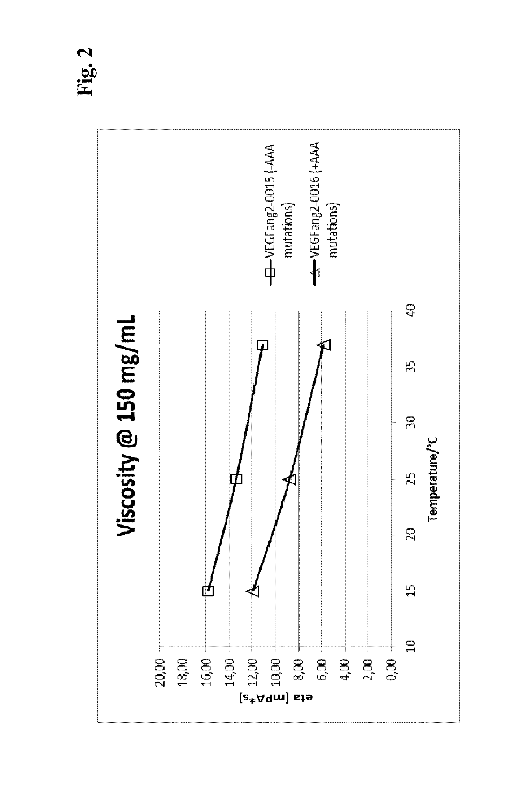
[0280]Viscosity measurement was essentially performed as described in (He, F. et al., Analytical Biochemistry 399 (2009) 141-3). Briefly, samples are concentrated to various protein concentrations in 200 mM arginine succinate, pH 5.5, before polystyrene latex beads (300 nm diameter) and Polysorbate 20 (0.02% v / v) are added. Samples are transferred into an optical 384-well plate by centrifugation through a 0.4 μm filter plate and covered with paraffine oil. The apparent diameter of the latex beads is determined by dynamic light scattering at 25° C. The viscosity of the solution can be calculated as η=η0(rh / rh,0) (η: viscosity; η0: viscosity of water; rh: apparent hydrodynamic radius of the latex beads; rh,0: hydrodynamic radius of the latex beads in water.

[0281]To allow comparison of various samples at the same concentration, viscosity-concentration data were fitted with the Mooney equation (Equation 1) (Mooney, Co...

example 3

Binding to VEGF, Ang2, FcgammaR and FcRn

VEGF Isoforms Kinetic Affinity Including Assessment of Species-Crossreactivity

[0288]Around 12000 resonance units (RU) of the capturing system (10 μg / ml goat anti human F(ab)′2; Order Code: 28958325; GE Healthcare Bio-Sciences AB, Sweden) were coupled on a CM5 chip (GE Healthcare BR-1005-30) at pH 5.0 by using an amine coupling kit supplied by the GE Healthcare. The sample and system buffer was PBS-T (10 mM phosphate buffered saline including 0.05% Tween20) pH 7.4. The flow cell was set to 25° C.—and the sample block set to 12° C.—and primed with running buffer twice. The bispecific antibody was captured by injecting a 50 nM solution for 30 sec at a flow of 5 μl / min. Association was measured by injection of human hVEGF121, mouse mVEGF120 or rat rVEGF164 in various concentrations in solution for 300 sec at a flow of 30 μl / min starting with 300 nM in 1:3 dilutions. The dissociation phase was monitored for up to 1200 sec and triggered by switching...


PUM

PropertyMeasurementUnit
Viscosityaaaaaaaaaa

Abstract

The present invention relates to bispecific antibody against human vascular endothelial growth factor (VEGF/VEGF-A) and against human angiopoietin-2 (ANG-2) of human IgG1 or IgG4 subclass with mutations I253A, H310A, and H435A, methods for their production, pharmaceutical compositions containing said antibodies, and uses thereof.

Description

CROSS REFERENCE TO RELATED APPLICATIONS[0001]This application claims the benefit of priority under 35 USC 119(a) to European patent application number 12 176299.1, filed 13 Jul. 2012, the disclosure of which is incorporated herein by reference in its entirety.SEQUENCE LISTING[0002]The instant application contains a Sequence Listing which has been submitted in ASCII format via EFS-Web and is hereby incorporated by reference in its entirety. Said ASCII copy, created on Jul. 10, 2013, is named P4996_Sequence_listing.txt and is 120,465 bytes in size.FIELD OF THE INVENTION[0003]The present invention relates a method for the reduction of the viscosity of an antibody (including a bispecific antibody) of human IgG1 or human IgG4 subclass, to bispecific antibodies against human vascular endothelial growth factor (VEGF / VEGF-A) and against human angiopoietin-2 (ANG-2), methods for their production, pharmaceutical compositions containing said antibodies, and uses thereof.BACKGROUND OF THE INVEN...

Claims


Application Information

Patent Timeline
IPC IPC(8): C07K16/22
CPCC07K2317/31C07K16/2863C07K2317/94A61K2039/505C07K2317/73C07K16/468C07K2317/56C07K2317/76C07K2317/732C07K2317/52C07K2317/90C07K16/22C07K2317/92A61K2039/54C07K2317/565C07K2317/33C07K2317/35C07K2317/21A61P27/06A61P27/02
Inventor DUERR, HARALDHERTING, FRANKKLEIN, CHRISTIANREGULA, JOERG THOMASRUETH, MATTHIASSTUBENRAUCH, KAY-GUNNAR
Owner ROCHE GLYCART AG
Why Eureka
Try Eureka