Blockstent device and methods of use

a block stent and catheter technology, applied in the field of medical devices, can solve the problems of limiting the fixation of the device, balloon migration and occlusion of non-target vessel segments, higher cost and longer treatment time, etc., to facilitate folding, compression or expansion, the effect of reducing the leakage of fluid

Active Publication Date: 2014-01-16
METACTIVE MEDICAL
View PDF36 Cites 41 Cited by
  • Summary
  • Abstract
  • Description
  • Claims
  • Application Information

AI Technical Summary

Benefits of technology

[0006]The walls of blockstents can be formed from a variety of expandable, rigid materials, preferably metals. The metal used to make the wall of a blockstent can be selected from the group consisting of gold, platinum, silver, titanium, vanadium, aluminum, nickel, tantalum, zirconium, chromium, silver, gold, silicon, magnesium, niobium, scandium, platinum, cobalt, palladium, manganese, molybdenum, alloys thereof, and / or combinations thereof. Other metals can be used so long as they are safe to use as an implanted medical device, can be formed into thin walls, and can be expanded from a compressed state and remain expanded in the body, holding their shape under typical conditions. Preferably, the blockstent is made of a ductile metal such as gold, platinum, silver, alloys thereof, and / or combinations thereof. In a fully expanded form, the blockstent can be configured in a variety of sizes and shapes, depending on the size and shape of the blood vessel to be treated, with preferable forms including a cylinder with rounded, hemispherical, or flat ends. Available shapes include, but are not limited to, cylindrical or oblong. Preferably, the blockstent can have an expanded diameter ranging from about 2 mm to about 30 mm. The oblong blockstent can have an expanded length of between about 5 mm to about 60 mm. The blockstent wall has a width, or thickness ranging from about 3 μm to about 180 μm. Such width allows for compression into a small volume and facilitates passage through blood vessels and catheters. For example, blockstents can be folded and compressed to a diameter small enough to pass through 3Fr to 12Fr catheters, such that small, medium, and large diameter blood vessels can be treated, or maneuvered through small vessels, including but not limited to cerebral arteries.
[0009]The surface of the blockstent wall can be configured to increase local thrombus formation and tissue growth into the blockstent wall in order to secure the blockstent in place and reduce the risk of blockstent migration. The wall of the blockstent can further be configured to release solutions that can include drugs, pharmacologically active molecules, or pharmacologic compositions, such as those that would increase the formation of local thrombus, stimulate cell proliferation or the production of extracellular matrix, or increase the rate or extent of tissue growth, such as tissue growth into pores, or around projections, of the wall of the blockstent.
[0014]In one embodiment, the blockstent has an interior layer located on the interior surface of the central layer or wall. The interior layer may be made from the same materials as the central layer, or can be made of different materials. The interior layer may be comprised gold, platinum, silver, alloys thereof, or combinations thereof. The interior layer may also be comprised of polymer, plastic, latex, rubber, an elastomer, fiber material, and combinations thereof. The interior layer can have a thickness ranging between about 0.1 μm to about 59 μm. Preferably, the interior layer may be an elastomeric coating that strengthens the wall, reduces the leaking of fluid from the blockstent during expansion, or facilitates folding, compression, or expansion of the blockstent.
[0015]In another embodiment, the blockstent may include two or more metal regions joined by a flexible polymer and / or elastomer joint. The joint allows for better maneuverability and increased trackability as the blockstent is advanced to the desired location. In other embodiments, the blockstent may include three or more metallic regions that are joined through two or more flexible joints.
[0016]The blockstent wall defines an opening that allows for the passage of fluid. An attachment between the blockstent and delivery device is formed whereby the void of the blockstent defined by the inner surface of the wall can be joined in fluid communication with the lumen of a hollow cylindrical member of the delivery device which is configured to allow for the proximal end of the lumen to accept a fluid source and for fluid to pass from the fluid source, through the lumen of the hollow cylindrical member of the delivery device, and into the void of the compressed blockstent, resulting in expansion of the blockstent.
[0028]The shape and size of the blockstent may be modified after expansion. For example, prior to separation from the delivery catheter, withdrawing fluid from the void of the blockstent can reduce the size of the blockstent. Also prior to separation, a force can be applied to the blockstent through the delivery catheter by advancing the delivery catheter forward or pulling the delivery catheter back, thus modifying the shape of the blockstent. After separation, an external force can be applied to the blockstent by inflating the balloon portion of a balloon catheter adjacent to the blockstent to modify the shape of the blockstent or push a portion of the blockstent towards a blood vessel. In certain embodiments, this can reduce the amount of blockstent that protrudes from the blood vessel into the lumen of the adjacent parent, or native, vessel. Also, the opening of the expanded blockstent can be sealed through a variety of methods, or left open.

Problems solved by technology

There are two major drawbacks to the use of detachable balloon catheters for blood vessel occlusion.
First, the balloons are made of polymers that generally resist tissue incorporation that limits fixation of the devices where they are placed.
Unfortunately, there is a substantial rate of balloon and valve failure, resulting in deflation.
Without tissue incorporation, balloon deflation can lead to balloon migration and occlusion of non-target vessel segments.
There are several major drawbacks to the use of metal coils and basket structures for blood vessel occlusion.
First, numerous coils are usually required to occlude the blood vessel segment, resulting in higher costs and longer treatment times. Second, coil placement is difficult to control, often resulting in coil placement in non-target vessel segments.
Third, coils only partially fill the blood vessel.
The accumulation of thrombus and scar tissue is required to occlude the blood vessel, a process that takes weeks to occur and is sometimes incomplete, often resulting in incomplete occlusion or recanalization and a failed treatment.
Although only a single basket structure is usually required to occlude a blood vessel segment, and the devices are generally easier to control, these devices only partially fill the blood vessel and require the accumulation of thrombus and scar tissue to occlude the blood vessel.
As with coils, this process that takes weeks to occur and is sometimes incomplete, often resulting in incomplete occlusion or recanalization and a failed treatment.

Method used


Image

  • Blockstent device and methods of use
  • Blockstent device and methods of use
  • Blockstent device and methods of use

Examples


Embodiment Construction

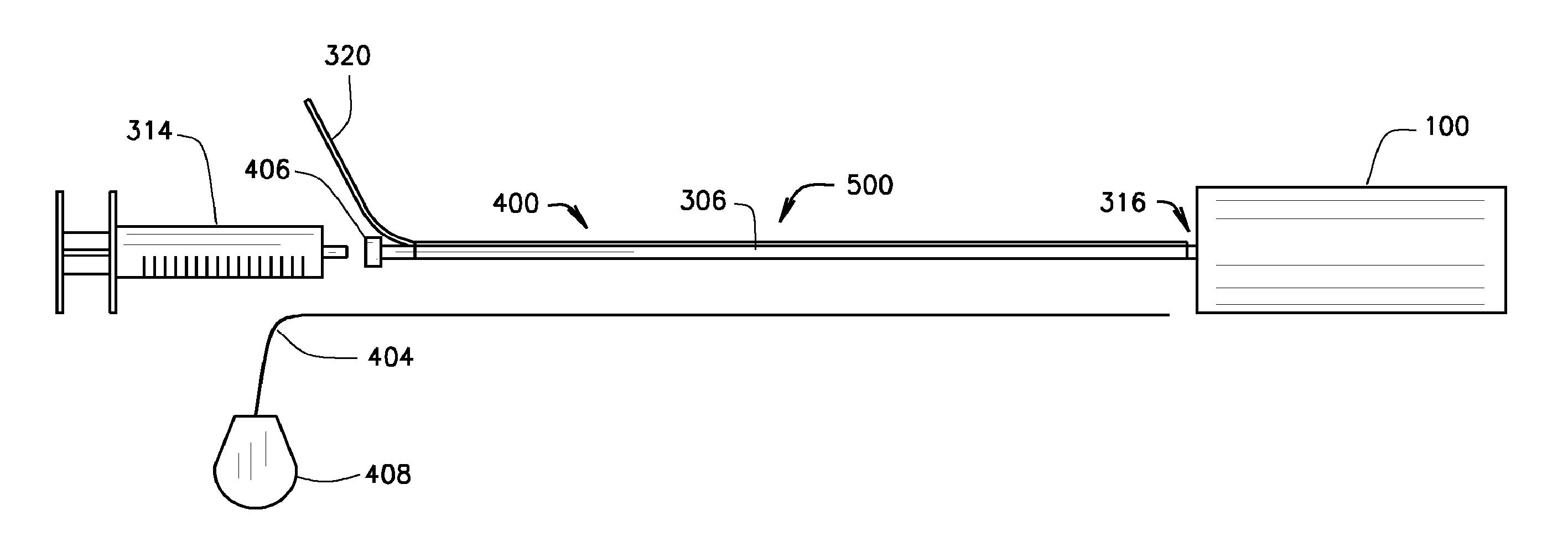
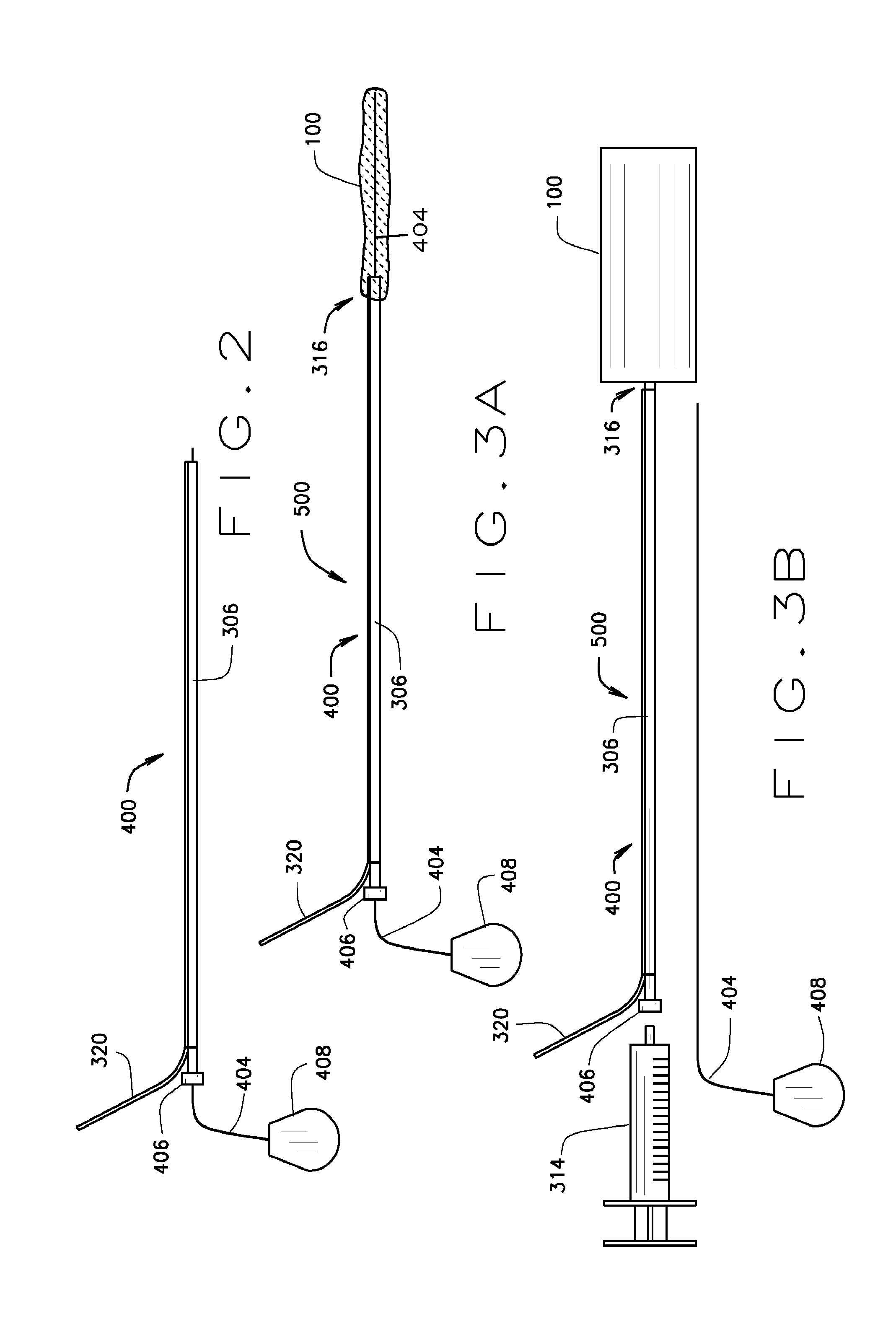
[0064]The present invention relates to a medical device comprising an expandable metal structure known as a “blockstent” and a delivery catheter. The blockstent is a thin-walled stent-like, cylindrical, device that can be expanded into a semi-rigid form that can remain in the body for an extended period. Specifically, the blockstent is configured for use in occluding segments of arteries, veins, and other biological conduits. The delivery catheter is configured to deliver the blockstent to a blood vessel and to provide a pathway, through a cylindrical member or lumen, for fluid to move into the central void of the blockstent, in order to expand it and fill at least a portion of the lumen of the blood vessel.

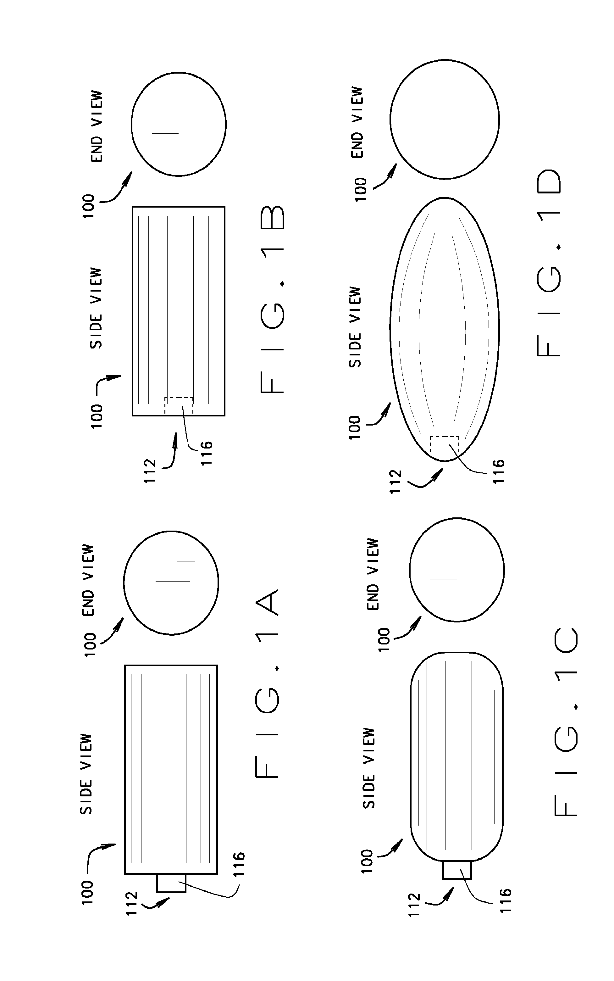
[0065]A cylindrical embodiment of the blockstent 100 with flat ends is shown in FIG. 1A in an expanded state. This embodiment has an external proximal neck 116 that defines an opening 112 for the passage of fluids, liquids, gases, or solids into the central void of the blockstent...


PUM

PropertyMeasurementUnit
diameteraaaaaaaaaa
lengthaaaaaaaaaa
thicknessaaaaaaaaaa

Abstract

What is disclosed is a medical device comprising a compressed, cylindrical or oblong, thin-walled, expandable metal structure (a “blockstent”) and a flexible, elongated device (a “delivery catheter”) to position the compressed blockstent into the lumen of a blood vessel segment to be treated, and methods of use for occlusion of treated blood vessel segments. A blockstent can be made with ductile metals such as gold, platinum, or silver such that the blockstent will conform to the shape of the lumen of the treated blood vessel segment during expansion and allow for the shape of the blockstent to be permanently changed by the application of an external force. The surface of the blockstent can be configured to promote local thrombus on the external surface of the blockstent and to promote the growth of tissue into the wall of the blockstent in order to occlude the treated blood vessel and fix the blockstent in place. The wall of the blockstent can also be configured to release drugs or pharmacologically active molecules such as those that promote thrombosis, cell proliferation, extracellular matrix deposition to promote this thrombus formation and tissue growth.

Description

CROSS REFERENCE TO RELATED APPLICATIONS[0001]This application claims priority to U.S. Provisional Application No. 61 / 433,305 filed on Jan. 17, 2011, which is incorporated by reference in its entirety.FIELD OF THE PRESENT DISCLOSURE[0002]The present disclosure relates to a medical device comprising a blockstent and a delivery catheter for the treatment of blood vessel segments of the vascular system. The present disclosure also relates to various forms of blockstents and delivery catheters, and methods of their manufacture. The present disclosure further relates to methods of occluding blood vessel segments using the various medical devices, whereby the blockstent ultimately remains in the blood vessel segment. Blockstents are cylindrical, thin-walled expandable metal structures comprised of a stent-like device and designed to fill the lumen of a blood vessel segment. Blockstents are configured for: attachment to delivery catheters, compression, advancement through the vascular syste...

Claims


Application Information

Patent Timeline
Patent Type & Authority Applications(United States)
IPC IPC(8): A61F2/958A61M25/10
CPCA61F2/958A61M25/1029A61B17/12031A61B17/12109A61B17/12131A61B17/12136A61B17/12113A61M31/00A61F2/06Y10T29/49826A61B2017/12054A61B2017/1205A61B2017/12068A61B2017/12063A61B2017/12059A61B2017/00526A61B2017/00893A61M2210/12A61B2090/037A61M2210/005A61F2250/0098A61B2090/3966A61B17/1214A61B17/12172A61B17/12177A61B17/12181A61B2017/00004A61B2017/00849A61B2017/00867A61B2017/00884A61B2017/12077A61B2017/22038
Inventor FRANANO, F. NICHOLASSTEPHENSON, KATHERINE J.
Owner METACTIVE MEDICAL
Why Eureka
Try Eureka