Image-guided removal and thermal therapy of breast cancer

a breast cancer and image-guided technology, applied in the field of post-surgical treatment of breast cancer, can solve the problems of inconvenient patient treatment, high cost of breast irradiation therapy, and still burdensome patients

Inactive Publication Date: 2014-01-23
DOWLATSHAHI KAMBIZ
View PDF25 Cites 24 Cited by
  • Summary
  • Abstract
  • Description
  • Claims
  • Application Information

AI Technical Summary

Benefits of technology

[0009]Methods, apparatus and compositions are disclosed to remove cancerous tissue and ablate at least a 1 cm wide zone of tissue surrounding the site of removal of cancerous tissue, such as a breast tumor, by heating for a short time using a “hot balloon.”

Problems solved by technology
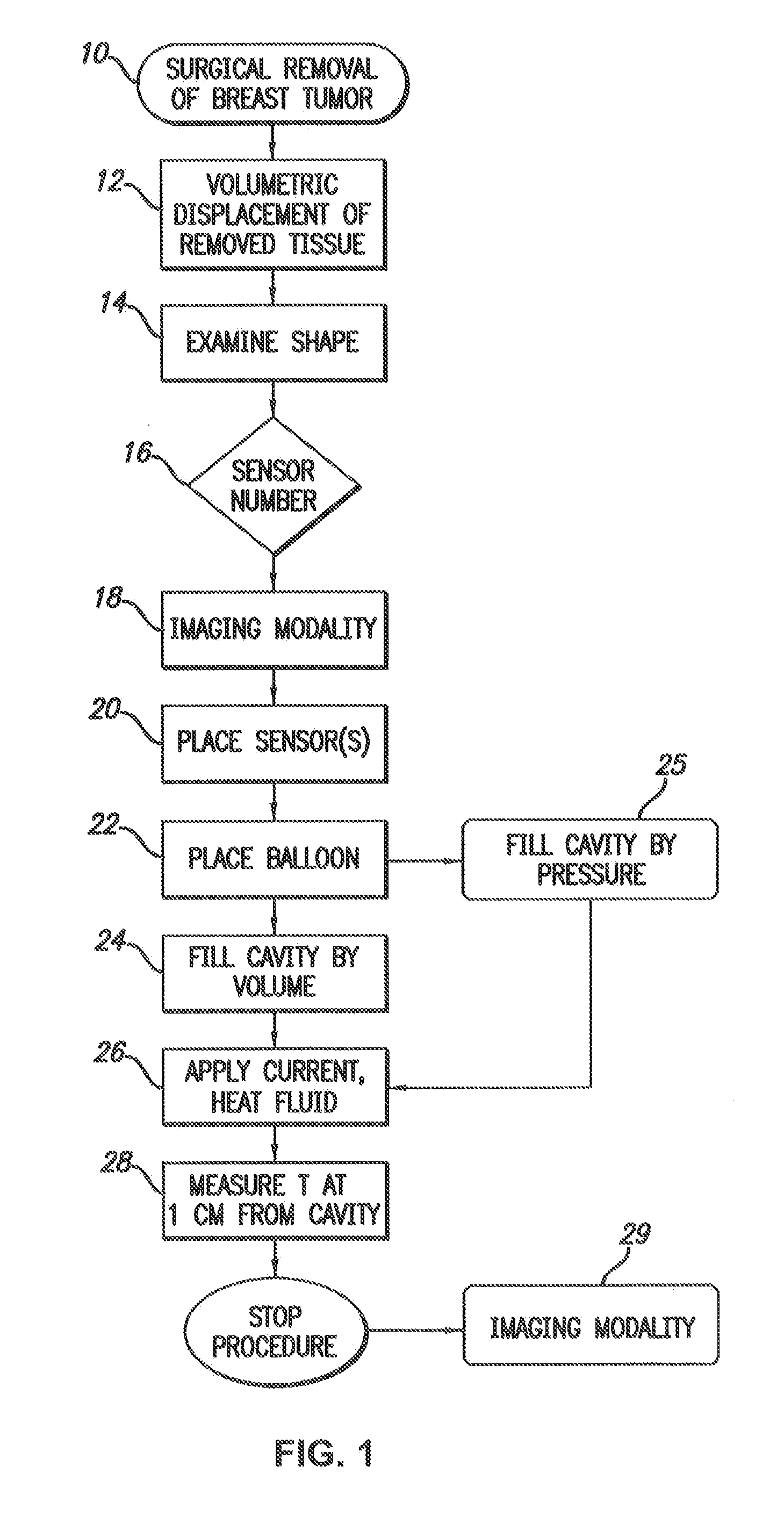
Although this approach shortens the treatment time and encourages more women to seek a breast saving operation, it is still burdensome for the patient to carry a balloon in the breast for a week receiving treatment twice a day.
Furthermore, the cost of the breast irradiation therapy remains high; an item which is part of the burgeoning healthcare budget.
This is inconvenient for the patient, and has the attendant risks and costs of radiation.
In particular, a patient with recurrent breast cancer who has already undergone radiation therapy, is unable to undergo further radiation therapy.

Method used


Image

  • Image-guided removal and thermal therapy of breast cancer
  • Image-guided removal and thermal therapy of breast cancer
  • Image-guided removal and thermal therapy of breast cancer

Examples


example 1

Thermal Ablation of the Goat Mammary Gland as a Model for Post-Lumpectomy Treatment of Breast Cancer

[0057]Background:

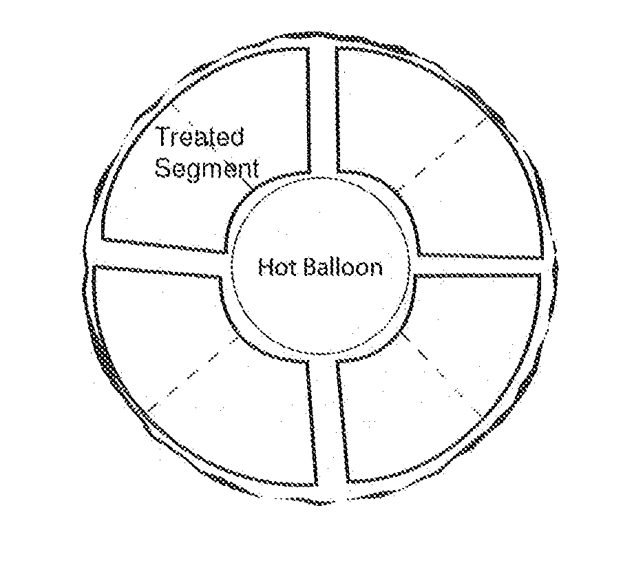
[0058]Partial breast irradiation (PBI) post-lumpectomy for carcinoma deploying the Mammosite balloon catheter is practiced as alternative to the whole breast irradiation in selected patients. A one centimeter shell of targeted breast tissue surrounding the lumpectomy cavity may be effectively treated with a balloon inflated with hot fluid instead of irradiation.

[0059]Methods:

[0060]45-50 kilogram Nubian-Cross goats in post-partum phase were selected. Under endo-tracheal anesthesia, the mammary glands measuring 4-7 cm in diameter were visualized by ultrasound and 2-3 cc of the gland were excised for histological documentation and creation of a space for the balloon. A ThermaChoice® deflated balloon (Ethicon) was inserted into the space and the incision was closed ensuring one cm thick tissue coverage. The balloon was inflated with 15-20 cc of 5% dextrose and pre-heated ...

example 2

Necrosis in a Cow Udder by Thermal Therapy

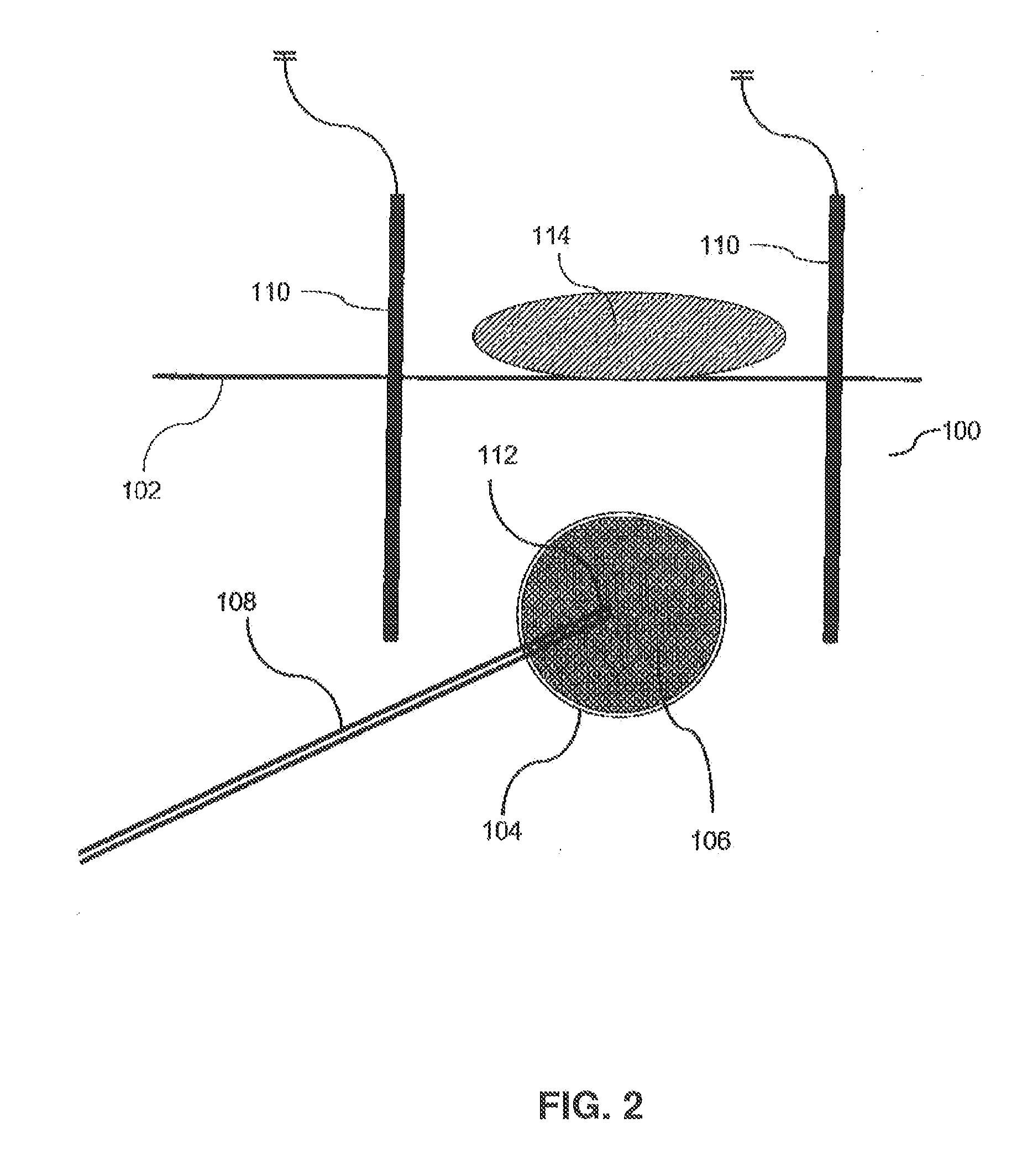
[0075]Fresh, resected bovine udders were obtained from a local slaughterhouse and tested ex vivo using the thermal therapy system. A deflated balloon was passed into the tissue through a small stab incision and was inflated with 30-50 cc of 5% dextrose in water. Thermal sensor needles were inserted into the tissue 1 cm from the balloon surface. The fluid in the balloon was heated to 95° C. The actual treatment commences after the temperature reaches 95° C. and finished promptly at 2 minute intervals. After the temperature of the sensors reached 60° C., the heating element was turned off and the fluid removed. The deflated balloon was then removed from the tissue. A deep incision was made in the tissue to the point of contact between the balloon and the tissue and the cavity was examined visually and under the microscope.

[0076]Analysis of the data included measuring the diameter of the cavity and measuring the thickness of tissue necrosis ver...

example 3

Experimental Model on a Stereotactic Table

[0077]A sheep udder placed in a sealed plastic bag was firmly held within the compression plates of the stereotactic table. Initial scout view was taken and a spot in the center of the udder was selected.

[0078]A powder was then created by boiling an egg, removing the shell, and crushing the shell into a powder. The powdered egg shell was then inserted into the sheep udder through an incision and the incision was thereafter closed. The powdered egg shell 250 can be seen in FIG. 9, which depicts images of the sheep udder taken at angles of 15 degrees from right and left sides of the stereotactic table. The images discussed with respect to this example were all taken in a similar manner. The powdered egg shell simulates microcalcifications seen in mammography of human breast as early sign of developing breast cancer. Again, a scout view of the “target” followed by stereo images were taken to determine its dimensions and coordinates within the u...


PUM


Abstract

A method of removing undesirable cells and killing cells surrounding a cavity caused by the removal of the undesirable cells includes the step of inserting a probe through an incision made in the skin of a patient. The method further includes the steps of removing undesirable cells using the probe and using image-guided technology and inserting a balloon into the cavity formed by the removal of the undesirable cells. Still further, the method includes the steps of conforming the balloon to a shape of the cavity and providing sufficient fluid to fill the cavity based on the volume and reaching a temperature in the fluid to achieve a temperature in the tissue surrounding the cavity that kills cells surrounding the cavity.

Description

[0001]This patent application is a continuation-in-part of U.S. Ser. No. 11 / 753,510, which is a non-provisional claiming priority under 35 U.S.C. §119(e) to U.S. Provisional Patent Application No. 60 / 808,064, filed May 24, 2006, and U.S. Provisional Patent Application No. 60 / 915,852, filed May 3, 2007, and further claims the benefit of U.S. Provisional Patent Application No. 61 / 792,127, filed Mar. 15, 2013, wherein all of such applications are expressly incorporated herein by reference in their entirety.BACKGROUND OF THE INVENTION[0002]Methods, apparatus and compositions are described for treating post-surgical breast cancer by applying heat to tissue surrounding a cavity remaining after surgical removal of cancerous tissue, thereby ablating remaining transformed cells and preventing metastasis of the transformed cells. Applications include use after lumpectomy and other forms of excision of small tumors detected by mammography.[0003]Widespread annual screening mammography has resul...

Claims


Application Information

Patent Timeline
Patent Type & Authority Applications(United States)
IPC IPC(8): A61B18/04
CPCA61B18/04A61B34/20A61B2017/22051A61B2018/0022A61B2018/00333A61B2018/00577A61B2018/00791A61B2018/046
Inventor DOWLATSHAHI, KAMBIZ
Owner DOWLATSHAHI KAMBIZ
Why Eureka
Try Eureka