Time Domain-Based Methods for Noninvasive Brain-Machine Interfaces

a brain-machine interface and time domain technology, applied in the field of brain computer interface systems, can solve the problems of long training time required by users to achieve satisfactory performance, inherent risks of surgery and gradual degradation of signal integrity, and the design strategy of controlling such prostheses, etc., to achieve the effect of reducing training tim

Active Publication Date: 2014-02-27
UNIV OF MARYLAND
View PDF2 Cites 71 Cited by
  • Summary
  • Abstract
  • Description
  • Claims
  • Application Information

AI Technical Summary

Benefits of technology

[0017]The present invention is directed to a BCI system that continuously decodes observed, imagined or actual movements from EEG signals with substantially reduced training time by a subject. The acquired signals may be utilized in a BCI system or a brain machine interface (BMI) for controlling a machine. The term machine as used herein refers to a device, tool or mechanism designed to serve a specific purpose or goal. Thus a machine may be a computer cursor on a display screen (e.g., as provided in a BCI), a robotic limb (e.g., a hand, arm or leg), a motorized wheelchair, a videogame, a digital phone, a switch, an automobile, or a motorized assistive device.
[0018]For example, using the disclosed noninvasive BCI system and observational learning, subjects were able to accomplish two-dimensional control of a cursor with performance levels comparable to those of invasive BCI systems. Compared to other studies of noninvasive BCI systems, training time was substantially reduced, requiring only a single session of decoder calibration (˜20 minutes) and subject practice (˜20 minutes). Further, standardized low-resolution brain electromagnetic tomography was utilized to reveal that the neural sources that encoded observed cursor movement may implicate a human mirror neuron system (MNS). These findings offer the potential to continuously control complex devices such as robotic arms with one's mind without lengthy training or surgery.
[0020]According to another aspect of the present invention, a noninvasive BMI system is provided that decodes the kinematics of natural hand movements from EEG signals. The disclosed method controls each degree of freedom in a prosthetic or orthotic device by simultaneously decoding multiple desired kinematic parameters from multi-channel noninvasive neural activity. The decoded signals thus serve as new signals for controlling neuromotor prostheses. Hand velocity was continuously extracted from signals collected during a three-dimensional (3D) center-out reaching task. To assure a realistic task, subjects were not cued. Rather, the subjects chose which target to acquire and when to initiate movement. Since EEG coupled with our decoding method facilitated the investigation of sensor contributions to decoding with high temporal resolution, the location of salient sensors across time lags was examined. Using standardized low-resolution brain electromagnetic tomography (sLORETA) (Pascual-Marqui (2002), supra, Methods Find Exp. Clin. Pharmacol., 24 (Suppl D): 5-12), the sources most involved in encoding hand velocity were estimated. Moreover, the relationship between decoding accuracy and movement variability was investigated.
[0021]According to one disclosed method, hand movement intentions are deciphered from neural data streams in humans. The inputs to the method include neural signals from the human brain in the form of noninvasive (EEG or MEG) or invasive (ECoG and multi-unit activity) data, and the outputs are the desired kinematic parameter trajectories (e.g., finger joint angles and velocities). The decoded outputs may be used to simultaneously control multiple degrees of freedom in a prosthetic or orthotic hand for medical (e.g., regaining hand function after an upper-limb amputation, atrophy, or paralysis), rehabilitation training (e.g., assistive or resistive movement therapies), and motor performance enhancement (e.g., remote control of robotic actions) applications in which an exoskeleton is not being permanently integrated with the body. Moreover, as the neural signals acquired noninvasively using scalp electroencephalography (EEG) and processed according to the disclosed methods in this invention are correlated with meaningful behavioral variables (e.g., sequence of spatial positions or spatial configurations of hand joint angles during grasping), confluence between the brain and the exoskeleton may be achieved. Thus, the disclosed method for noninvasive decoding of human (multi-joint) hand gesture patterns from noninvasive scalp EEG signals alone can achieve BMI confluence.
[0022]According to another aspect of the present invention, a noninvasive BMI system is provided that decodes the kinematics of human bipedal locomotion from EEG signals. The desired decoded signals, such as the predicted joint positions of the lower extremities, may be utilized for the control of wearable lower-limb robots or “exoskeletons” for medical (e.g., for restoration of walking and running after spinal cord injury, amyotrophic lateral sclerosis or traumatic brain injury), rehabilitation training (e.g., assistive or resistive movement therapies), and motor performance enhancement (e.g. military or industrial) applications in which the exoskeletons are not being permanently integrated with the body. In another implementation, the decoded neural signals may be utilized to remotely control a robot (e.g., a walking robot). Moreover, as the neural EEG signals are acquired and processed according to the disclosed methods, such data may be correlated with meaningful behavioral variables (e.g., sequence of spatial positions or spatial configurations of legs during walking), and confluence between the brain and the exoskeleton may be achieved. Thus, the disclosed methods for noninvasive decoding of human (multi-joint) bipedal locomotion patterns from noninvasive scalp EEG signals alone may achieve brain-machine confluence.

Problems solved by technology

However, designing strategies to control such prostheses, especially from neural activity, remains a challenge.
Some drawbacks of invasive BCI systems are the inherent risks of surgery and gradual degradation of signal integrity.
However, a limitation of conventional noninvasive BCI systems for two-dimensional control of a cursor, in particular those based on sensorimotor rhythms, is the lengthy training time required by users to achieve satisfactory performance.
However, little work has been done to continuously decode natural, multi-joint limb kinematics from neural signals acquired noninvasively.
However, such MEG systems are immobile and therefore unsuitable for practical BCI systems.
Researchers have not demonstrated continuous decoding of limb kinematics from EEG-based systems.
Further, it is generally thought that the signal-to-noise ratio, the bandwidth, and the information content of neural data acquired via noninvasive scalp EEG are insufficient to extract sufficiently detailed information about natural, multi-joint movements of the upper limb (Lebedev et al.
The problem of decoding hand gestures, which involves simultaneously decoding multiple finger joint angles, is relatively complex due to the many degrees of freedom involved.
Moreover, the methods used in many prior studies have involved invasive procedures required to measure neural signals.
Other studies have indicated that, in the case of noninvasive EEG signals, signal averaging must be used over many movement attempts to extract a usable signal and the extracted signal cannot be employed to reproduce a time-varying arm trajectory.
While the evolution of exoskeleton hardware has proceeded at a rapid pace, controlling these devices has lagged significantly behind.
However, such devices are not scalable as a human-machine solution for minimizing the time between thought and action as well as for patients with severe injuries who need some assistance just to lift or move their limbs.
Unfortunately, this strategy has fails to work for patients with severe motor impairment who do not generate enough neuromuscular activity to be captured by sEMG.
Moreover, sEMG patterns are severely compromised by neurological disease, injury or noise during movement rendering them difficult to decode accurately.
However, such invasive technology is not likely to be an acceptable solution for exoskeletons for use by humans.

Method used


Image

  • Time Domain-Based Methods for Noninvasive Brain-Machine Interfaces
  • Time Domain-Based Methods for Noninvasive Brain-Machine Interfaces
  • Time Domain-Based Methods for Noninvasive Brain-Machine Interfaces

Examples


example 1

Methods and Materials

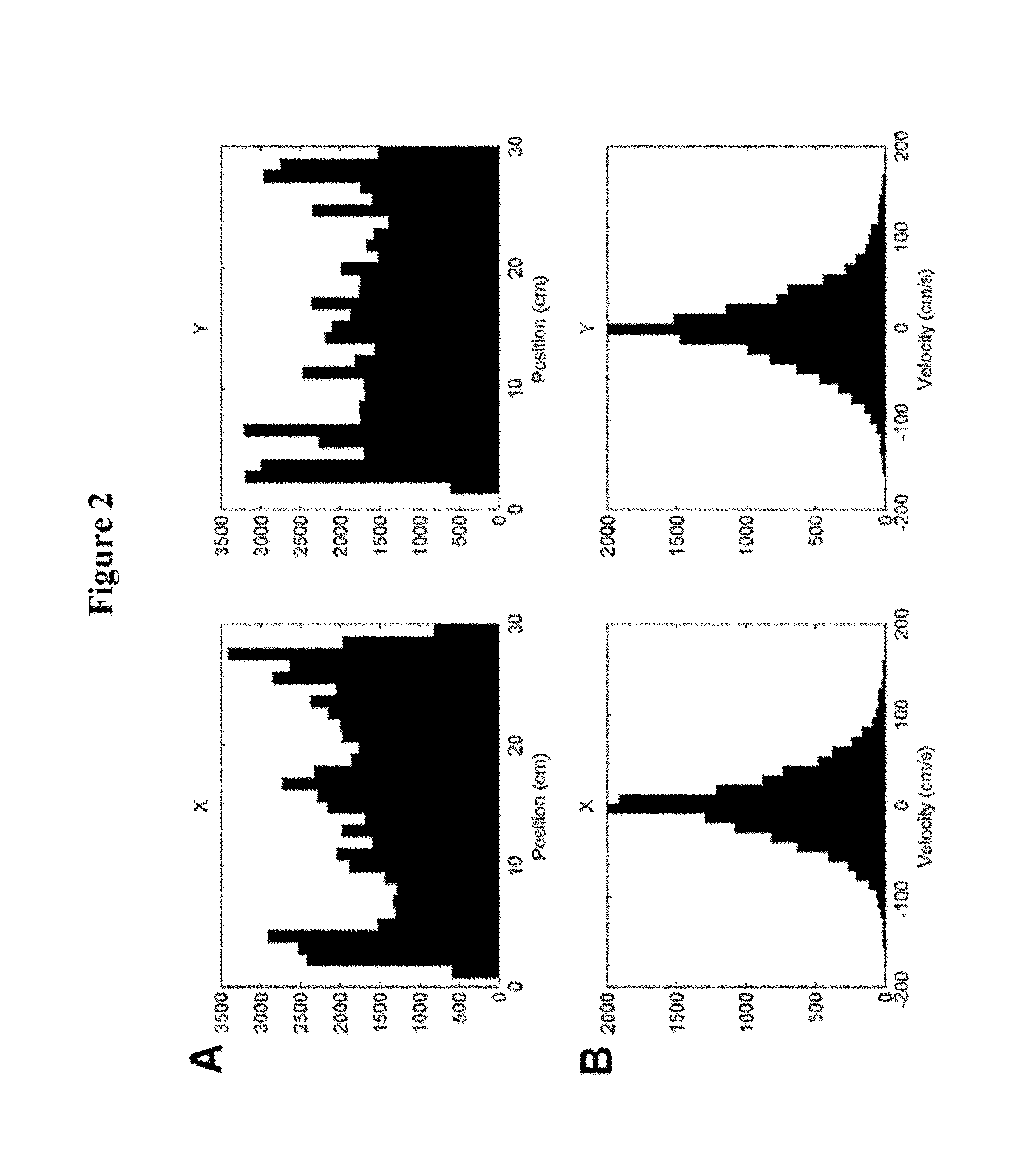
[0071]Five healthy, right-handed subjects performed a three-phase task: calibration, practice, and target acquisition. None of the subjects had previously participated in a BCI study. In all phases, the subjects EEG signals were acquired while they sat upright in a chair with hands resting in their laps at arm's length away from a computer monitor that displayed a workspace of dimensions 30×30 cm and a cursor of diameter 1.5 cm (0.20% of workspace). The subjects were instructed to remain still and relax their muscles to reduce the introduction of artifacts into the EEG recordings.

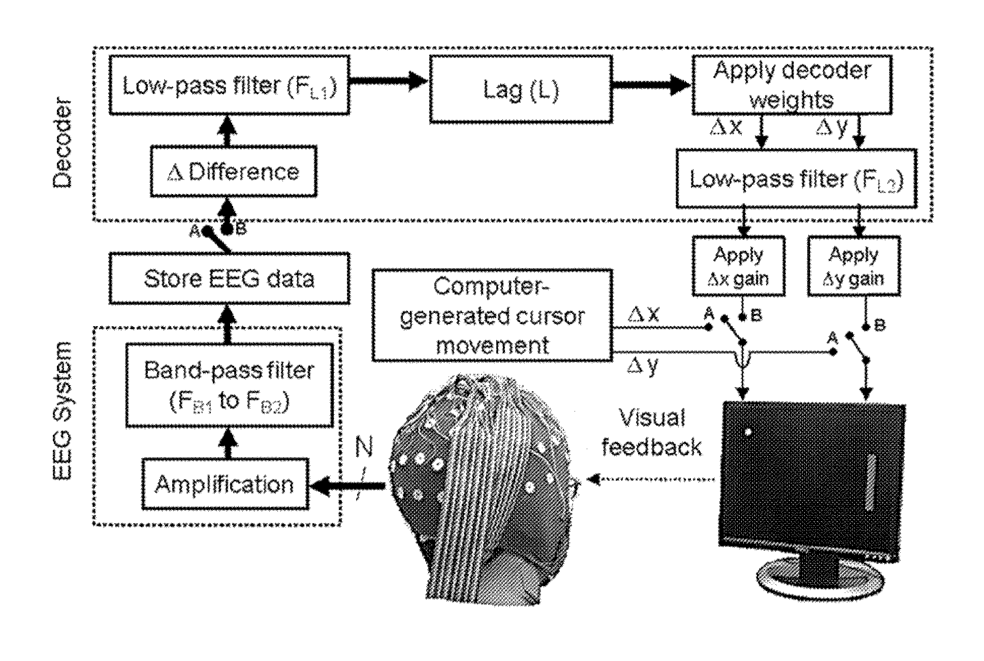
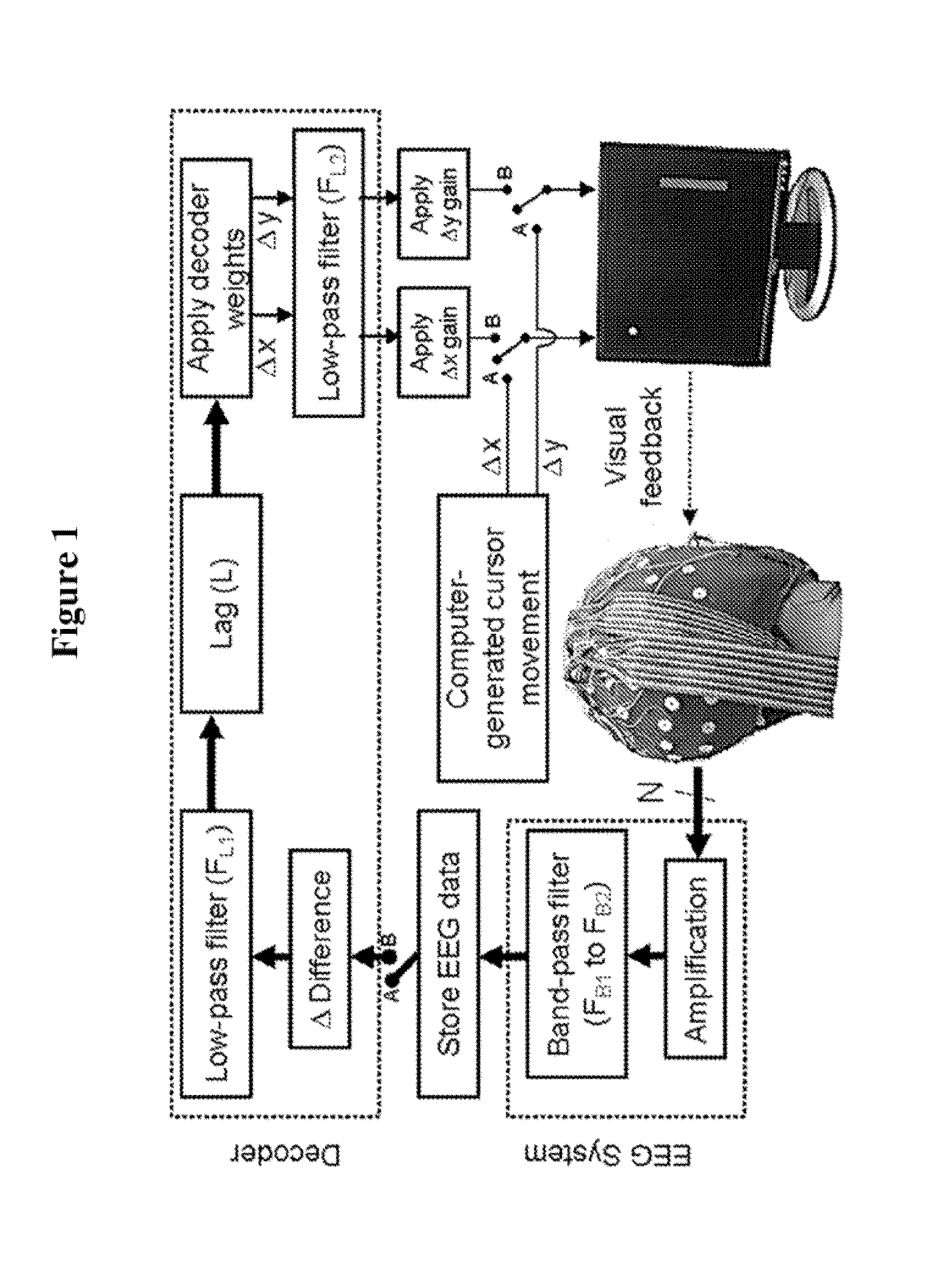
[0072]A diagram of data processing flow for the EEG-based BCI system according to the first embodiment is illustrated in FIG. 1. When the switches are in position A, the system is in an observation / calibration mode. In the observation / calibration mode, a subject observes a replay of a pilot subject's cursor movements on a computer screen while data from a plurality N (e.g. 34) of EEG se...

example 2

Methods and Materials

[0127]Referring to FIG. 10, five healthy, right-handed subjects sat upright in a chair and executed self-initiated, center-out reaches to self selected push-button targets near eye level. The subjects were instructed to attempt to make uniformly distributed random selections of the eight targets without counting. Subjects were further instructed to fixate on an LED on the center target throughout data collection and to only blink when their hand was resting at the center target. To ensure the minimization of eye movements, a researcher monitored the subjects' eyes during data collection, and the correlation between electroocular activity and hand kinematics was analyzed off-line (available at www.jneurosci.org as supplemental material). For each subject, the experiment concluded after each target was acquired at least 10 times.

[0128]A 64-sensor cap for sensing EEG signals (e.g., such as available from Electro-Cap International Inc. of Eaton, Ohio) was placed on ...

example 3

Methods and Materials

[0159]Five healthy, right-handed subjects sat upright in a chair and executed self-initiated, center-out grasping tasks. In particular, the subjects were seated behind a table with five objects (calculator, CD, espresso cup, zipper and a beer mug) arranged in front of them in a semicircle with an approximate radius of 30 cm. The initial position of the hand was palm down and flat on the table at the center of the semicircle. On the presentation of an auditory “go” cue (100 ms tone at 2 kHz), the subjects were instructed to select, reach out and grasp any of the five objects. Subjects kept a steady grasp on the objects until an auditory “stop” cue (200 ms tone at 1 kHz) was presented 5 s after the “go” cue, on hearing which they returned their hand to the resting initial position.

[0160]The time until the presentation of the “go” cue for the next trial was Gaussian distributed with a mean of 7 s and standard deviation of 1 s. Five blocks of 12 minutes each were re...


PUM


Abstract

A noninvasive brain computer interface (BCI) system includes an electroencephalography (EEG) electrode array configured to acquire EEG signals generated by a subject. The subject observes movement of a stimulus. A computer is coupled to the EEG electrode array and configured to collected and process the acquired EEG signals. A decoding algorithm is used that analyzes low-frequency (delta band) brain waves in the time domain to continuously decode neural activity associated with the observed movement.

Description

CROSS-REFERENCE TO RELATED APPLICATIONS[0001]This application is based on U.S. Patent Application Ser. No. 61 / 331,664, filed May 5, 2010, and U.S. Patent Application Ser. No. 61 / 369,128, filed Jul. 30, 2010, which applications are both incorporated herein by reference in their entireties and to which priority is claimed.STATEMENT REGARDING FEDERALLY SPONSORED RESEARCH OR DEVELOPMENT[0002]The U.S. Government has a paid-up license in this invention and the right in limited circumstances to require the patent owner to license others on reasonable terms as provided for by the terms of N000140910126 awarded by the Office of Naval Research.FIELD OF THE INVENTION[0003]The present invention relates to brain computer interface systems which continuously decode observed, intended or self-generated movements from non-invasively acquired neural signals.BACKGROUND OF THE INVENTION[0004]Developments in the fields of robotic prosthesis have made it possible to build devices that mimic a human in i...

Claims


Application Information

Patent Timeline
Patent Type & Authority Applications(United States)
IPC IPC(8): A61F2/72A61B5/04A61B5/0478A61B5/0484
CPCA61F2/72A61B5/0478A61B5/04012A61B5/04842A61B5/246A61B5/378A61B5/398G06F3/013G06F3/015A61B5/291A61B5/316
Inventor CONTRERAS-VIDAL, JOSE L.BRADBERRY, TRENT J.GENTILI, RODOLPHE J.AGASHE, HARSHAVARDHAN
Owner UNIV OF MARYLAND
Why Eureka
Try Eureka