

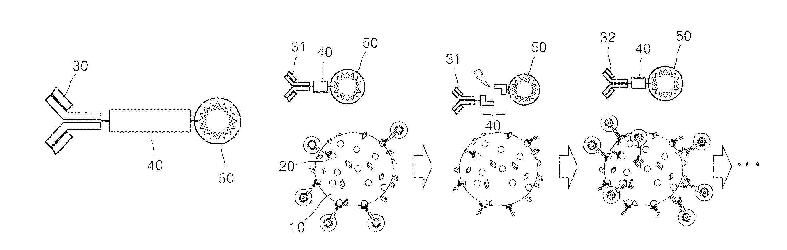
Method of sequential and multiple immunostaining for detection of various antigens in the same specimens
a technology of immunostaining and specimens, applied in the field of sequential and multiple immunostaining for detection of various antigens in the same specimen, can solve the problems of small size, inability to guarantee the identification of any cancer cells present in tissues, and inability to accurately identify the metastasis of a given cancer to another part of the body
- Summary
- Abstract
- Description
- Claims
- Application Information
AI Technical Summary
Benefits of technology
Problems solved by technology
Method used
Image

Examples
example 1
Cancer Cell Labeling Via Antibody-Photocleavable (PC) Linker-Complex
[0044]10 μL of self-manufactured 50 nmol heterobifunctional photocleavable linker (Chemical formula shown below) in DMF and 15 μL of 50 nmol dye solution (Fluorescein PEG Thiol (MW 5000), Rhodamine PEG Thiol, (MW 5000), Nanocs Inc.) in phosphate buffer (50 mM, pH 5) were reacted at room temperature for 30 minutes.
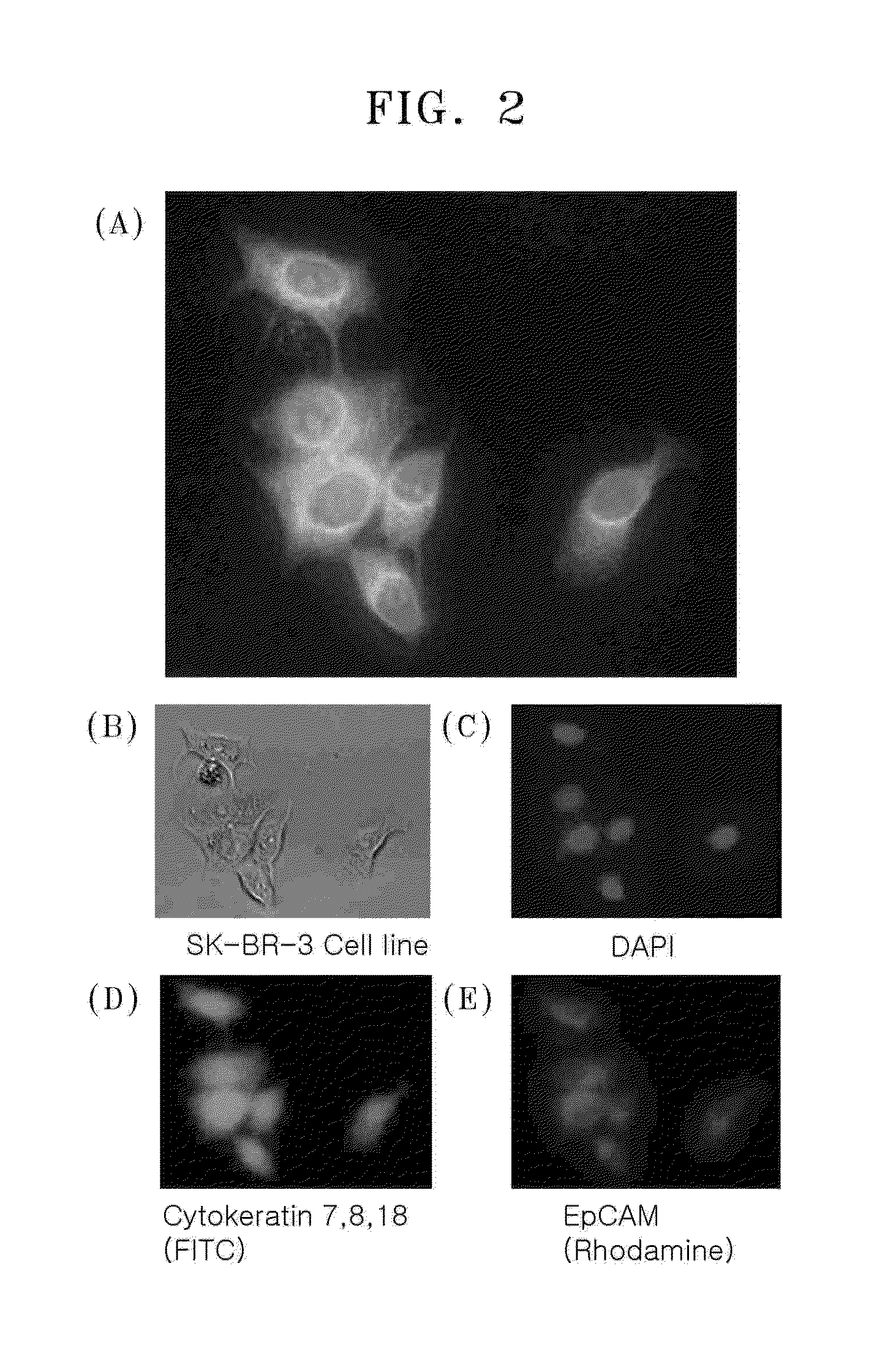
[0045]100 μL of 10 nmol anti-cytokeratin 7,8,18 antibody or anti-EpCAM antibody in 1× PBS solution was added, and the mixture was reacted overnight at 4° C. Then, unreacted dyes were removed using Amicon® Ultra centrifugal filter (Millipore, MW 30,000) and the resulting product was concentrated 5-fold. 5 μL of the obtained antibody-PC linker-complex was added to a PBS buffer solution with 1% BSA. The mixture was added to SK-BR3 breast cancer cells and stirred at 15 rpm for 1 hour. Binding between SK-BR3 and the complex was confirmed by fluorescence microscope (Olympus IX81).
[0046]FIG. 2A shows a merged imag...
example 2
Verification of Dye Dissection According to Light Exposure
[0047]BT474 breast cancer cells were fixed with 4% paraformaldehyde solution for 10 minutes and then treated with 0.2% Trition-X 100 for 10 minutes to increase permeability of the cells. After adding 1% BSA solution, the cells were stained with an anti-cytokeratin antibody—PC linker—FITC complex or anti-IGFR antibody—PC linker—Rhodamine complex at room temperature for 60 minutes. Then, the cells were washed with 1× PBS and observed under fluorescence microscope while increasing the intensity of irradiation (J) of light at a wavelength of 365 nm.
[0048]FIG. 3 shows the change in fluorescence signal of FITC and Rhodamine dyes bound to BT474 breast cancer cells according to light exposure. It was confirmed that as the intensity of irradiation increased, the intensity of fluorescence signal decreased and, thus, the ratio of dye being removed increased.
example 3
Staining of Reset Cells
[0049]SK-BR3 breast cancer cells were fixed with 4% paraformaldehyde solution for 10 minutes and then treated with 0.2% Trition-X 100 for 10 minutes to increase permeability of the cells. After adding 1% BSA solution, the cells were stained with an anti-cytokeratin antibody—PC linker—FITC complex or anti-IGFR antibody—PC linker—Rhodamine complex at room temperature for 60 minutes. Then, the cells were washed with 1× PBS, exposed to light (20 J) at 365 nm, and then respectively stained with anti-EpCAM antibody—PC linker—FITC complex or anti-EGFR antibody—PC linker—Rhodamine complex at room temperature for 60 minutes, and the intensity of fluorescence signal was measured according to each dye complex. The cells in the control group were subject to the same process as described above in regard to fixing the cells, increasing permeability of the hcells, and BSA blocking, followed by staining the cells with an anti-EpCAM antibody—PC linker—FITC complex or anti-EGFR...
PUM
| Property | Measurement | Unit |
|---|---|---|
| Fluorescence | aaaaa | aaaaa |
Abstract
Description
Claims
Application Information