
Methods for screening inhibitors of tumor associated protein aggregation

Active Publication Date: 2014-06-19
VLAAMS INTERUNIVERSITAIR INST VOOR BIOTECHNOLOGIE VZW +2
View PDF1 Cites 9 Cited by
  • Summary
  • Abstract
  • Description
  • Claims
  • Application Information

AI Technical Summary

Benefits of technology

The patent text discusses the discovery of a new way to treat cancer caused by mutated p53 proteins. These mutations can lead to the formation of aggregates, which can inactivate other proteins and promote tumor growth. The patent describes a screening method to find compounds that can prevent the aggregation of p53 and other proteins in the tumor. This method can help to develop new treatments for p53-related cancers. The patent also suggests that this concept can be extended to other tumor-suppressor proteins, such as PTEN and p16, and even to wild-type proteins in certain tumor contexts.

Problems solved by technology

Although favorable, this would only functionally restore a limited number of mutants.
Despite the therapeutic potency of strategies stabilizing structural p53 mutants, it is noted that, in murine models, prolonged treatment might favor the development of p53-resistant tumors or result in premature aging in some murine models.
However, it should be remarked that adenovirus-based gene therapy has several draw-backs: it is not expressed for long-term, has a limited packaging capacity to express other genes, and it spreads slowly and works poorly when injected intravenously.
But, more importantly, adenoviruses, even inactivated, can prompt an immune response, which already resulted in the death of a patient treated with adenovirus gene therapy.
Accordingly, it has recently been shown that disruption of the mutant p53 and p73 complex by small peptides consequently restores p73 activity.61 Notwithstanding this potential, such a strategy is severely complicated by the occurrence of many isoforms of p63 and p73 and the residing unclearness about the exact role of p63 and p73 in tumor progression.

Method used


Image

  • Methods for screening inhibitors of tumor associated protein aggregation
  • Methods for screening inhibitors of tumor associated protein aggregation
  • Methods for screening inhibitors of tumor associated protein aggregation

Examples


example 1

Structurally Destabilized p53 Mutants Aggregate In Vitro

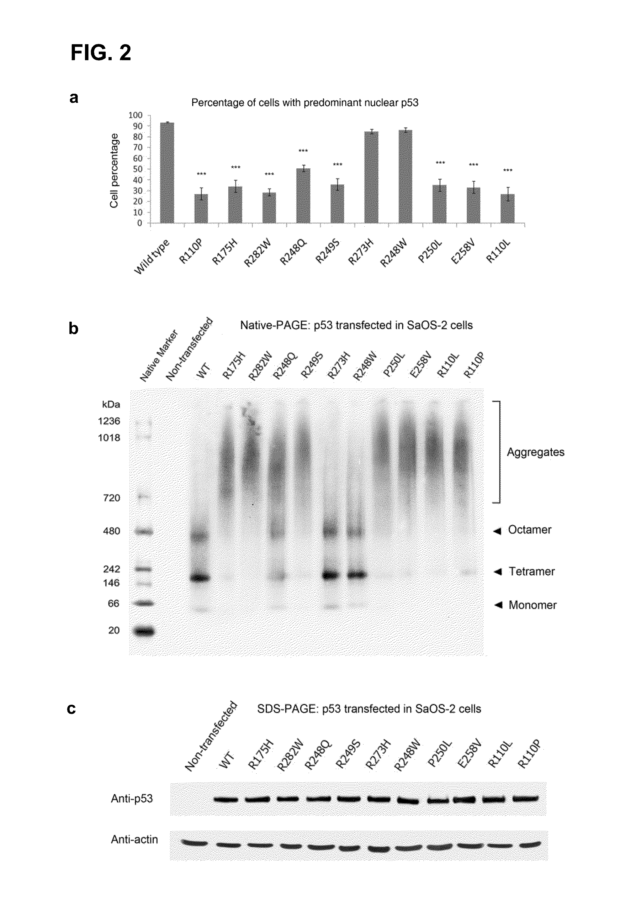
[0155]In order to investigate the effect of contact and structural mutations in p53 on their cellular distribution, wild-type and mutant p53 in the human osteosarcoma SaOS-2 cell line, which is devoid of endogenous p53, was first transiently over-expressed. Immunofluorescence revealed a predominant nuclear distribution of wild-type p53 and of the DNA-contact mutants R248W and R273H. In contrast, p53 mutants R175H, R282W, R249S, R248Q, P250L, E258V, R110L and R110P showed reduced nuclear staining, with a compensatory increase of cytoplasmic staining (data not shown; and FIG. 2, panel a), with the latter regularly containing “punctate” cytoplasmic spots. A punctate staining suggested the assembly of mutant p53 into large aggregates within the perinucleus, and was consistent with an impaired nuclear import of p53.14

[0156]To further investigate the aggresomal nature of the observed inclusions, several strategies were adopted. (i) ...

example 2

Analysis of the Aggregation Propensity of p53

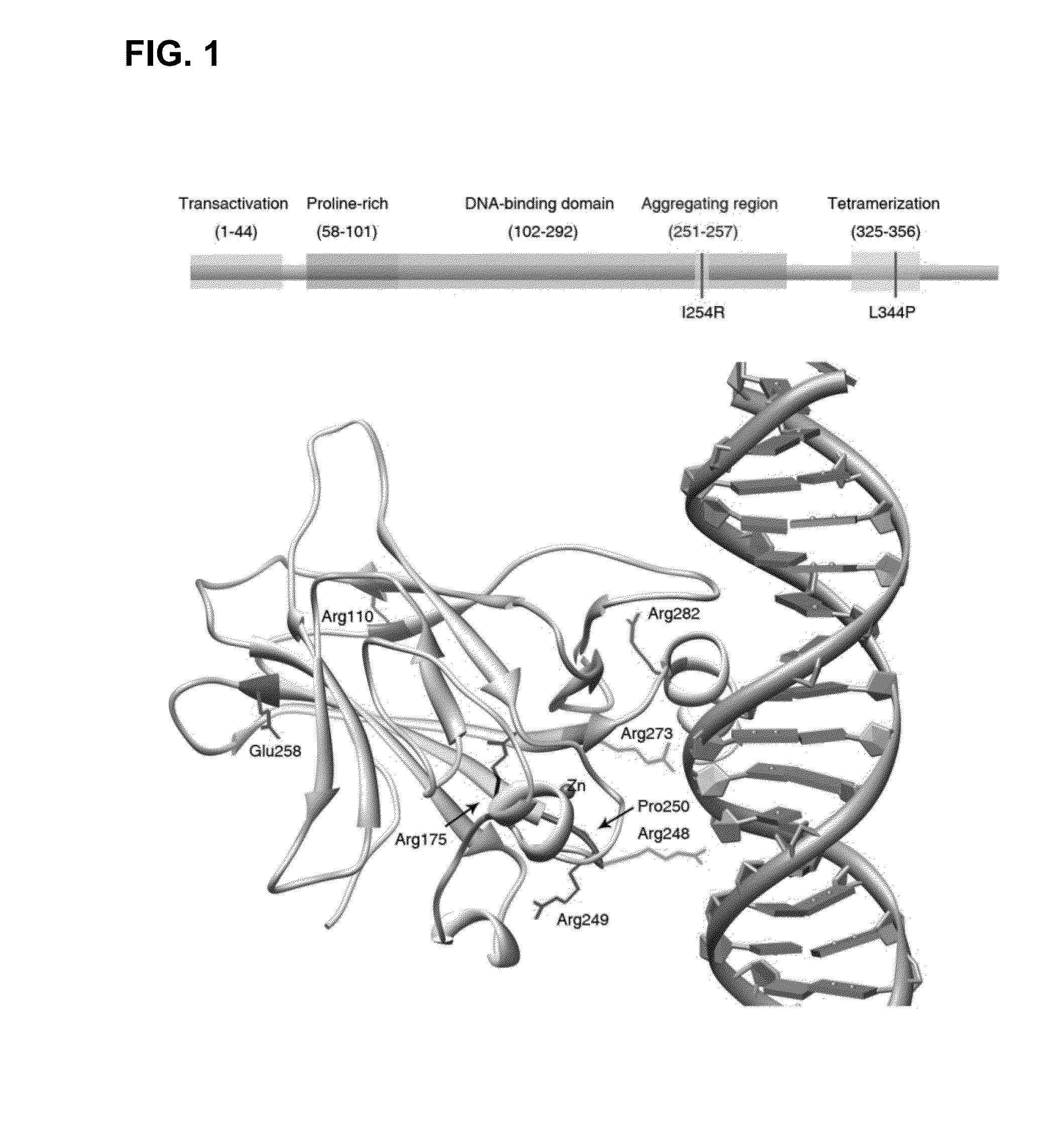
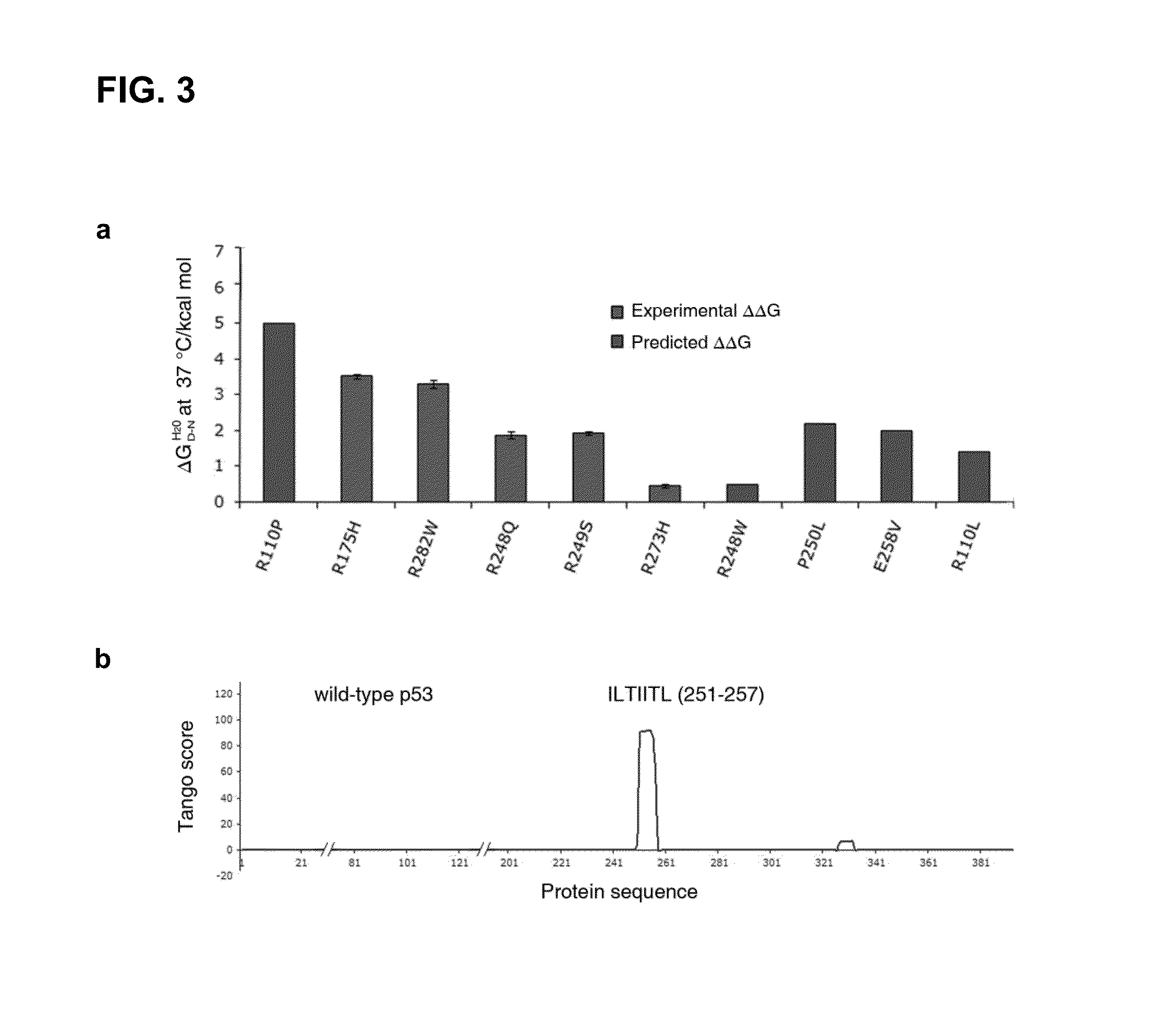
[0157]To better understand why structurally destabilized mutations in p53 would induce aggregation, TANGO,17 an algorithm to predict protein aggregation sequences, was used to identify regions in the protein that would be prone to aggregation. Here, an aggregation-nucleating segment was identified that spans residues 251 to 257 (ILTIITL; SEQ ID NO:2) in the hydrophobic core of the p53 DNA-binding domain (DBD). In the native structure, these residues form a 13-strand that is an integral part of the hydrophobic core of the p53 DBD (FIG. 1). Mutations that destabilize the tertiary structure of the DBD are, therefore, likely to increase the exposure of regions that are normally buried in the hydrophobic core,18 such as the aggregation-nucleating region, and, therefore, also prone to trigger aggregation of the p53 protein by assembly of the aggregation-nucleating stretch into an intermolecular β-sheet-like structure (FIG. 3).

[0158]To retrieve ...

example 3

Interfering with the Aggregation Propensity of p53

[0160]To further reveal the significance of this aggregating zone, an in silico analysis was performed using TANGO, searching for additional mutations that would abrogate the aggregation propensity of that zone. Several mutations with such characteristics were identified and introduced into the p53 expression vectors. An example of such mutation is residue 254, present in the middle of the aggregation nucleus, which were mutated from a hydrophobic to a positive charge (I254R) (FIG. 5, panel a). By introducing this additional mutation, suppressed aggregation of these p53 mutants was expected.

[0161]Indeed, when analyzing the double mutant p53E258V / I254R and p53R175H / I254R proteins, on BN-PAGE after transient over-expression in SaOS-2 cells, both failed to aggregate; instead, they were efficiently degraded as indicated by the significant reduction in overall mutant protein levels compared to the wild-type (FIG. 5, panel b). Importantly,...


PUM

PropertyMeasurementUnit
Fractionaaaaaaaaaa
Dynamic viscosityaaaaaaaaaa
Fluorescenceaaaaaaaaaa

Abstract

The disclosure relates to the fields of protein aggregation diseases including cancer. More specifically, it concerns a screening method for identifying compounds that inhibit or disrupt co-aggregation of one or more member proteins of a disease-related protein aggregome, in particular, a tumor-associated protein aggregome. Further, disclosed are agents and compounds identified by the screening method that can be applied to prevent or to treat protein aggregation diseases, such as cancer.

Description

CROSS-REFERENCE TO RELATED APPLICATIONS[0001]This application is a national phase entry under 35 U.S.C. §371 of International Patent Application PCT / EP2012 / 055291, filed Mar. 26, 2012, designating the United States of America and published in English as International Patent Publication WO 2012 / 130785 A1 on Oct. 4, 20121, which claims the benefit under Article 8 of the Patent Cooperation Treaty and under 35 U.S.C. §119(e) to U.S. Provisional Patent Application Ser. No. 61 / 465,892, filed Mar. 25, 2011.STATEMENT ACCORDING TO 37 C.F.R. §1.821(c) or (e)—SEQUENCE LISTING SUBMITTED AS A TXT AND PDF FILES[0002]Pursuant to 37 C.F.R. §1.821(c) or (e), a file containing a TXT version and a PDF version of the Sequence Listing has been submitted concomitant with this application, the contents of which are hereby incorporated by reference.TECHNICAL FIELD[0003]The disclosure relates to the fields of biotechnology, medicine, and protein aggregation diseases including cancer. More specifically, it c...

Claims


Application Information

Patent Timeline
IPC IPC(8): G01N33/50C12Q1/68
CPCC12Q1/68G01N33/5008G01N33/5011G01N2333/4748G01N2500/10
Inventor SCHYMKOWITZ, JOOSTROUSSEAU, FREDERICDE SMET, FREDERIK
Owner VLAAMS INTERUNIVERSITAIR INST VOOR BIOTECHNOLOGIE VZW
Learn More