Method and system for evaluating stability of cardiac propagation reserve

a technology of propagation reserve and stability, applied in the field of methods, systems and apparatus for detecting the risk of ventricular arrhythmia, can solve the problems of unclear stratification strategy, no reliable clinical test that predicts, ep testing is an invasive procedure, and carries a non-negligible death risk, so as to prevent the accurate assessment of instabilities, increase heart rate, and reduce the effect of index

Inactive Publication Date: 2014-08-28
DUKE UNIV +1
View PDF5 Cites 0 Cited by
  • Summary
  • Abstract
  • Description
  • Claims
  • Application Information

AI Technical Summary

Benefits of technology

[0051]An advantage of the present invention over the Starobin-Chernyak method is that their aggregate index depends on the slope of the steady state restitution curve, which is not reliable indicator of stability of propagation [Ideker 2002]. For example, the magnitude of this index can vary due to flattening of restitution curves during myocardial ischemia [Franz 2003], which prevents the accurate assessment of instabilities. Indeed, the index decreases at higher heart rates when propagation of the excitation wave is more prone to instabilities caused by enhanced interactions with spatial inhomogeneities during ischemia. Our initial model-based investigation of SoPR shows that the present invention will be free from this limitation.
[0052]Other major non-invasive methods of assessing an individual's susceptibility to cardiac arrhythmias include different types of assessment of the QT and / or RR interval variability, both spatial and temporal (U.S. Patents by Chamoun U.S. Pat. No. 5,020,540, 1991; Wang No. U.S. Pat. No. 4,870,974, 1989; Kroll et al. U.S. Pat. No. 5,117,834, 1992; Henkin et al. U.S. Pat. No. 5,323,783, 1994; Xue et al. U.S. Pat. No. 5,792,065, 1998; Lander U.S. Pat. No. 5,827,195, 1998; Lander et al. U.S. Pat. No. 5,891,047, 1999; Hojum et al. U.S. Pat. No. 5,951,484, 1999). These methods are different from the present invention because they are linked to specific types of spatial and temporal inhomogeneities. Thus, in contrast to SoPR, they cannot predict instabilities that are developing in structurally homogeneous cardiac muscle.
[0053]The present invention is explained in greater detail in the drawings herein and the specification set forth below.

Problems solved by technology

Yet, despite years of research, the risk stratification strategies are unclear and there are still no reliable clinical tests that predict who is susceptible to this potentially lethal heart rhythm disorders [Kusmirek & Gold 2007].
EP testing is an invasive procedure and carries a non-negligible risk of death.
Thus, the risk to the patient is well below the risk of current EP testing.
However, alternans is only one possible route to a fatal arrhythmia (FIG. 1).

Method used


Image

  • Method and system for evaluating stability of cardiac propagation reserve
  • Method and system for evaluating stability of cardiac propagation reserve
  • Method and system for evaluating stability of cardiac propagation reserve

Examples


example 1

Reaction-Diffusion Model

[0101]Model Formulation.

[0102]We implement a modified Chernyak-Starobin-Cohen (CSC) reaction-diffusion model to fit experimental QT and DI intervals. The state variables of the CSC model are membrane potential u(x,t) and recovery variable v(x,t). Both u and v are dimensionless and take values between 0 and 1. The governing equations are:

∂u∂t=∂2u∂x2-(u,v)+P(x,t)and∂v∂t=ɛ(ζu+vr-v),(1)

where membrane current i(u,v) is given by:

i(u,v)=λu for u<v and (u−1) for u≧v.  (2)

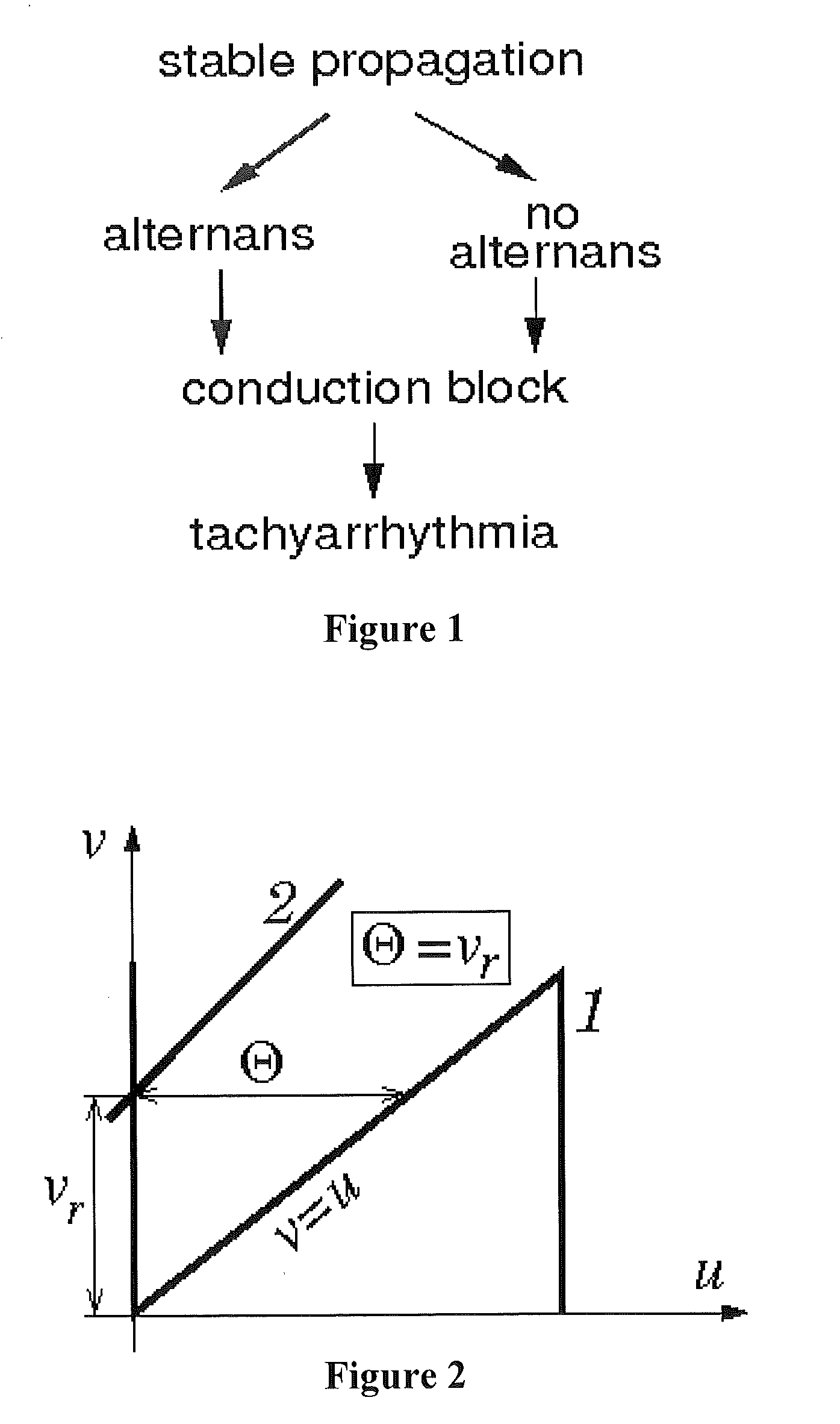
In (1-2), ε, λ and vr are model parameters and P(x,t) specifies the pacing pulses. At rest, u and v are equal to 0 and vr, respectively. The potential a quickly increases to 1 during the upstroke, stays near 1 during the action potential (AP), and afterwards returns to zero. The recovery variable v moves slowly toward 1 during AP and starts decreasing after AP ends. For a solitary pulse, v eventually returns to vr.

[0103]Parameter vr and the Critical Excitation Threshold.

[0104]Parameter vr has doub...

example 2

Data Collection, Processing and Construction of Restitution Portraits

[0107]The digitized original QT and RR interval signals can be recorded either during Cornell-type gradual exercise protocols or during electrophysiological (EP) studies with gradual electrical pacing. After resampling, these sequences can be used to determine the RPs. Custom software removes electrical noise and filters fluctuations and trends from the digitized signals using low-pass and high-pass frequency thresholds. The same software package can eliminate pacing stimuli without distortion of the filtered signals by implementing Daubechies 4th order wavelet [Starobin 2007].

[0108]For data acquired from the exercise test, we implement an adaptive least-mean square (LMS) algorithm to estimate QT interval fluctuations that are physiologically related to DI [Varadarajan 2009]. Using the output of the LMS filter, we compute a cross-correlation, CC, of this signal with DI fluctuations:

CC(n)=1QTlmsDI∑j=n-pn+pQTlms(j)DI...

example 3

Customizing Parameters of the CSC Model to RPs of Individual Patients

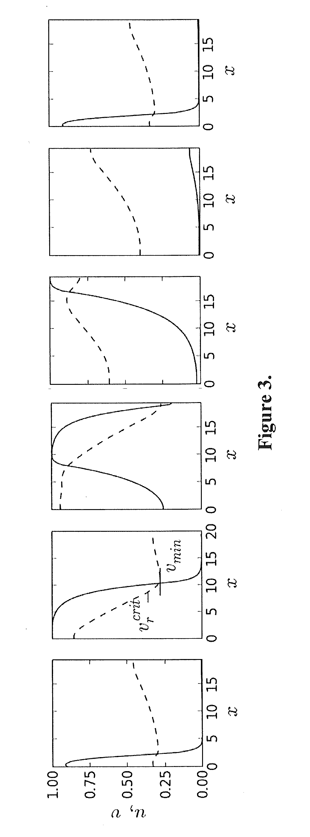
[0110]To produce results comparable to experiments, the 1D cable with the CSC model was stimulated at the left end with pacing period T, and APD and DI values were measured in the middle of the cable (FIG. 3). For all optimization runs, dimensionless APD, DI, and T values obtained from the model were converted to units directly comparable to the experimental QT, TQ, and RR intervals using the following scaling:

QT_=CmσNaAPD,TQ_=CmσNaDI,RR_=CmσNaT,(5)

where Cm and σNa are characteristic values of membrane capacitance and sodium membrane conductance. A bar over a symbol indicates the data predicted by the model.

[0111]In order to determine the SoPR values for each patient, we choose parameters of the CSC model so that the RP from the model matches the RP measured in the EP study. The CSC model (1-3) has six parameters: ε, ζ, τ, α, and β. Determining all six parameters simultaneously with a massive least-squares fit to t...


PUM


Abstract

A method of determining the susceptibility to ventricular arrhythmias in a subject, comprises the steps of: (a) collecting (e.g., by surface EKG or intracardiac EKG) at least one QT and DI interval data set from the subject during a stage of gradually increasing heart rate or a stage of gradually decreasing heart rate; (b) determining (e.g., by applying low- and high pass filtering) low-frequency QT-DI interval trends and high-frequency QT-DI fluctuation signals in said at least one QT and DI interval data set; (c) finding a plurality of correlated and anti-correlated portions between said high-frequency QT-DI fluctuation signals; (d) determining corresponding regression lines for said correlated and anti-correlated portions; (e) finding a plurality of (or in some embodiments all) steady state QT-DI points designated by intersections between said low frequency QT-DI trends and said corresponding regression lines; (f) fitting action potential durations computed from a rate dependent reaction-diffusion model to corresponding ones of said steady state QT-DI points to give (i) a model excitation threshold and (ii) a minimal level of refractoriness at a plurality of (or in some embodiments all of) said steady state QT-DI points; (g) at the steady state QT-DI point corresponding to the highest heart rate in said QT and DI interval data set, determining the difference between said minimal level of refractoriness and a model critical excitation threshold for a stable solitary pulse corresponding to the rate dependent reaction diffusion model of step (f) to give a reserve of refractoriness (RoR); (h) fitting action potential durations computed from a rate dependent reaction-diffusion model to said correlated and anti-correlated portions to give a rate of adaptation of each model excitation threshold to a corresponding steady state value at a plurality of (or in some embodiments all of) said steady state QT-DI points; (i) at the steady state QT-DI point corresponding to the highest heart rate in said QT and DI interval data set, determining the inverse of said rate of adaptation to give a reserve of memory (RoM); (j) combining said reserve of refractoriness (RoR) and said reserve of memory (RoM) to produce a metric of stability-of-propagation reserve (SoPR) in said subject, a higher value of SoPR indicating lower susceptibility to ventricular arrhythmias in said subject. Systems and apparatus for carrying out the method are also described.

Description

FIELD OF THE INVENTION[0001]The present invention concerns methods, systems and apparatus for detecting risk of ventricular arrhythmias in a subject.BACKGROUND OF THE INVENTION[0002]Each year approximately 310,000 Americans die of sudden cardiac death (SCD) from ventricular tachyarrhythmias [Lloyd-Jones 2009]. The first preventive step toward reducing mortality from SCD is to identify individuals at risk for developing arrhythmias. Yet, despite years of research, the risk stratification strategies are unclear and there are still no reliable clinical tests that predict who is susceptible to this potentially lethal heart rhythm disorders [Kusmirek & Gold 2007].[0003]Currently, the most reliable method of assessing vulnerability to cardiac arrhythmias is EP testing, in which a provocative intracardiac pacing protocol is applied to the patient with an aim of inducing an arrhythmia episode [Daubert 2006]. EP testing is an invasive procedure and carries a non-negligible risk of death. In ...

Claims


Application Information

Patent Timeline
Patent Type & Authority Applications(United States)
IPC IPC(8): A61B5/0452A61B5/00A61B5/04A61B5/363
CPCA61B5/0452A61B5/04012A61B5/4884A61B5/363A61B5/349A61N1/362A61B5/316A61B5/024A61B5/486
Inventor STAROBIN, JOSEPH M.VARADARAJAN, VIVEKNEU, WANDA KRASSOWSKAIDRISS, SALIM F.
Owner DUKE UNIV
Why Eureka
Try Eureka