Method for Consistent and Verifiable Optimization of Computed Tomography (CT) Radiation Dose

a computed tomography and radiation dose technology, applied in tomography, instruments, applications, etc., can solve the problems of inability to provide image quality measurements for clinical examinations, patient at risk without added diagnostic quality, and low radiation doses, so as to achieve minimal disruption in work flow

Inactive Publication Date: 2014-09-18
CHILDRENS HOSPITAL MEDICAL CENT CINCINNATI
View PDF1 Cites 75 Cited by
  • Summary
  • Abstract
  • Description
  • Claims
  • Application Information

AI Technical Summary

Benefits of technology

[0066]The system can report, in electronic or print format, expected image quality measures of scan protocols to an individual or organization, internal or external to the organization. The system can report outside an organization, for example, if hospitals are one day required to report this to a regulatory agency. The system may perform the above actions, including image processing, data storage, analysis, and monitoring in an automated fashion, for minimal disruption in the work flow.

Problems solved by technology

Radiation doses that are too low produce images of inadequate quality.
Radiation doses that are too high place the patient at risk without added diagnostic quality.
Therefore, the problem of ALARA in CT may be viewed as an image quality optimization problem in addition to a radiation dose optimization problem.
To date, image quality measurements for clinical examinations are typically not available to radiologists.
The limiting image quality characteristic for low-contrast applications such as CT of the abdomen / pelvis tends to be that of low contrast detectability (LCD), which incorporates both spatial and contrast resolution estimates.
Yet radiologists are still currently limited in many respects, including the following:a) Image quality is difficult to quantify, even by visual inspection; thus, standard image quality metrics have not been well established.b) Image quality depends on both scan parameters and patient size; a quantitative model of how these factors interact to impact image quality has not been well established.c) Even if it could be measured, without a predictive model, image quality can only be assessed after the scan has been completed, limiting its usefulness.d) Minimum image quality thresholds have not been established.
It would be unreasonable to expect that these individuals would dedicate a large amount of their time to ensure image quality / dose optimization.
Thus, even if a method of quantifying image quality is developed, unless it is integrated into the current workflow with minimal disruption and is easy to use, it is unlikely to experience widespread adoption.
Many of the current challenges surrounding CT dose optimization are related to the problem of image quality and dose verification.
Without an automated system, radiologists who believe or claim that they optimize dose can only verify this at great expense, if at all, due to the difficulty in acquiring, analyzing, aggregating, and reporting data from individual scans.

Method used


Image

  • Method for Consistent and Verifiable Optimization of Computed Tomography (CT) Radiation Dose
  • Method for Consistent and Verifiable Optimization of Computed Tomography (CT) Radiation Dose
  • Method for Consistent and Verifiable Optimization of Computed Tomography (CT) Radiation Dose

Examples


Embodiment Construction

[0103]The system provides quantitative image quality assessment to achieve As Low As Reasonably Achievable (ALARA) on a consistent basis for all CT scanners in an organization. The system in the embodiments described below allows Computed Tomography (CT) scanners to incorporate scanner parameters and patient size into calculations to accurately determine the minimum radiation dose necessary to achieve diagnostic image quality. Finally, the embodiments below provide a comprehensive, transparent system to automate, analyze, and monitor the aggregate history of CT scans to ensure proper dosing and image quality across an organization.

Estimation Models

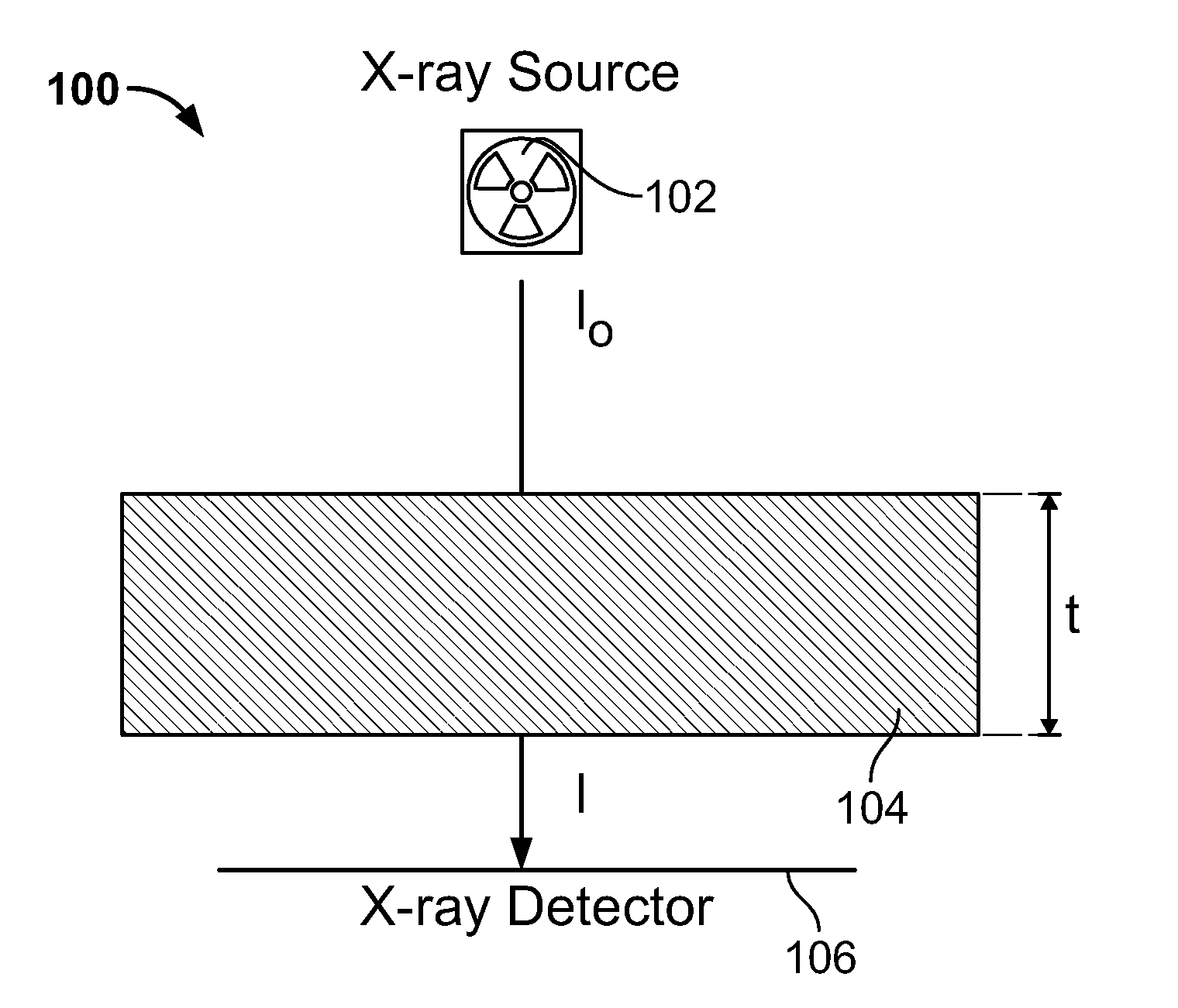
[0104]FIG. 1 depicts the basic schematic of a CT device 100 having an X-ray source 102, and an X-ray detector 106 where I0 is the initial intensity of the X-ray source, I is the Intensity at the X-ray detector after passing through t, a thickness of an object 104; The Beer-Lambert law describes attenuation characteristics of an x-ray beam ...


PUM


Abstract

A system and a method is disclosed for consistently and verifiably optimizing computed tomography (CT) radiation dose in the clinical setting. Mathematical models allow for estimation of patient size, image noise, size-specific radiation dose, and image quality targets based on digital image data and radiologists preferences. A prediction model estimates the scanner's tube current modulation and predicts image noise and size-specific radiation dose over a range of patient sizes. An optimization model calculates specific scanner settings needed to attain target image quality at the minimum radiation dose possible. An automated system processes the image and dose data according to the mathematical models and stores and displays the information, enabling verification and ongoing monitoring of consistent dose optimization.

Description

CROSS-REFERENCE TO RELATED APPLICATIONS[0001]This application claims the benefit of U.S. Provisional Application No. 61 / 541,671, filed Sep. 30, 2011, which is hereby incorporated by reference.FIELD OF THE INVENTION[0002]The current disclosure relates to a Computed Tomography (CT) method for optimizing radiation dose. The current disclosure also relates to an integrated system to provide consistent CT radiation dose optimization based on individual patients throughout an organization by minimizing deviation from quantitative image quality targets.BACKGROUND OF THE INVENTION[0003]Hospitals are beginning to monitor CT radiation dose in response to issued health guidelines. When undergoing medical radiation exposure, including that of computed tomography (CT), the guiding principle is that of As Low As Reasonably Achievable (ALARA). Thus, imagers desire to use the minimum radiation dose necessary to achieve diagnostic image quality. Radiation doses that are too low produce images of ina...

Claims


Application Information

Patent Timeline
Patent Type & Authority Applications(United States)
IPC IPC(8): A61B6/00
CPCA61B6/5258A61B6/032A61B6/405A61B6/463A61B6/488A61B6/544A61B6/545A61B6/583A61B6/563F04C2270/041G16H40/63
Inventor LARSON, DAVID B.
Owner CHILDRENS HOSPITAL MEDICAL CENT CINCINNATI
Why Eureka
Try Eureka