Portable Blood Count Monitor

a portable, blood cell technology, applied in the field of medical devices, systems and methods, can solve the problems of manual counting being subject to sampling errors, certain abnormal cells in the blood may not be identified correctly, and the current cost of healthcare in the united states is a large and rapidly growing burden on the national economy. , to achieve the effect of convenient setup

Inactive Publication Date: 2014-09-18
RGT UNIV OF CALIFORNIA +1
View PDF7 Cites 14 Cited by
  • Summary
  • Abstract
  • Description
  • Claims
  • Application Information

AI Technical Summary

Benefits of technology

[0016]Aspects of the disclosure provide improved methods, systems, and devices for performing complete blood count and other blood and blood cell analysis procedures. The disclosure includes an inexpensive and transportable personal blood count monitor (PBCM) that can be taken home by a patient, easily set up, and routinely used to obtain and report specific blood count information, including critical blood cell count information. This information can be readout by the patient or communicated to another monitoring location or doctor. The personal blood count monitor can count red blood cells, white blood cells, platelets, and selected subsets of the aforementioned cells, as well as hemoglobin levels and hematocrit. Methods used by the personal blood count monitor for such counting are also disclosed, as are medical procedures performed by the patient to use the personal blood count monitor to track and analyze their blood count and chemotherapy progress.
[0017]Many of the blood count devices disclosed herein are easily transportable, easy to configure, and do not require advanced training or education to use. While many of the blood count devices disclosed herein are intended for home use for a patient, these devices may also be used in a hospital, a clinic, a doctor's office, or various other locations. Trained physicians, such as various oncologists (in medical oncology and hematology, radiation oncology, surgical oncology, gynecology oncology, pediatric oncology, etc.), infectious disease specialists, rheumatologists, transplantation teams, and physicians who need to follow a patient's red blood cell count (general surgeons, gastroenterologist, primary care providers, emergency medicine practitioners, orthopedic surgeons, otolaryngologists, neurosurgeons, etc.) may also use the blood count device. Many of the blood count devices disclosed herein use a relatively small amount of blood, for example, two to five microliters or in some cases even less, versus current blood count devices which can require 10 to 5,000 microliters. Many of the blood count devices disclosed herein can be calibrated against a traceable standard before a blood count measurement is performed.
[0018]Use of the blood count devices disclosed herein can have many benefits, including personalized myelosuppression profiles, personalized optimal chemotherapy dosing, avoidance of unnecessary and expensive white blood cell growth factor usage, decreased emergency visits, decreased unplanned hospitalizations for febrile neutropenia, decreased debility and death from treatment related infections or complications thereof, improved response rates to chemotherapy, etc.
[0019]Also disclosed are methods to be used by the patient to use the personal blood count monitor to track and analyze their blood count and chemotherapy progress. The personal blood count monitor may be taken home by the patient and used on the fifth, seventh, ninth, and eleventh days of each cycle of chemotherapy patient. The personal blood count monitor can be used by the patient or care-giver to perform a complete blood count or other blood count and report the results to a doctor's office where the results can be used to evaluate the patient's bone marrow response to the chemotherapy. The results can be manually reported via telephone or e-mail by the patient or automatically reported to a remote location using, for example, telemetry, a wide area network (WAN), the Internet, a mobile phone, or through e-mail that may be secure and encrypted. Generally, such automatic reporting can be achieved through a HIPAA certified secured transmission. A computerized system that receives the test results may save the results to a file that can become of permanent part of the patient's medical record. If the patient develops a fever at any time during a chemotherapy cycle, an additional blood count can be taken and reported. Based on the results of the blood count measurements, the doctor can decide on a course of treatment. For example, this treatment could range from having the patient go to an emergency room for evaluation and therapy, to prescribing antibiotics over the telephone, to starting the patient on white blood cell grown factors (e.g., filgastrim, pegfilgastrim), to having the patient take over the counter medications, and continuing to monitor the blood count. The costs of the various options may vary dramatically, and the use of the blood count monitor can be instrumental in providing the optimum medical care at the lowest cost. The personal blood count monitor can permit the doctor to optimize the chemotherapy treatment for a patient on a personalized basis and ultimately reduce the cost of the overall therapy.
[0021]The sampling chambers may come preloaded with at least one of a surfactant, a dying agent, a lysing agent, a dry form reagent, a liquid reagent, or a predetermined volume of a diluent. Alternatively or in combination, the slide may comprise one or more reagent chambers for storing any one of the aforementioned agents. In some embodiments, the inner surfaces of the sampling chambers may be configured to be hydrophilic, for example, by being coated with a hydrophilic substance. Such hydrophilicity may facilitate the spreading out of very small volumes of liquid in the sampling chambers.
[0031]The analysis of white blood cells may comprise one or more steps of lysing red blood cells, staining the white blood cells, taking images of the white blood cells, and analyzing the images to determine white blood cell number in addition to subpopulation numbers and percentages. The red blood cells in the second chamber will typically be lysed with a lysing agent before the image of the second sampling chamber may be analyzed to facilitate the counting of white blood cells. The lysing agent may be selected from the group comprising SDS, saponins, snake venom, quaternary ammonium salts, triton-X, and the like. The dying agent may comprise a nucleic acid staining agent. To determine the number or type of white blood cells in the blood sample, the white blood cells in the second sampling chamber may be stained with the nucleic acid staining agent and a relationship between fluorescence at a first color with fluorescence at a second color for individual white cells may be determined. White blood cells may then be typed based on this relationship, i.e., particular types of white blood cells may fall within particular ranges of fluorescent intensity at the first and second wavelengths or wavelength ranges. Additional dimensions of analysis may also be implemented to differentiate cell types and properties. For example, fluorescent intensities at other wavelengths or wavelength ranges as well as forward and side light scattering may be measured. Also, platelets may also be identified as small, dim objects with low fluorescent intensities. Thus, the number of platelets may also be determined along with the number of white blood cells. The nucleic acid staining agent may comprise one or more of acridine orange, thiozole orange, acridine red, 7-AAD, LDS 751, and hydroxystilbamidine. Fixatives may be added to improve the staining of platelets.

Problems solved by technology

The current cost of healthcare in the United States is a large and rapidly growing burden on the national economy.
However, certain abnormal cells in the blood may not be identified correctly, requiring manual review of the instrument's results and identification of any abnormal cells the instrument could not categorize.
Manual counting can be subject to sampling error because so few cells are counted compared with automated analysis.
Although automated analyzers give fast, reliable results regarding the number, average size, and variation in size of red blood cells, they often do not detect the shapes of the cells.
As discussed above, current methods of performing CBC require a visit to a clinic or a laboratory which can be inconvenient, if not unhealthy, to rural patients.
Such methods also involve the use of expensive and cumbersome equipment with limited options for portable use and operations.
As discussed above, certain abnormal cells in the blood may not be identified correctly using this method.
Such blood count monitors, however, can be less than ideal in many cases.
For example, such monitors may only be able to count and analyze one type of cell.
Also, the image analysis algorithms used may be less than optimal and may be prone to sampling error if too few samples are taken.
Ease of use by unskilled operators is also a short-coming of current devices.

Method used


Image

  • Portable Blood Count Monitor
  • Portable Blood Count Monitor
  • Portable Blood Count Monitor

Examples


Embodiment Construction

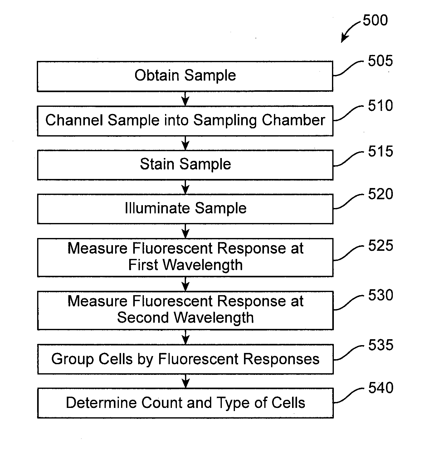
[0050]Aspects of the invention provide improved devices, systems, and methods for performing blood count measurements. Various aspects of the invention described herein may be applied to any of the particular applications set forth below or for any other types of biological analysis systems. It shall be understood that different aspects of the invention can be appreciated individually, collectively, or in combination with each other.

[0051]1. System Overview

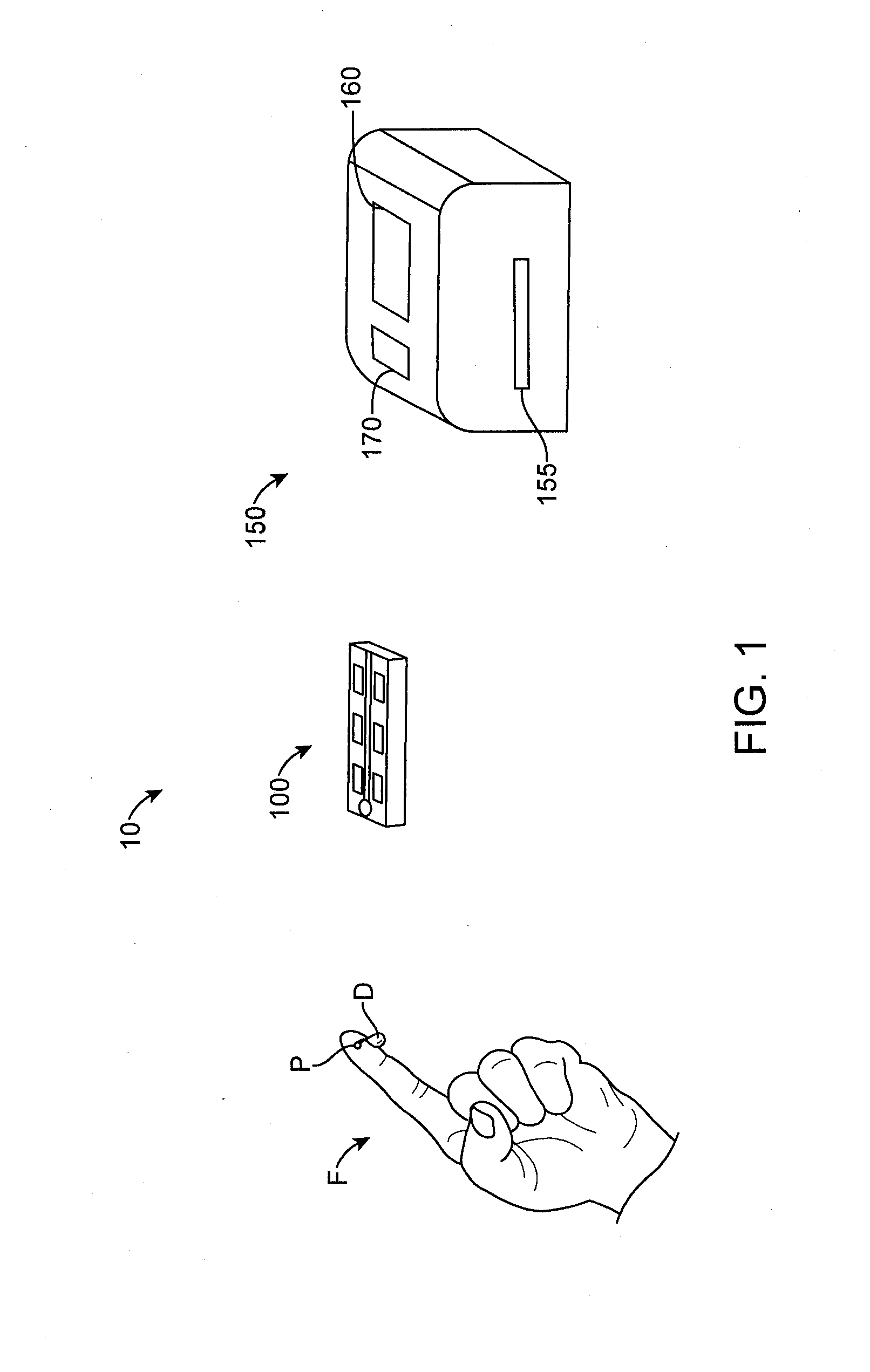
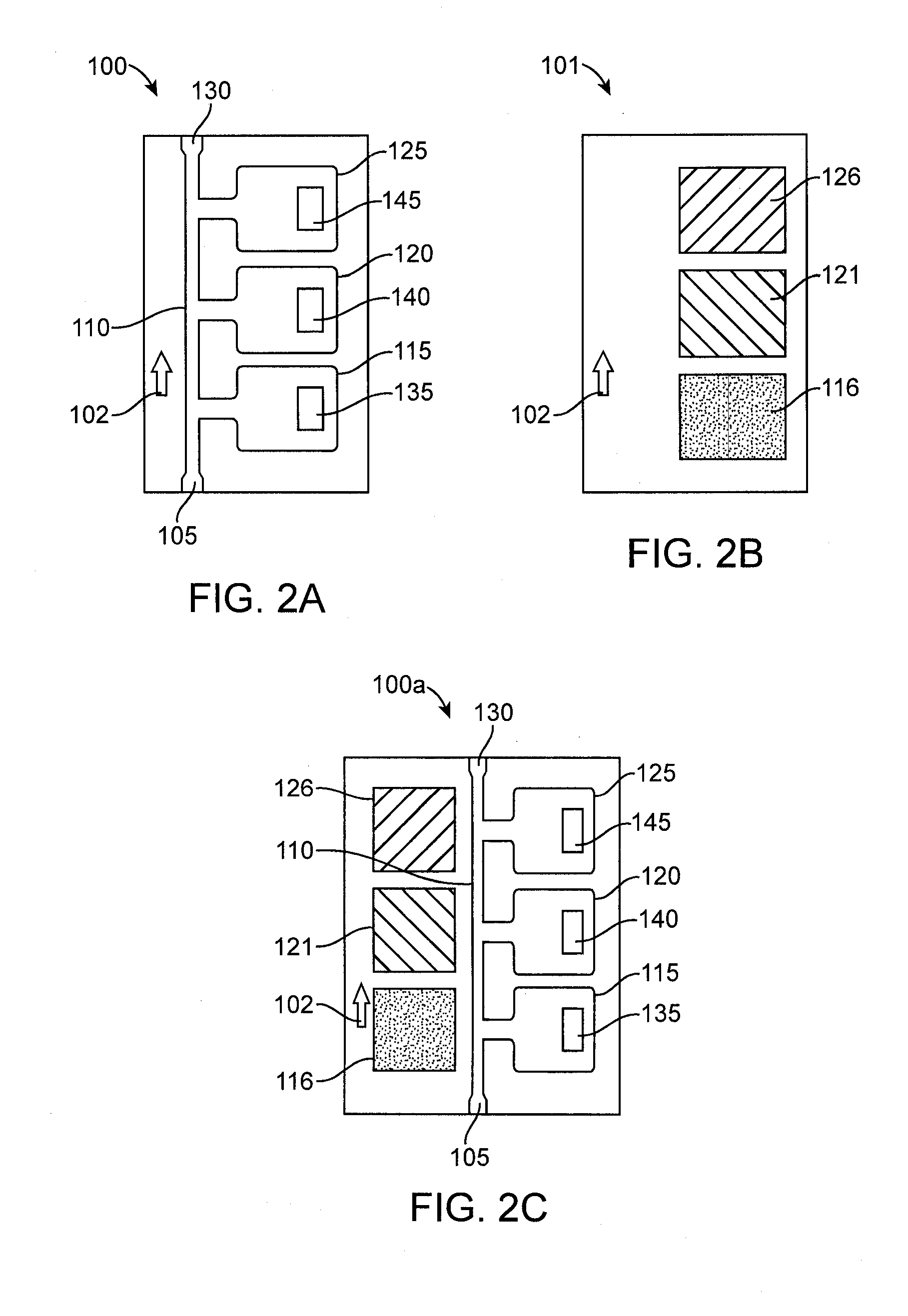
[0052]FIG. 1 shows a system 10 for performing blood count analysis. The system 10 comprises a blood collection and holding slide 100 and an automated portable slide analyzer 150. The automated portable slide analyzer 150 comprises a display 160 and a control panel 170.

[0053]The slide 100 will typically be relatively low in cost, be optically clear, and may comprise two optically clear glass, plastic, or polycarbonate substrates that may be separated by 4 to 100 microns with one hole for the insertion of blood. As shown in FIG. 1, ...


PUM


Abstract

Devices, systems, and methods are disclosed for determining the number and type of blood cells in a blood sample. The blood sample is collected and held in a slide. In the slide, the blood sample is separated and channeled into at least two sampling chambers, one for red blood cells, another for white blood cells, and optionally yet another for platelets. The sampling chambers have wetting agents, lysing agents, staining agents, or the like therein to mix with the blood and facilitate cell count. The slide is placed in a portable slide analyzer where the sampling chambers are illuminated and images of the sampling chambers are taken. These images are converted into electronic form and sent by a communications module of the slide analyzer to a remote external location where the images are analyzed to determine the number and type of blood cells in the blood sample.

Description

CROSS-REFERENCE[0001]This application claims the benefit of U.S. Provisional Application No. 61 / 780,732, filed on Mar. 13, 2013, the contents of which is incorporated herein by reference in its entirety.STATEMENT AS TO FEDERALLY SPONSORED RESEARCH[0002]This invention was made with partial government support under an Acceleration of Innovation Research grant awarded by the National Science Foundation (NSF Accelerating Innovation Research Grant No. 1127888, entitled “Creation of an Ecosystem for Biophotonic Innovation” and dated Aug. 1, 2011 to July 31, 2013), as well as from the Center for Biophotonics Science and Technology, a designated NSF Science and Technology Center managed by the University of California, Davis, under Cooperative Agreement No. PHY0120999. The government has certain rights in the invention.BACKGROUND OF THE INVENTION[0003]The present disclosure relates to medical devices, systems, and methods. More specifically, the present disclosure relates to point-of-care h...

Claims


Application Information

Patent Timeline
Patent Type & Authority Applications(United States)
IPC IPC(8): G06K9/00
CPCG06K9/00134G01N33/5094G01N33/80
Inventor SMITH, ZACHARYGAO, TINGJUANLANE, STEPHENWACHMANN-HOGIU, SEBASTIANDWYRE, DENISHEIFETZ, LAURENCEHOOD, JAMESMATTHEWS, DENNISTATSUKAWA, KEITH
Owner RGT UNIV OF CALIFORNIA
Why Eureka
Try Eureka