Method and quality control molecular based mouse embryo assay for use with in vitro fertilization technology

a applied in the field of method and quality control molecular based mouse embryo assay for use with in vitro fertilization technology, can solve the problems of reducing embryo viability and development impairment, difficult to assess the impact of suboptimal environment using morphology as a marker, and posing significant laboratory challenges.

Inactive Publication Date: 2014-10-09
FUJIFILM IRVINE SCI INC
View PDF0 Cites 2 Cited by
  • Summary
  • Abstract
  • Description
  • Claims
  • Application Information

AI Technical Summary

Benefits of technology

[0023]The invention disclosed herein further includes a method for enhancing the sensitivity of embryo assay using embryo development to the blastocyst stage. The method includes providing a transgenic embryo comprising at least one reporter gene operably linked to at least one embryonic pluripotency marker; incubating the transgenic embryo under culture conditions / test items; and evaluating embryo development morphologically and via the expression of said embryonic marker from one-cell to blastocyst and gastrulation stages.

Problems solved by technology

It is the initially crude nature of homeostatic regulation in the early embryo and its subsequent development through later stages of pre-implantation development that pose significant challenges in the laboratory.
Perturbations to the environment surrounding the embryo during development in culture, relative to “normal” conditions encountered in the reproductive tract, result in reduced embryo viability and impaired development.
However, it is often difficult to assess the impact of suboptimal environment using morphology as a marker.
In certain instances, embryos can develop to apparently morphologically normal blastocysts but at the cellular level, these embryos can be compromised and have a reduced capacity to implant and produce a successful term pregnancy.
However, there are a number of limitations of this assay which are often overlooked.
For example, the assay can only detect conditions which are grossly and harshly embryo toxic.
The MEA cannot detect or differentiate growth promoting or inhibiting factors at a very early stage in development.

Method used


Image

  • Method and quality control molecular based mouse embryo assay for use with in vitro fertilization technology
  • Method and quality control molecular based mouse embryo assay for use with in vitro fertilization technology
  • Method and quality control molecular based mouse embryo assay for use with in vitro fertilization technology

Examples


example 1

OCT-4

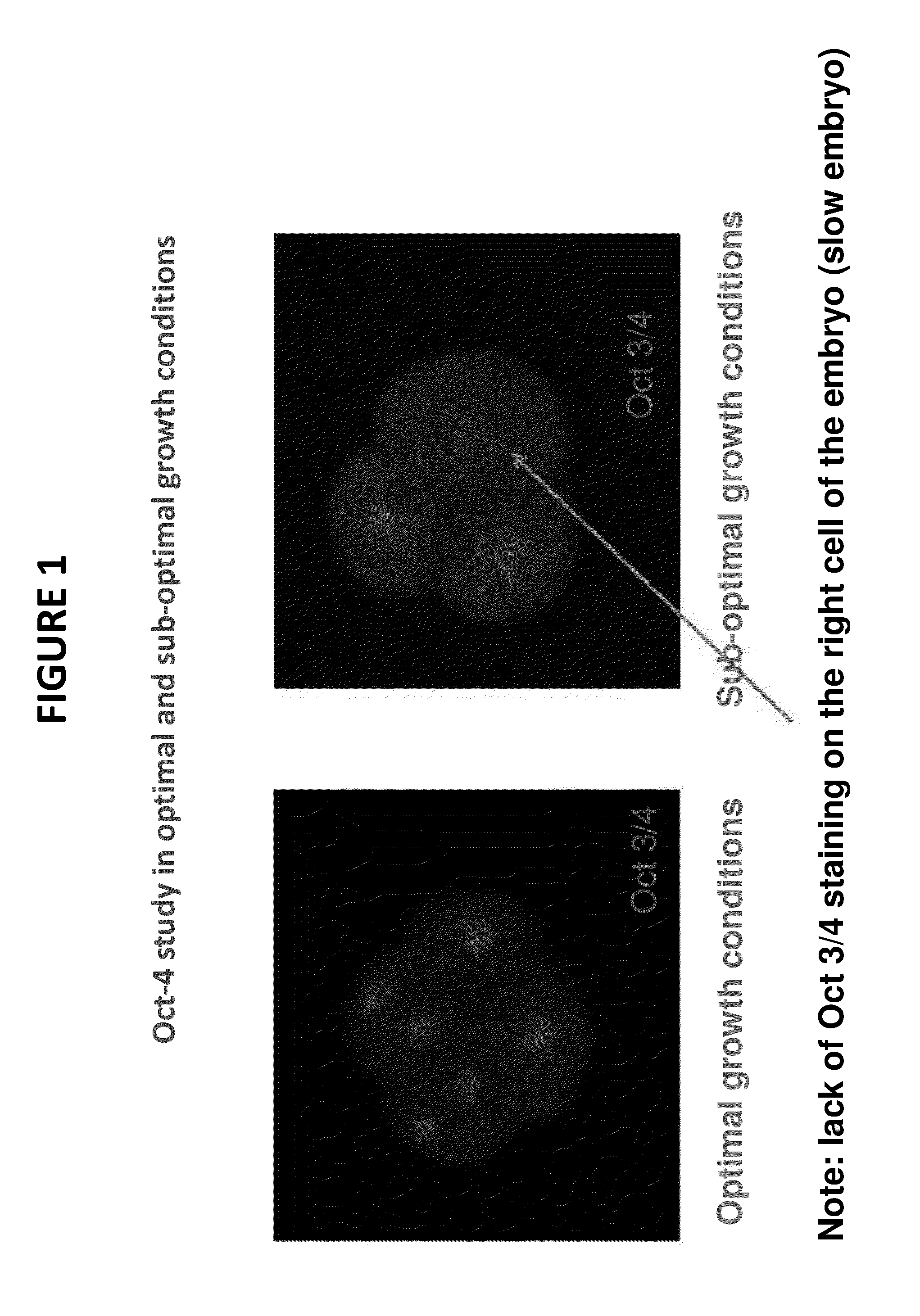
[0054]FIG. 1 is a photograph from the fluorescence microscope showing one embodiment of a molecular-based mammalian embryo assay for use in quality control. A mouse embryo with the pluripotency regulator Oct-4 visualized with red fluorescence tag. The 2-cell embryo is cultured in vitro. After approximately 48 hours, the embryos were stained and evaluated via fluororescence microscopy. As is evident from FIG. 1, the embryo on the left, which was incubated under optimal growth conditions, is well developed with uniform staining. By contrast, the blastocyst on the right was incubated in sub-optimal growth conditions. A lack of Oct-4 staining is observed on the right cell of the embryo on the right, demonstrating that the sub-optimal growth conditions result in slower embryonic development.

example 2

OCT-4

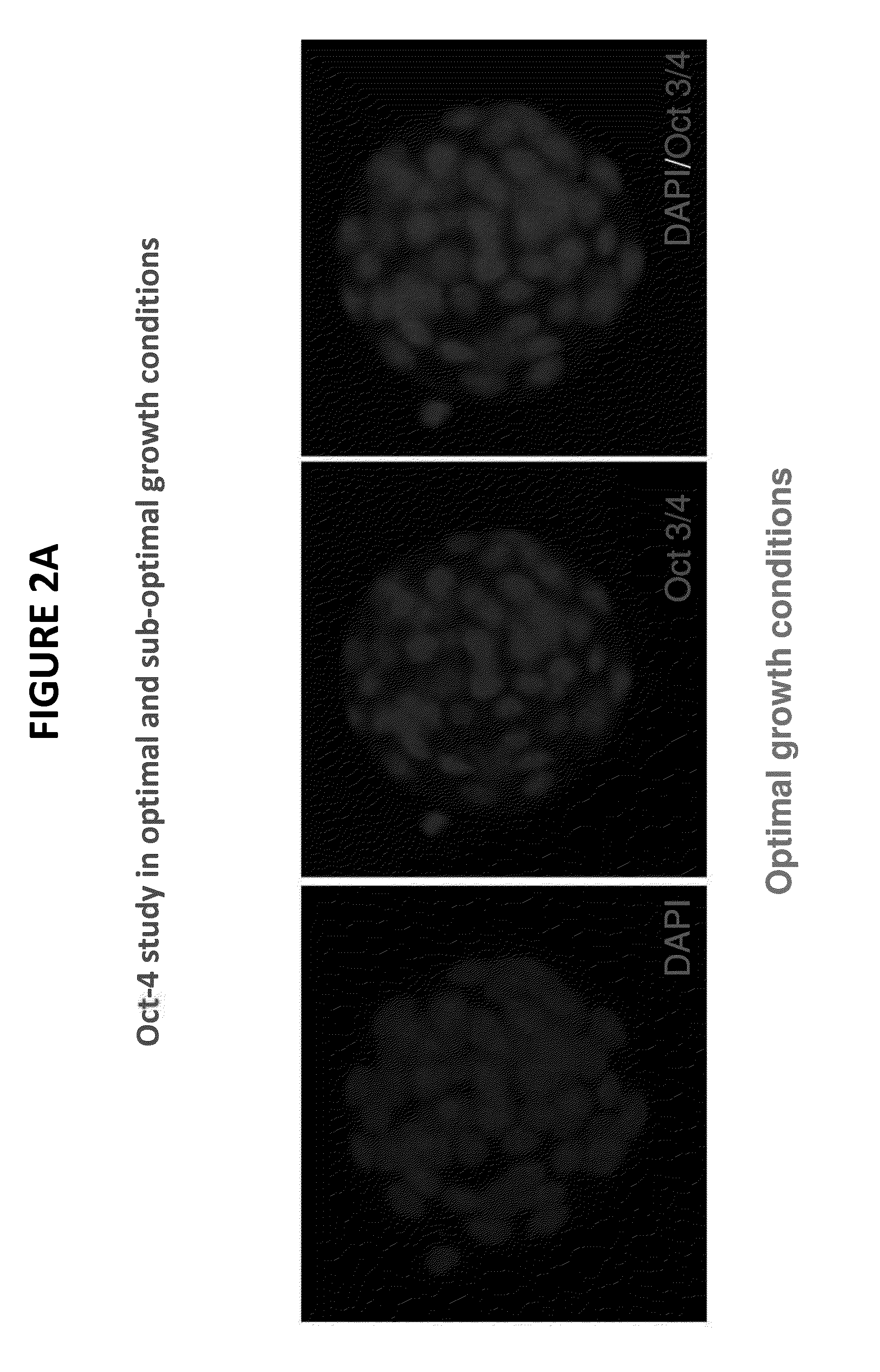
[0055]FIGS. 2A and 2B illustrate photographically a molecular-based mouse embryo assay at the blastocyst stage. The embryos were cultured in vitro for 96 hours and stained and observed via a fluorescence microscope. In FIG. 2A, under optimal growth conditions, normal embryo development is observed as demonstrated by the uniform staining. By contrast, in FIG. 2B, the embryos were incubated under sub-optimal growth conditions. A lack of Oct-4 staining on some mural trophectoderm cells is observed in the embryos as noted by the arrows on FIG. 2B. Without the use of the presently claimed technology, the sub-optimal culture conditions would not be evident or identifiable as sub-optimal when using the conventional MEA standard QC protocol because the blastocyst appears to be developing at a normal rate. However, by observing the slow growth in the center and left pictures of FIG. 2B, it is clear that blastocyst development is qualitatively less uniform and less optimal than in the em...

example 3

SOX-2

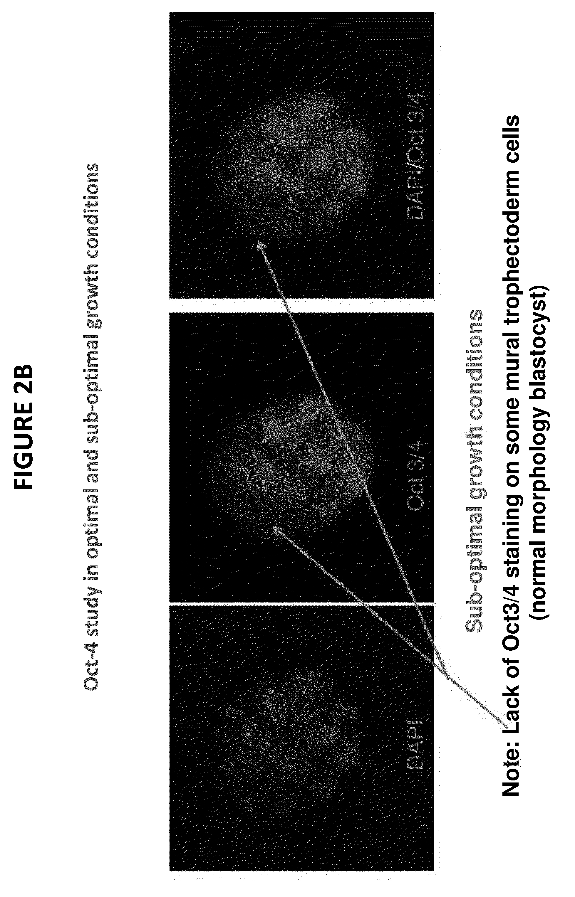
[0056]FIGS. 3A, 3B, and 3C further demonstrate the superior QC features of the claimed invention. FIGS. 3A-3C are fluorescence microscopy photographs of control murine Embryos were incubated to the blastocyst stage in vitro, stained, and observed microscopically. After 96 hours of culture, the image on the left of FIG. 3A shows an embryo that was fixed and stained with DAPI and observed microscopically to assess growth. The staining pattern observed in the embryo is uniform and evidences normal, healthy blastocyst development under optimal conditions. The center image in FIG. 3A shows the embryo having the SOX-2 viability marker stained with green fluorescence and DAPI. Notice the uniform staining as well as the well-defined differentiation of the blastocyst.

[0057]Turning to FIG. 3B, blastocysts incubated in sub-optimal growth conditions are observed. The picture of FIG. 3B shows a mouse embryo incubated under sub-optimal culture conditions. After 96 hours, the embryo is fixed ...


PUM

PropertyMeasurementUnit
timeaaaaaaaaaa
timeaaaaaaaaaa
green fluorescentaaaaaaaaaa

Abstract

A method for qualitatively assessing products used in in vitro fertilization is provided. Also disclosed is an improved quality control assay for use in clinical Assisted Reproductive Technologies (ART).

Description

BACKGROUND OF THE INVENTION[0001]1. Field of the Invention[0002]The present invention relates to a method for assessing products used in in vitro fertilization. Also disclosed is a quality control assay for use in clinical Assisted Reproductive Technologies (ART).[0003]2. Description of the Related Art[0004]The in vitro fertilization (IVF) laboratory plays a fundamental role in the delivery of treatment to infertile couples. Ensuring proper Quality Control (QC) in the IVF laboratory is critical to the success of any IVF program as the environment of the laboratory can alter the quality of the embryos produced. An optimal culture medium and a stable environment are necessary for the successful development of human embryos in vitro. The ultimate role of the embryology laboratory is to maintain the inherent viability of the gametes and embryos in an environment outside the female reproductive tract. The dynamic nature of pre-implantation embryo development is unique because, unlike som...

Claims


Application Information

Patent Timeline
Patent Type & Authority Applications(United States)
IPC IPC(8): C12Q1/68
CPCC12Q1/68A01K67/0275A01K2267/02A01K2267/0393
Inventor NI, HSIAO-TZUES-SLAMI, SAMIRAGILBERT, REBECCA SUSAN
Owner FUJIFILM IRVINE SCI INC
Why Eureka
Try Eureka