
Alpha toxin detection of gpi anchored proteins

a technology of gpi and toxin, which is applied in the field of alpha toxin detection can solve the problems of difficult prediction of gpi anchored protein annotation in mammalian protein databases, and often hampered experimental isolation and identification of gpi anchored proteins from mammalian cells

Inactive Publication Date: 2014-10-23
UNIV OF GEORGIA RES FOUND INC
View PDF0 Cites 4 Cited by
  • Summary
  • Abstract
  • Description
  • Claims
  • Application Information

AI Technical Summary

Benefits of technology

The patent text describes a method for detecting and targeting glycosylphosphatidylinositol (GPI)-anchored proteins (GPI-APs) in cancer cells using a toxin called alpha-toxin from Clostridium septicum. These GPI-APs are found on the surface of cancer cells and are associated with the growth and spread of cancer. The method involves exposing cells or tissues to alpha-toxin and detecting the complex formed between the toxin and the GPI-APs. This complex can be used as a diagnostic marker for cancer or to target the cells for therapy. The method can be used with biological samples such as cells, tissue, or blood from patients with cancer. The patent also describes a method for obtaining a GPI-AP protein by treating the complex with alpha-toxin to remove the toxin and isolate the GPI-AP. Overall, the patent provides a method for detecting and targeting cancer cells using a specific toxin and a biological sample from the patient.

Problems solved by technology

The predictive annotation of GPI anchoring in mammalian protein databases is difficult as there are no common consensus sequences that clearly indicate that a protein will receive a GPI anchor.
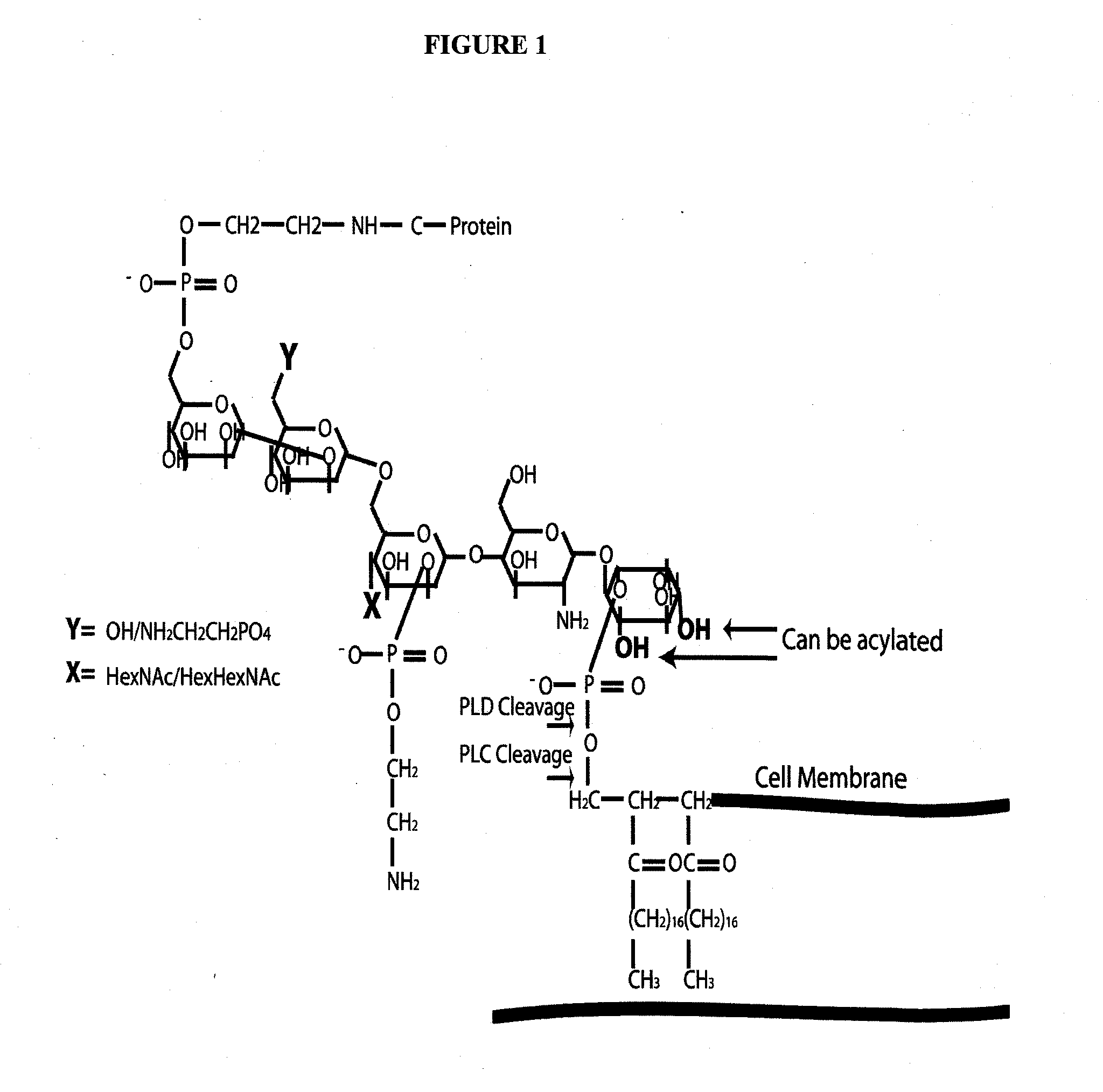
Furthermore, the experimental isolation and identification of GPI anchored proteins from mammalian cells is often hampered due to the lower expression levels of GPI anchored proteins in many cell types coupled with difficulty in extracting these proteins due to the presence of both lipid and glycan structures (FIG. 1A, core GPI structure).

Method used


Image

  • Alpha toxin detection of gpi anchored proteins
  • Alpha toxin detection of gpi anchored proteins
  • Alpha toxin detection of gpi anchored proteins

Examples


example 1

Method

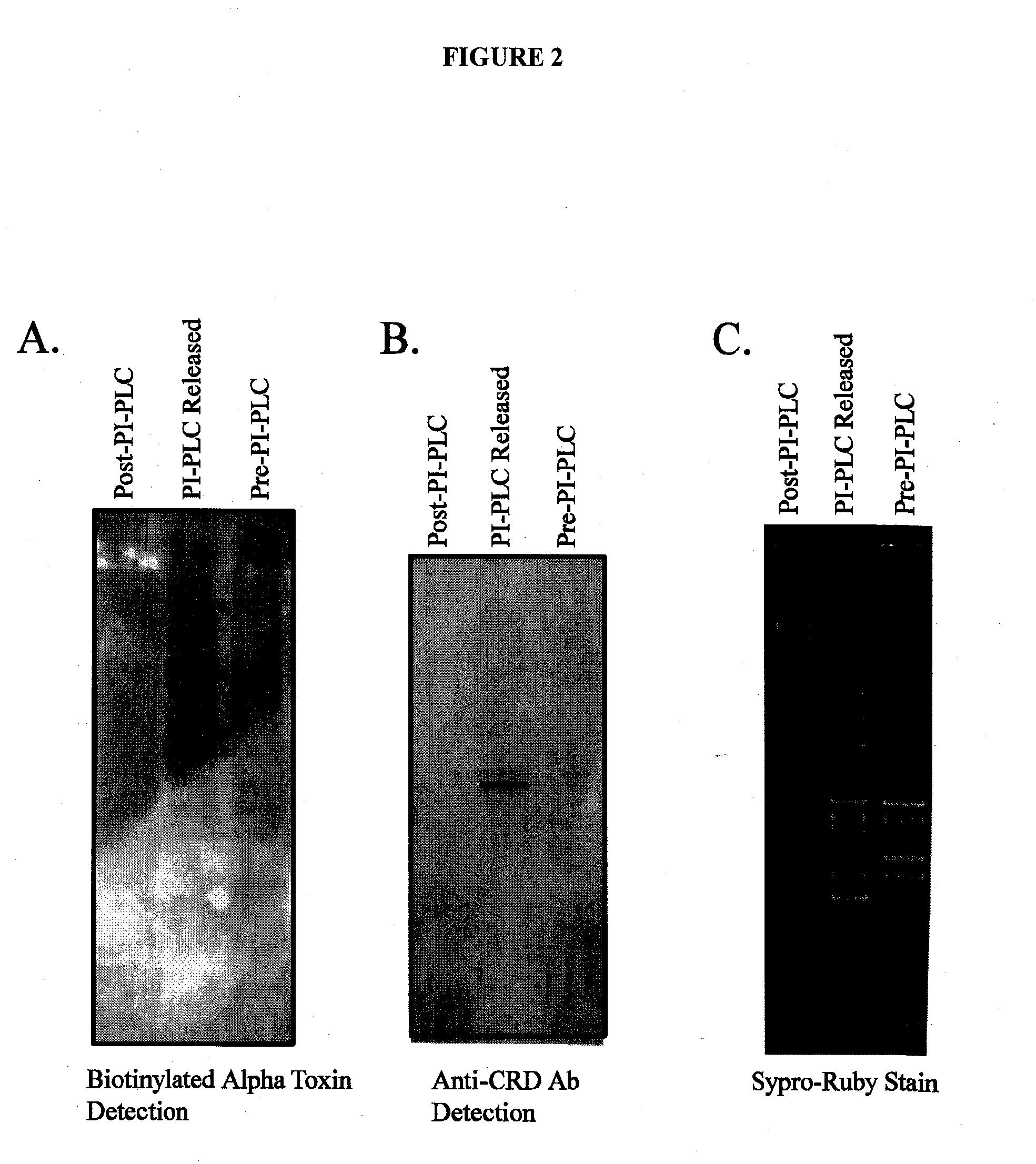
[0091]Alpha toxin was expressed and purified from the PET22 vector using E. Coli BL21 cells. The purified alpha toxin was labeled with biotin using the Pierce EZ-link sulfo-NHS-LC Biotin labeling reagent according to the manufacturers instructions. Membrane proteins were extracted from the invasive breast cancer cell line MDAMB231 using a triton X-114 phase separation protocol. Fractions of proteins from the extraction were sampled as follows: pre-PI-PLC fraction contained all membrane proteins solubilized in SDS-loading buffer, PI-PLC released containing the proteins that were released into the soluble phase after PI-PLC enzyme incubation, the post-PI-PLC fraction contained membrane proteins remaining that were solubilized in SDS-loading buffer. Proteins corresponding to twenty micrograms per fraction were separated by polyacrylamide gel electrophoresis on 4-12% Bis Tris gel. The gel was stained with Sypro-Ruby stain and imaged under a UV light source. Proteins on a duplicate...

example 2

[0094]The development of techniques to capture and enrich GPI-APs released from mammalian cells would greatly enhance the characterization of human GPI-APs. One very promising methodology is the use of carbohydrate binding proteins known as lectins. Toxins such as aerolysin from A. hydrophila and alpha toxin from C. septicum have been shown to act as GPI-specific lectins, binding to the conserved glycan core of the GPI anchor. Recent studies have shown that aerolysin requires interactions with the protein N-glycan as well as the GPI core [13]. Studies using alpha toxin from C. Septicum have demonstrated that this protein recognizes with very broad specificity the core GPI anchor structure [4]. Researchers have been utilizing these toxins to discover new CHO cell mutants with defects in GPI biosynthesis. Recently, a mutant form of the alpha toxin from C. Septicum that is non-toxic to cells was shown to have potential value as an agent to differentiate GPI-positive cells from GPI-nega...

example 3

[0098]To show that alpha toxin can be used to bind colon cancer GPI-APs, intact LS174T colon cancer cells were incubated with phospholipase-1×PBS (PIPLC) or 1 U / ml PI-PLC in 1×PBS (+PIPLC) for 1 hour at 37 C. Ten percent of the proteins released from the cells were loaded for each sample (lanes 1 and 2, see FIG. 7). The remaining 90% was incubated with alpha toxin beads (lanes 3 and 4 of FIG. 7). The captured proteins were separated on 4-12% Bis-Tris gel before being silver stained. FIG. 7 indicates that alpha toxin can specifically capture phospholipase released GPI anchored proteins. Alpha toxin Selectively captured proteins from the PIPLC treated cells. Equivalent levels of proteins were present in the reactions indicated by the 10% input.

Examples

Proteomic Identification of Glycosylphosphatidylinositol (GPI) Anchor-Dependent Membrane Proteins Elevated in Breast Carcinoma

Experimental Procedures (Second Set of References Applies)

[0099]Posttranslational addition of a glycosylphospha...


PUM

PropertyMeasurementUnit
Concentrationaaaaaaaaaa
Contentaaaaaaaaaa

Abstract

The present invention relates to a method for the purification, concentration and identification of glycosylphosphatidylinositol anchored proteins (GPI-APs) from a biological sample (cells, tissues and / or blood / serum) in a patient or subject, including a human patient or subject. A new method to separate GPI-anchored glycoproteins, a class of glycoproteins found in all animal cells and fluids including serum, from other glycoproteins and proteins for the purpose of identifying potential biomarkers for various diseases, including cancer, especially breast cancer, vaginal cancer, endometrial cancer, uterine cancer, cervical cancer, pancreatic cancer and prostate cancer. The method uses the alpha-toxin from Clostridium septicum to separate GPI-anchored glycoproteins for identification and optionally quantification. The GPI-APs so obtained may be used to raise antibodies for inclusion in an immunosorbent assay for the diagnosis or the monitoring of therapy of cancer in a patient.

Description

RELATED APPLICATIONS AND GRANT SUPPORT[0001]This application claims the benefit of priority of international application PCT / US2012 / 048581, filed 27 Jun. 2012, entitled “Alpha Toxin Detection of GPI Anchored Proteins”, which claims the benefit of priority of claims the benefit of priority of provisional application Ser. No. 61 / 512,976, filed 29 Jul. 2011 of identical title and Ser. No. 61 / 579,957, filed Dec. 23, 2011, also of identical title, all of said applications being incorporated by reference in their entirety herein.[0002]This invention was made with government support under grant nos. W81XWH-08-1-0565; BC075534, UO1CA128454 and P41RR018502 awarded by the United States Department of Defense. Consequently, the government has certain rights in the invention.FIELD OF THE INVENTION[0003]The present invention relates to a method for the purification, concentration and identification of glycosylphosphatidylinositol anchored proteins (GPI-APs) from biological samples (cells, tissues...

Claims


Application Information

Patent Timeline
IPC IPC(8): G01N33/68
CPCG01N33/6893C07K14/705G01N33/5011G01N33/57415G01N33/57492G01N2800/52C07K16/1282
Inventor PIERCE, JAMES MICHAELABBOTT, KAREN LYNN
Owner UNIV OF GEORGIA RES FOUND INC
Learn More