Methods and devices suitable for imaging blood-containing tissue

a blood-containing tissue and imaging technology, applied in the field of medical imaging, can solve the problems of insufficient contrast in visible-light color images to clearly distinguish small blood vessels

Inactive Publication Date: 2014-11-20
TEL HASHOMER MEDICAL RES INFRASTRUCTURE & SERVICES +1
View PDF2 Cites 1 Cited by
  • Summary
  • Abstract
  • Description
  • Claims
  • Application Information

AI Technical Summary

Problems solved by technology

Visible-light color images often have insufficient contrast to clearly discern smaller blood vessels.

Method used


Image

  • Methods and devices suitable for imaging blood-containing tissue
  • Methods and devices suitable for imaging blood-containing tissue
  • Methods and devices suitable for imaging blood-containing tissue

Examples


example 1

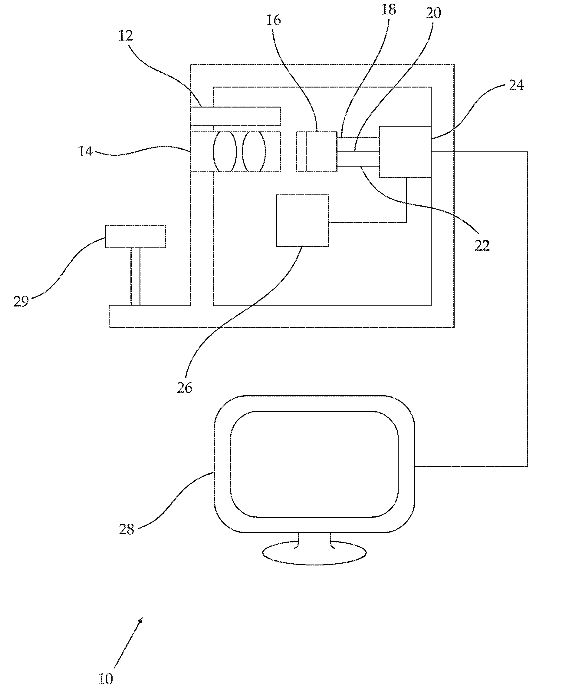
[0170]A first laboratory mouse was anesthetized. The skin covering the abdominal cavity was cut to define a loose flap, and the flap secured with needles to a flat surface, skin side down.

[0171]Using an SD-300 spectral imaging camera (ASI, Migdal Haemek, Israel) fitted with a halogen lamp illuminator and a cross-polarizing filter set, a spectral image of a portion of the inner surface of the flap was acquired between 400 nm and 800 nm. The acquired image is reproduced, in black-and-white, in FIG. 1A. In FIG. 1B, the spectrum of a portion of the image where a blood vessel was present (portion a) and the spectrum of the image where no blood vessel was present (portion b) are displayed.

example 2

[0172]A second laboratory mouse was anesthetized. The skin covering the abdominal cavity was cut to define a loose flap, and the flap secured with needles to a flat surface, skin side down.

[0173]Using an SD-300 spectral imaging camera (ASI, Migdal Haemek, Israel) fitted with a halogen lamp illuminator and a cross-polarizing filter set, a spectral image of a portion of the inner surface of the flap was acquired between 400 nm and 800 nm.

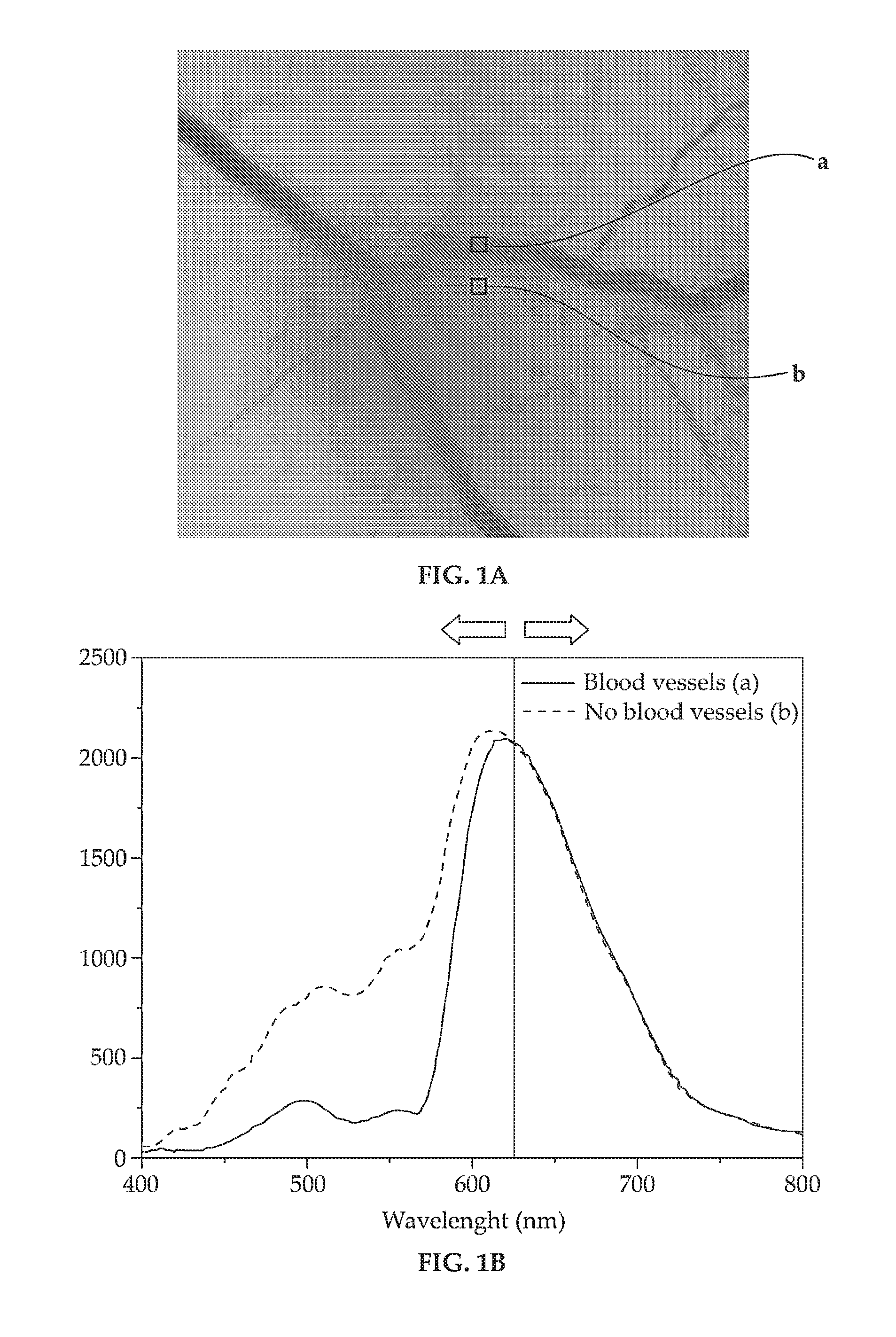
[0174]An RGB image including all the image data acquired between 400 nm and 800 nm is reproduced in black-and-white in FIG. 2A.

[0175]A red-free narrow band (520 nm-580 nm) monochrome image using data acquired between is reproduced in black-and-white in FIG. 2B. Although the contrast is superior to the equivalent RGB image, the spatial resolution appears to be similar.

[0176]An image generated in accordance with an embodiment of the method described herein where the first wavelength range was 400 nm to 600 nm and the second wavelength range was 600 nm t...


PUM


Abstract

Disclosed are methods and devices useful for imaging blood-containing tissue, for example for angiography, especially retinal angiography, whereby an image is generated by dividing pixels of an image acquired at some wavelength range by corresponding pixels of an image acquired at a different wavelength range. In some embodiments, a first wavelength range includes predominantly light having wavelengths between about 400 nm and about 620 nm and a second wavelength range includes predominantly light having wavelengths between about 620 nm and about 800 nm.

Description

RELATED APPLICATION[0001]The present application gains priority from UK Patent Application GB 1102209.2 filed 9 Feb. 2011.FIELD AND BACKGROUND OF THE INVENTION[0002]The invention, in some embodiments, relates to the field of medical imaging, and more particularly but not exclusively, to methods and devices suitable for imaging blood-containing tissue, for example for angiography, especially retinal angiography.[0003]Angiography is a technique for the in vivo imaging of blood vessels.[0004]In some instances, angiography is performed by administering a radio-opaque contrast agent in the blood vessels of a living subject and then acquiring an image of the blood vessels using an X-ray imaging modality. Such methods are useful, for example, to acquire a 3-dimensional representation of cerebral blood vessels.[0005]To avoid the use of an X-ray imaging modality, it is known to acquire a visible light image of shallow blood vessels in a bodily surface, for example, in the retina or mucosa su...

Claims


Application Information

Patent Timeline
Patent Type & Authority Applications(United States)
IPC IPC(8): A61B5/026A61B3/12
CPCA61B3/1241A61B5/0261A61B1/041A61B3/12A61B5/0075A61B5/0077A61B5/0084A61B5/489F04C2270/041G06T5/50G06T7/0012
Inventor KOSTENICH, GENADYORENSTEIN, ARIEBELKIN, MICHAEL
Owner TEL HASHOMER MEDICAL RES INFRASTRUCTURE & SERVICES
Why Eureka
Try Eureka