Implantable drug delivery device

a drug delivery and implantable technology, applied in the field of controlled time of release and rate of release, can solve the problems of large prosthesis devices, increased possibility of retinal trauma, and inability to produce adequate simulated vision to truly aid the visually impaired,

Inactive Publication Date: 2007-02-20
SECOND SIGHT MEDICAL PRODS
View PDF24 Cites 125 Cited by
  • Summary
  • Abstract
  • Description
  • Claims
  • Application Information

AI Technical Summary

Benefits of technology

[0043]It is an object of the invention to attach an electrode array body to the retina while avoiding or minimizing harmful stresses on the retina from the electrode array body.
[0044]It is an object of the invention to enable a surgeon to easily locate the mounting aperture for attachment of an electrode array body to the retina of an eye by a surgical tack.

Problems solved by technology

While some of these early attempts met with some limited success, these early prosthesis devices were large, bulky and could not produce adequate simulated vision to truly aid the visually impaired.
Such a device increases the possibility of retinal trauma by the use of its “bed of nails” type electrodes that impinge directly on the retinal tissue.
The retina is extraordinarily fragile.
In particular, retinal neurons are extremely sensitive to pressure; they will die if even a modest intraocular pressure is maintained for a prolonged period of time.
Furthermore, the retina, if it is perforated or pulled, will tend to separate from the underlying epithelium, which will eventually render it functionless.
Thus attachment of a conventional prosthetic retinal electrode device carries with it the risk of damage to the retina, because of the pressure that such a device could exert on the retina.
The penetrating electrodes are damaging to the delicate retinal tissue of a human eye and therefore are not applicable to enabling sight in the blind.
This cantilever configuration introduces complexity and it is very difficult to control the restoring force of the cantilever due to varying eye sizes, which the instant invention avoids.
This patent does not address reduction of damage to the retina and surrounding tissue or problems caused by excessive pressure between the retinal electrode array and the retina.
However, in many instances such drugs are not capable of being administered either orally or intravenously without the risk of various detrimental side effects.
For example, intravenous ganciclovir (GCV) is effective in the treatment of CMV retinitis in AIDS patients, but bone marrow toxicity limits its usefulness.
It is further limited by the risk of sepsis related to permanent indwelling catheters and the inability to receive concurrent therapy with zidovudine (AZT).
While these capillary openings in this construction are effective for releasing certain drugs to the eye, they add considerable complexity to the manufacture of the device because it is difficult to control the size of these openings in large-scale manufacturing using various polymers.
Nonetheless, as described in U.S. Pat. No. 4,014,335, certain problems have been identified with such devices such as the difficult task of sealing the margins of the membrane to form the container.
In addition, stresses and strains introduced into the membrane walls from deformation during manufacturing of those devices may cause the reservoir to rupture and leak.
However, there are many disadvantages associated with their use including the fact that it is often difficult to obtain the desired release rate of the drug.
If relatively high levels are required inside the eye, such devices are ineffective.

Method used


Image

  • Implantable drug delivery device
  • Implantable drug delivery device
  • Implantable drug delivery device

Examples


Embodiment Construction

[0062]The following description is the best mode presently contemplated for carrying out the invention. This description is not to be taken in a limiting sense, but is made merely for describing the general principles of the invention. The scope of the invention should be determined with reference to the claims.

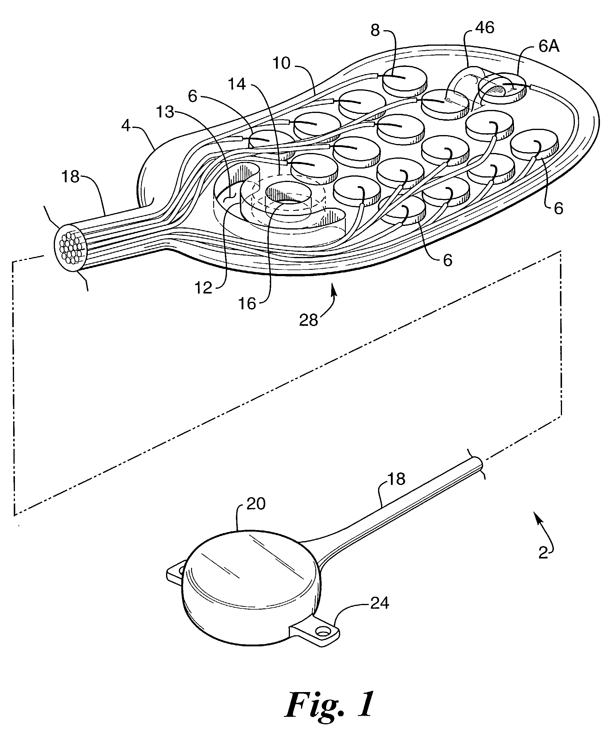
[0063]FIG. 1 provides a perspective view of a preferred embodiment of the retinal electrode array, generally designated 2, comprising oval-shaped electrode array body 4, a plurality of electrodes 6 made of a conductive material, such as platinum or one of its alloys, but that can be made of any conductive biocompatible material such as iridium, iridium oxide or titanium nitride, and single reference electrode 6A made of the same material as electrode 6, wherein the electrodes are individually attached to separate conductors 8 made of a conductive material, such as platinum or one of its alloys, but which could be made of any biocompatible conductive material, that is envelope...


PUM


Abstract

The invention is directed to an implantable device to enable delivery of drugs to the retina. The device minimizes stress to the retina by virtue of its softness and smooth shape that conform to the retina. Drugs are delivered by osmosis or by the device dissolving. It may be connected to an externally mounted pump and drug reservoir that control the amount of drug. It contains one or more holes that are positioned to deliver drugs to the desired location. Drugs may stimulate the retina to enable vision in blind patients. Drugs may be injected directly inside the eye by a trans-scleral pump and valve drug delivery device.

Description

CROSS REFERENCE TO RELATED APPLICATION[0001]This application is a continuation-in-part of U.S. patent application Ser. No. 09 / 783,236, filed on Feb. 13, 2001, entitled “IMPLANTABLE RETINAL ELECTRODE ARRAY CONFIGURATION FOR MINIMAL RETINAL DAMAGE AND METHOD OF REDUCING RETINAL STRESS.”FEDERALLY SPONSORED RESEARCH[0002]This invention was made with government support under grant No.[0003]R24EY12893-01, awarded by the National Institutes of Health. The government has certain rights in the invention.FIELD OF THE INVENTION[0004]This invention relates to a device and methods and more particularly to a controlled time of release and rate of release, drug delivery device, which may be implanted in a living body.BACKGROUND OF THE INVENTION[0005]In 1755, LeRoy passed the discharge of a Leyden jar through the orbit of a man who was blind from cataract and the patient saw “flames passing rapidly downwards.” Ever since, there has been a fascination with electrically elicited visual perception. Th...

Claims


Application Information

Patent Timeline
Patent Type & Authority Patents(United States)
IPC IPC(8): A61N1/00A61K9/22A61N1/05
CPCA61N1/0543A61F9/0017
Inventor GREENBERG, ROBERT
Owner SECOND SIGHT MEDICAL PRODS
Why Eureka
Try Eureka