
Method and Apparatus for Registering Image Data Between Different Types of Image Data to Guide a Medical Procedure

a technology of image data and image data, applied in the field of image processing, can solve the problems of high false negative rate of trus-guided biopsy for prostate cancer diagnosis, difficult or impossible manual correlating of image data between two types of image modalities, etc., and achieve the effect of improving diagnosis and/or treatmen

Inactive Publication Date: 2015-01-15
MADABHUSHI ANANT +1
View PDF4 Cites 16 Cited by
  • Summary
  • Abstract
  • Description
  • Claims
  • Application Information

AI Technical Summary

Benefits of technology

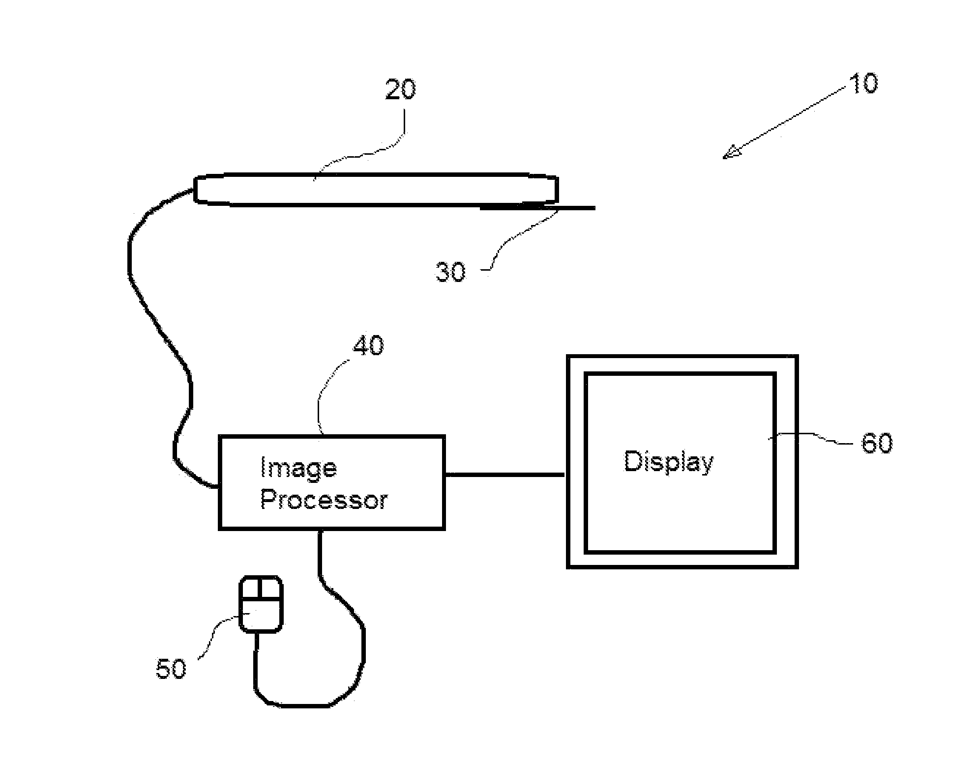
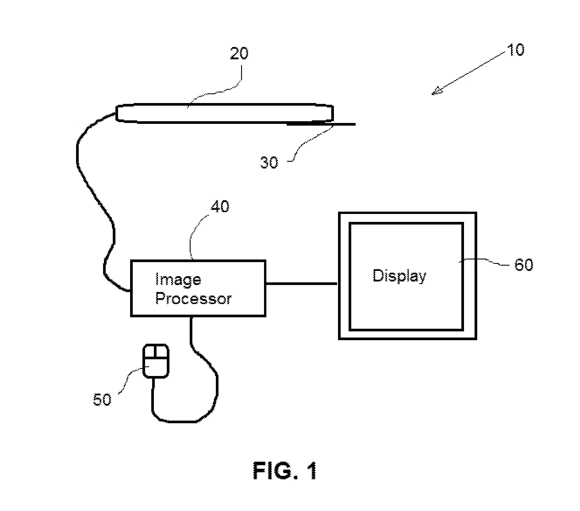
This patent describes a system that can combine different types of medical images, like MRI and ultrasound, to help doctors make better diagnostic and treatment decisions. The system automatically aligns the images, so that medical professionals don't have to manually adjust them. This can make the process easier and more accurate, which can improve the quality of care for patients.

Problems solved by technology

However, manually correlating the image data from the two types of image modalities can be difficult or impossible in many instances.
Hence TRUS-guided biopsy has a high false negative rate for prostate cancer diagnosis.

Method used


Image

  • Method and Apparatus for Registering Image Data Between Different Types of Image Data to Guide a Medical Procedure
  • Method and Apparatus for Registering Image Data Between Different Types of Image Data to Guide a Medical Procedure
  • Method and Apparatus for Registering Image Data Between Different Types of Image Data to Guide a Medical Procedure

Examples


example 1

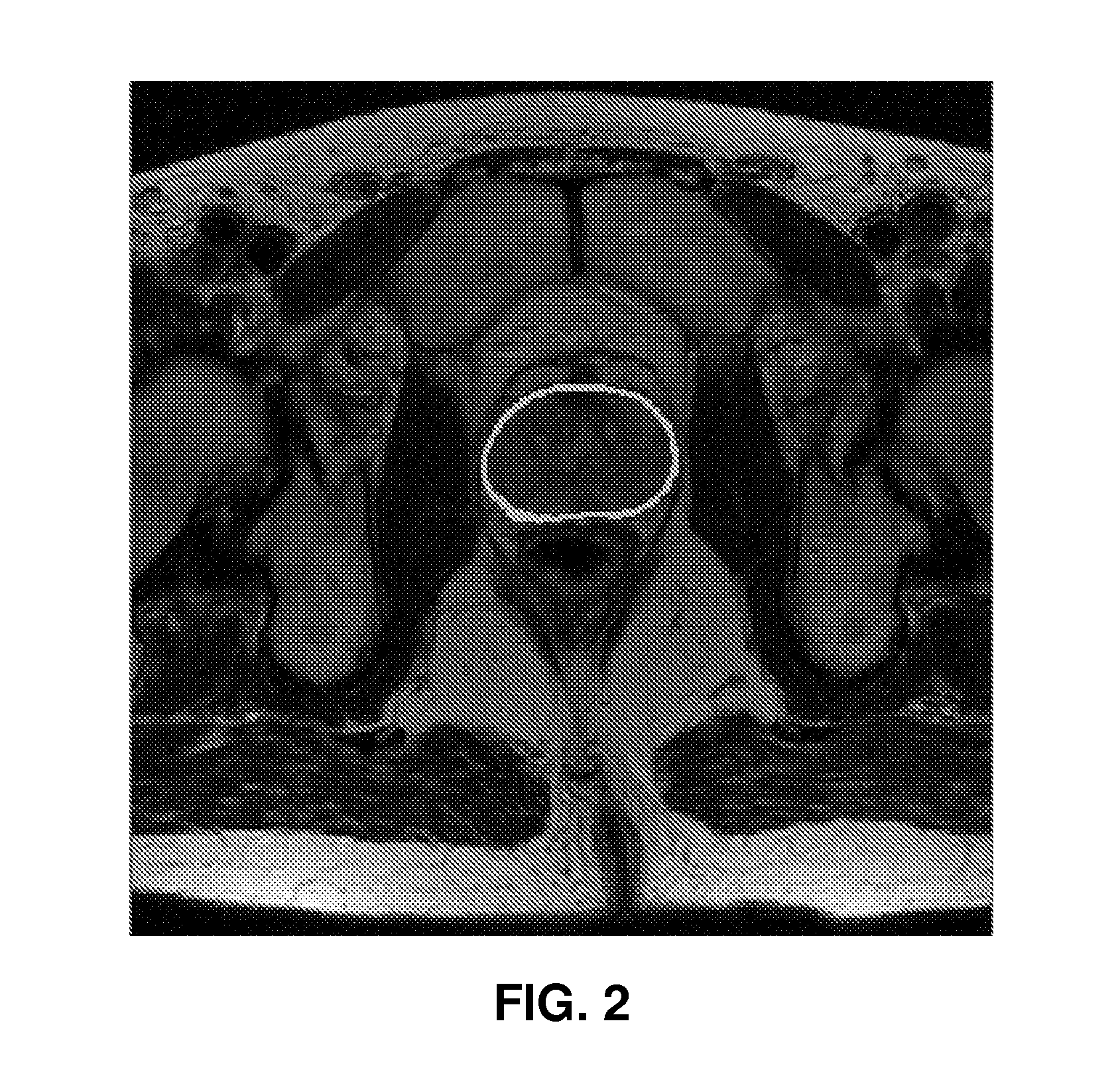
[0079]A T2 weighted MRI was acquired using a Siemens 1.5 T scanner and a pelvic phase array coil for seven patients. Three dimensional TRUS imagery was acquired using a bi-planar side-firing transrectal probe. A radiologist segmented each prostate and selected 3-5 landmarks on MRI and TRUS images. Registration was performed by segmenting the prostate in the MRI manually. The prostate location was then estimated for the TRUS image. The identified MRI portion is then registered with the estimated TRUS portion. The registration can be evaluated using any of a number of measurements or analytics. In the present instance, registration was evaluated using the sum of square differences of landmarks and prostate overlap. In the present instance, the measured overlap was 5.85±1.08 mm and an overlap of 73.6±4.7%

[0080]Automated Image Registration

[0081]In addition to the process described above, the process of registering the MRI and TRUS data can be semi-automated or fully automated. An automa...

experiment 1

tenuation Correction on Registration Accuracy

[0141]It is possible that subtle differences in intensity characteristics across the TRUS image can lead to a probabilistic model Pi(c) that does not accurately model the prostate location. Incorrect estimation of Pi(c) can therefore result in sub-optimal image registration. The effect of attenuation correction was evaluated on registration accuracy in terms of RMSE. The MAPPER process was evaluated with and without attenuation correction for D1.

[0142]FIG. 11 illustrates the quantitative results for Te, with and without attenuation correction for five texture features. Attenuation correction has two effects on the registration results: (1) attenuation reduces RMSE variance across studies and therefore gives a more robust image registration; and (2) attenuation lowers RMSE and, hence, provides a more accurate registration accuracy. The positive effects of attenuation correction on registration occur independent of which feature was used.

experiment 2

Regularization Weight

[0143]The regularization weight ∝ controls the relative importance of a smooth Te and accurately registering the prostate mask on MRI to the TRUS probabilistic model (i.e. maximizing Equation 10). To assess the sensitivity of the performance of the MAPPER process on the choice of ∝, the regularization weight ∝ was varied for {100, 1, 1 E-2, 1 E-4} and assessed RMSE for D1.

[0144]FIG. 12 illustrates the RMSE for each set of regularization parameters ∝ evaluated for two texture features, (a) Rayleigh and (b) variance. RMSE changes little with respect to ∝, even across a wide range of values, ∝ E {100, 1, 1E-2, 1E-4}.


PUM


Abstract

A method and apparatus are provided for registering image data between two different types of image data. The image data is obtained during two separate scanning processes. The first process is a high-resolution imaging process, such as a CT scan or an MRI. The second process is a lower resolution process, such as ultrasound. The image data is registered in real time so that the combined image data can be displayed during the second process to guide a medical professional during a procedure.

Description

PRIORITY CLAIM[0001]This application claims priority to U.S. Provisional App. No. 61 / 845,678 filed on Jul. 12, 2013. The entire disclosure of the foregoing application is hereby incorporated herein by reference.FIELD OF THE INVENTION[0002]The present invention relates to the field of image processing. In particular, this application relates to the field of registering portions of one image with a portion of one or more separate images. The images are registered in real time to guide a medical procedure, such as a biopsy or treatment.BACKGROUND[0003]In fields, such as the medical industry, it is useful to correlate image data from one image with image data from a separate modality. For instance, in the medical field, there are numerous types of images that may be used during diagnostics or treatment for a patient, including MRI, CT scans, ultrasound, x-rays and others. These various imaging modalities each have advantages and disadvantages. Furthermore, medical professionals may have...

Claims


Application Information

Patent Timeline
Patent Type & Authority Applications(United States)
IPC IPC(8): A61B8/08A61B10/02G01R33/28A61B18/20G01R33/48A61B8/00A61B10/04
CPCA61B8/463A61B8/5261G01R33/285A61B2018/2005A61B10/0233A61B5/0035A61B10/04A61B2010/045G01R33/4814A61B5/055A61B18/20A61B8/0841G01R33/5608A61B6/032A61B6/5247A61B8/12G06T2207/10088G06T2207/10132G06T2207/20076G06T2207/20221G06T2207/30081G06T7/33G06T7/35
Inventor MADABHUSHI, ANANTSPARKS, RACHEL
Owner MADABHUSHI ANANT
Learn More