
Cohesive robot-ultrasound probe for prostate biopsy

a robot and ultrasonic technology, applied in the field of diagnostic screening, can solve the problems of high false-negative rate, significant shortcoming and high false-negative rate, and difficult to account for and quantify

Active Publication Date: 2015-03-05
THE JOHN HOPKINS UNIV SCHOOL OF MEDICINE
View PDF5 Cites 12 Cited by
  • Summary
  • Abstract
  • Description
  • Claims
  • Application Information

AI Technical Summary

Benefits of technology

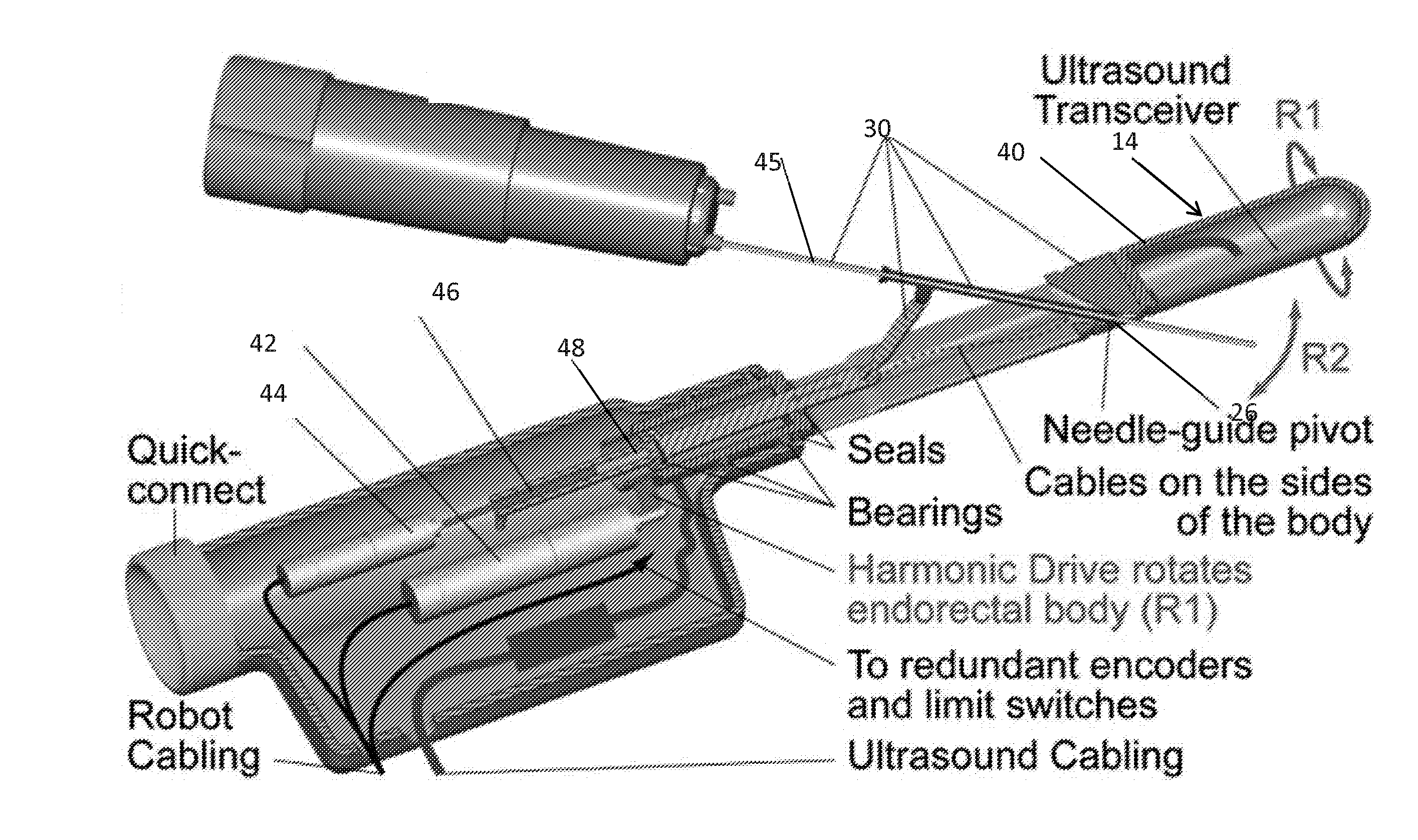
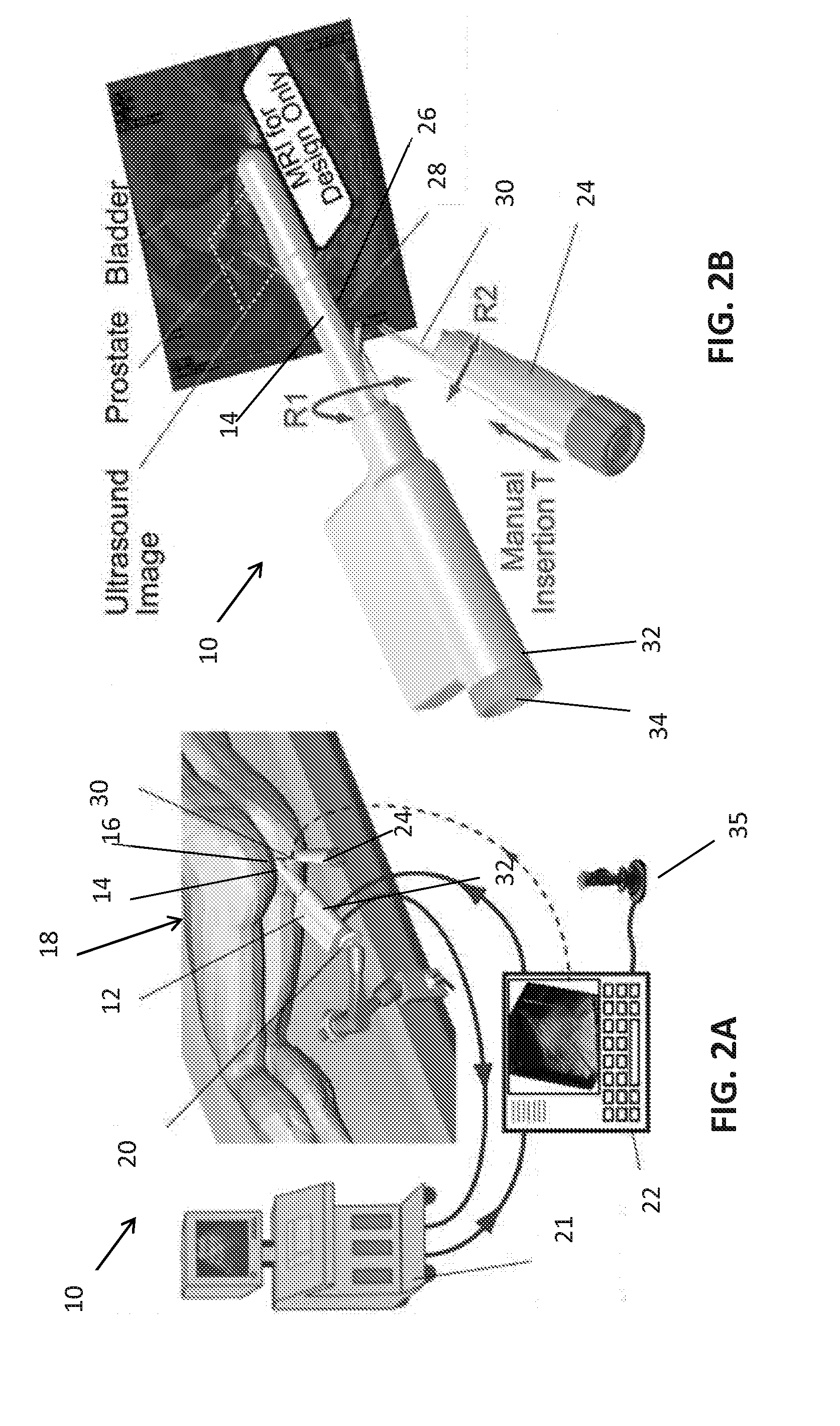
This patent describes a device for taking biopsies that includes a handle with a first motor to rotate the probe and a needle guide pivot adjacent to the channel. There is also a second motor for pivoting the biopsy needle within the channel, and a spacer to adjust the depth of the needle for better control. The technical effect of this device is improved accuracy and efficiency in taking biopsies.

Problems solved by technology

Surgery and radiation therapy can achieve excellent cancer control, but both surgery and radiation therapy are associated with adverse effects and an increased burden to our healthcare system.
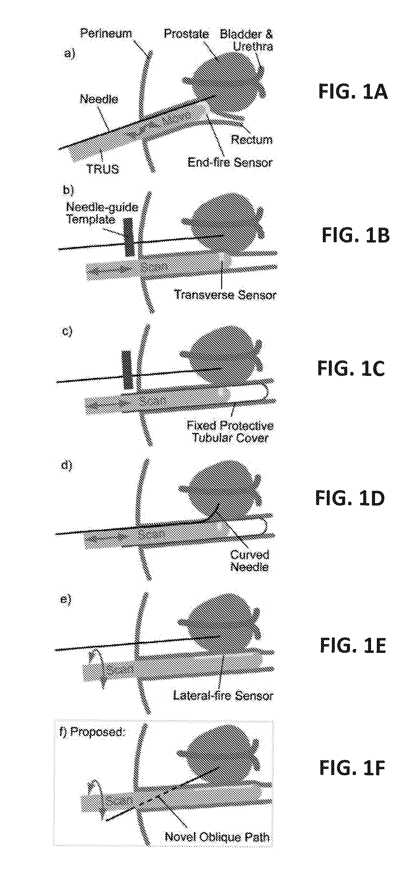
The current transrectal ultrasound-guided (TRUS) prostate biopsy, however, has significant shortcoming and high false-negative rates, largely related to targeting inaccuracies.
These biopsy navigation devices address several targeting error components but are commonly affected by prostate deformations, which are difficult to account for and to quantify.
These errors may be relatively large.
In fact, these errors can be larger than the radius of a “clinically significant” PCa tumor, and therefore, these errors alone can deteriorate the targeting plan.
Sensor array probes produce faster 3D acquisition but image quality and resolution tend to be lower because of space constraints, limiting the ability to guide the biopsy, because anatomic landmarks are more difficult to distinguish.
Internal motion scanning probes preserve 2D image quality but have longer acquisition times, making it difficult to guide the intervention in real-time.
However, these systems generally require the use of a transperineal biopsy path or cause deflections of the prostate.
The transperineal path is rarely used for biopsy because it causes more discomfort for the patients.
But, for most of the biopsy patient population-at-large, the transperineal biopsy is logistically and economically unfeasible.
The resulting images are distorted, and the volume is skewed and not entirely reliable if rendered in 3D.
Deflections are very difficult to quantify and correct.
This is a very common but difficult task with subjective planning, navigation, and quality control.
Among the problems is that prostate deflections are inexorable because the sensor must keep in contact with the rectum for the sonic waves to propagate.
The schematic shows a simple indentation, but in reality deflections may be complex.
While freehanded, it is nearly impossible to maintain the state of deflection while imaging and taking the biopsy.
Navigation and / or 3D are very helpful for the physician, but prostate deflections and derived skewed image problems remain.
This is also the case for TRUS-MRI fusion based on freehanded TRUS, deflections further deteriorating fusion accuracy.
Transrectal needle access is unfeasible.
However, that was problematic because core biopsy needles have internally moving parts which may jam if bent.
But perhaps more difficult to account are targeting errors.
When a needle is curved the amount of resistance encountered at the needle point contributes to its curvature making targeting uncertain when passing the heterogeneous tissues.
An apparent problem with the lateral-fire rotary scan approach is that it can't be used for transrectal biopsy.
For this approach however, the needle must cross the shaft of the probe, which is not possible with current probes.
However, this probe does not have a lateral-fire sensor.
Moreover, the needle-guide is locked to the probe thus inducing deformations when aligning for biopsy.
However, this is problematic because the needle may injure the anus and cause significant discomfort, and because the needle will lie outside the ultrasound plane making it difficult to monitor.

Method used


Image

  • Cohesive robot-ultrasound probe for prostate biopsy
  • Cohesive robot-ultrasound probe for prostate biopsy
  • Cohesive robot-ultrasound probe for prostate biopsy

Examples


Embodiment Construction

[0031]The presently disclosed subject matter now will be described more fully hereinafter with reference to the accompanying Drawings, in which some, but not all embodiments of the inventions are shown. Like numbers refer to like elements throughout. The presently disclosed subject matter may be embodied in many different forms and should not be construed as limited to the embodiments set forth herein; rather, these embodiments are provided so that this disclosure will satisfy applicable legal requirements. Indeed, many modifications and other embodiments of the presently disclosed subject matter set forth herein will come to mind to one skilled in the art to which the presently disclosed subject matter pertains having the benefit of the teachings presented in the foregoing descriptions and the associated Drawings. Therefore, it is to be understood that the presently disclosed subject matter is not to be limited to the specific embodiments disclosed and that modifications and other ...


PUM


Abstract

An embodiment in accordance with the present invention provides a device and method for a transrectal ultrasound (TRUS) guided prostate biopsy. The device includes an ultrasound wand equipped with a lateral fire ultrasound sensor. The ultrasound wand also defines a channel having an oblique path though the wand. The channel accommodates a biopsy needle, which can be pivoted as well inserted through the channel to different depths in order to perform a biopsy of the prostate. The device therefore uses only three degrees-of-freedom of movement in order to obtain the biopsy samples. Additionally, the ultrasound wand can be used to generate a three-dimensional image of the prostate.

Description

CROSS REFERENCE TO RELATED APPLICATIONS[0001]This application claims the benefit of U.S. Provisional Patent Application No. 61 / 622,130 filed on Apr. 10, 2012, which is incorporated by reference, herein, in its entirety.FIELD OF THE INVENTION[0002]The present invention relates generally to diagnostic screening. More particularly, the present invention relates to a device for performing a prostate biopsy.BACKGROUND OF THE INVENTION[0003]Prostate cancer (PCa) is the most frequently diagnosed non-skin malignancy for men in the United States. Studies have shown that it is necessary to treat 48 men to prevent one death from PCa, suggesting that significant overtreatment exists. On the other hand, in 2010 more than 32,000 men died of PCa. Surgery and radiation therapy can achieve excellent cancer control, but both surgery and radiation therapy are associated with adverse effects and an increased burden to our healthcare system. Alternative management options evolved, such as active surveil...

Claims


Application Information

Patent Timeline
IPC IPC(8): A61B10/02A61B8/12A61B19/00A61B8/08
CPCA61B10/0241A61B19/2203A61B8/12A61B8/0841A61B8/4245A61B34/30A61B2017/00274A61B2017/3413A61B2018/00547A61B2090/3782F04C2270/0421
Inventor STOIANOVICI, DANHAN, MISOPPETRISOR, DORUKIM, CHUNWOO
Owner THE JOHN HOPKINS UNIV SCHOOL OF MEDICINE
Learn More