Illumination System for Endoscopic Applications

a technology of endoscopic applications and liquid core fibers, which is applied in the field of liquid core fibers for endoscopic applications, can solve the problems of extremely vulnerable and high cost of liquid core fibers, and achieve the effects of improving the treatment of bladder cancer and/or tumors, and improving the diagnosis of bladder cancer

Inactive Publication Date: 2015-03-26
DANMARKS TEKNISKE UNIV +1
View PDF11 Cites 13 Cited by
  • Summary
  • Abstract
  • Description
  • Claims
  • Application Information

AI Technical Summary

Benefits of technology

[0032]Bladder cancer is identified and resected during endoscopic examination of the bladder through the urethra. A new kind of photodynamic diagnosis (PDD) of bladder cancer (BC) was developed in 2001 where hexaminolevulinate or 5-aminolevulinic acid (5-ALA) is used as precursor to the dye. The aminoacid 5-ALA is metabolized to protoporphyrin IX (PPIX) in the malignant cells which fluoresces at approx. 635 nm when excited with e.g., blue light. During endoscopic examination using a white light source as examination light, the malignant areas become visible as clear red areas when the light is shifted to a blue excitation light source. PDD improves the diagnoses and thus the treatment of bladder cancer substantially. The recurrence rate of bladder cancer within one year after tumor surgery is reduced from 47% to 30% when using PDD.
[0033]A problem with this procedure is that yellow urine in the bladder is also excited by the blue light to generate strong green fluorescence. This confounds the fluorescence and the vision in the bladder becomes heavily impaired by the green fluorescence and thus the diagnosis of the malignant tissue. This problem may be reduced by washing the bladder with glycine or saline prior to examination, but the subsequent examination is still a race against time because new urine may be accumulated in the bladder during examination which may make it difficult to identify all bladder tumors. A further object of the invention is therefore to improve the diagnosis of bladder cancer and / or tumors. This object is achieved by a cystoscopic system for photodynamic diagnosis of bladder cancer comprising an endoscopic tube accommodating at least one optical transmission path adapted to guide excitation light and examination light to a region of examination, and an optical relay system adapted to image the region of examination and accommodating an optical band-rejection filter adapted to attenuate at least said excitation light, wherein the excitation light is adapted to excite fluorescence of a photosensitive compound and the wavelength of the excitation light source is greater than 460 nm, more preferably greater than 500 nm and less than 550 nm.

Problems solved by technology

Bulky liquid core fibers with diameter of up to 8 mm may be used in the endoscope to transport the light, but such liquid core fibers are very costly and extremely vulnerable.

Method used


Image

  • Illumination System for Endoscopic Applications
  • Illumination System for Endoscopic Applications
  • Illumination System for Endoscopic Applications

Examples


Embodiment Construction

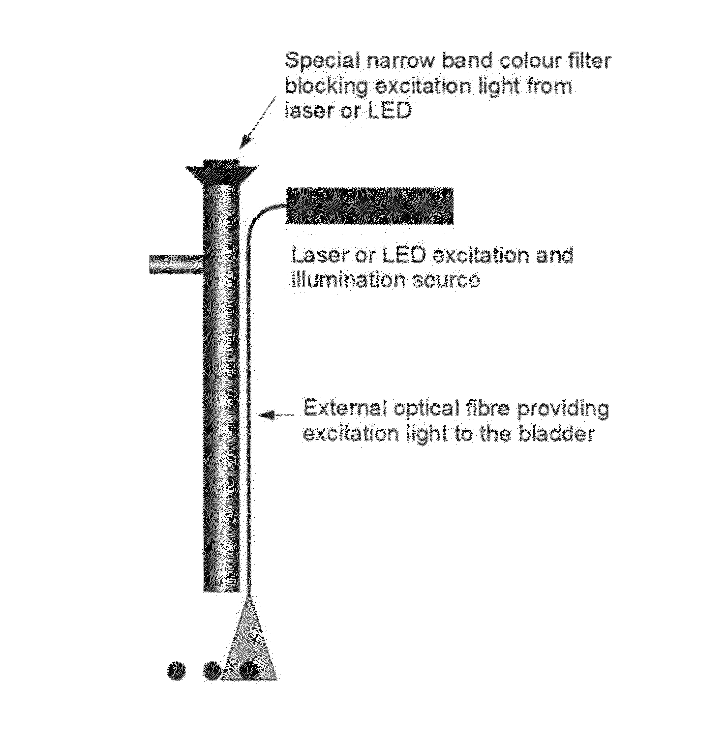
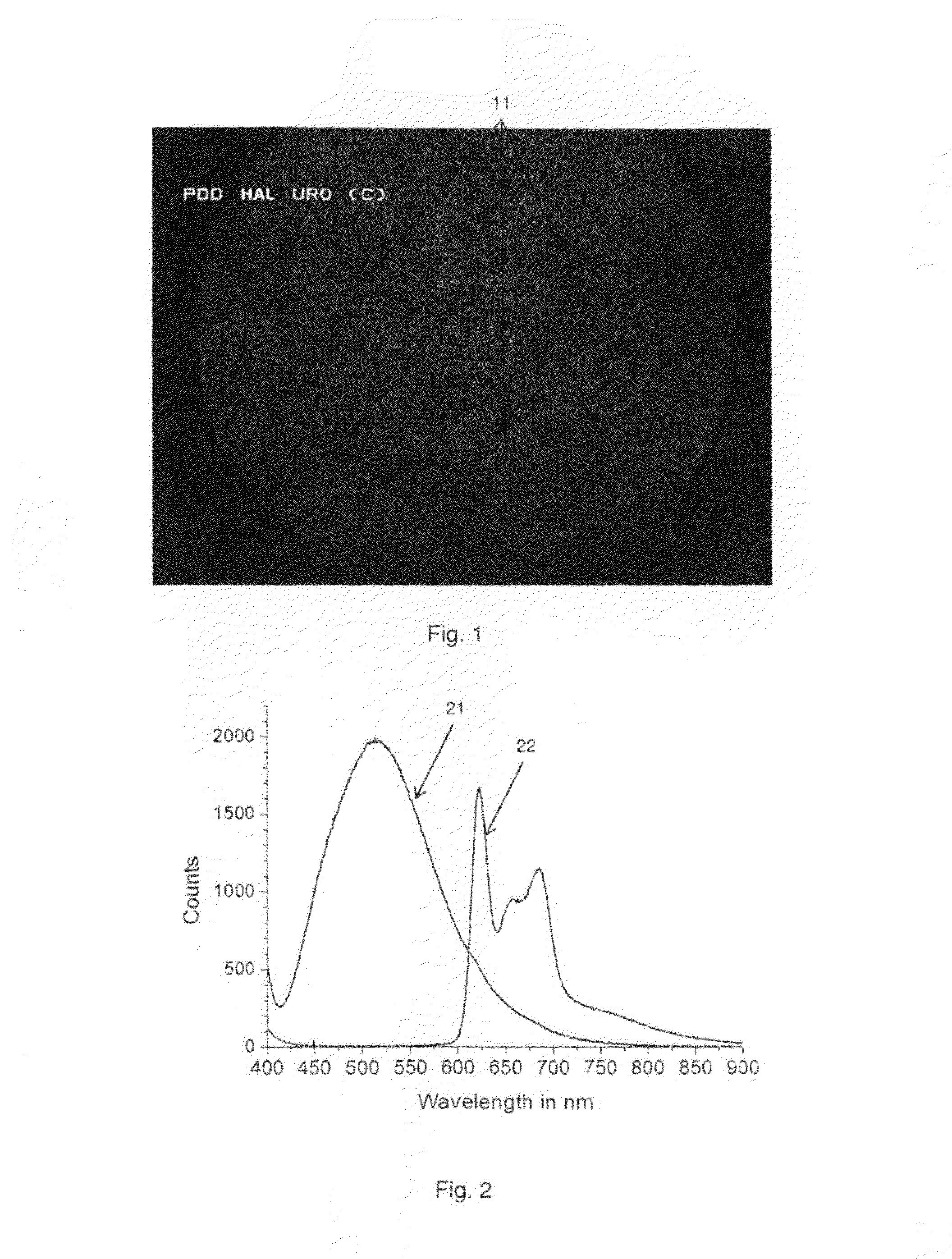
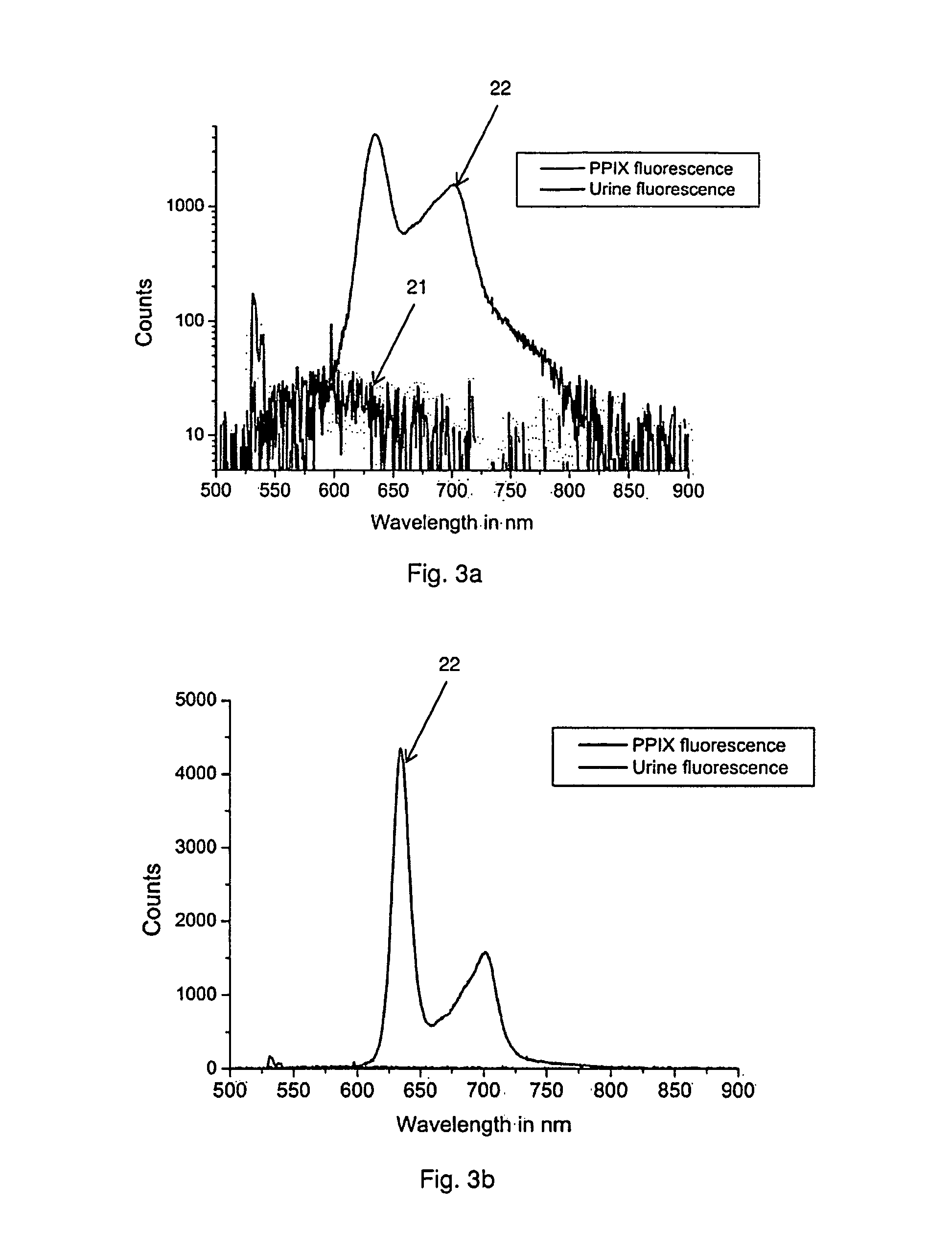
[0038]As stated above the present disclosure relates to an endoscope for examining a body cavity comprising tissue, the endoscope comprising a source of light consisting of a substantially monochromatic light source having a predefined central wavelength between 400 and 550 nm, means for guiding light from the substantially monochromatic light source towards at least a part of the tissue, and at least one band-rejection filter adapted to attenuate at least said central wavelength, wherein the substantially monochromatic light source is configured to generate autofluorescence in the irradiated tissue such that the irradiated tissue is observable, and wherein the endoscope is configured to display at least a part of the irradiated tissue through said band-rejection filter.

[0039]The present disclosure also relates to a method for illuminating at least a part of the inner surface of a body cavity of a subject for a viewer, the body cavity comprising tissue, the method comprising the ste...


PUM


Abstract

The present disclosure relates to an illumination system for endoscopic applications comprising at least one substantially monochromatic light source having a predefined central wavelength between 400 and 500 nm or between 500 and 550 nm, an optical transmission path adapted to guide light emanating from the light source to an endoscopic region of examination, and an optical band-rejection filter, wherein the illumination system is adapted to illuminate at least a part of the region of examination by generating autofluorescence in surrounding tissue, and the band-rejection filter is adapted to attenuate at least said light source wavelength to a viewer and wherein said light source is the single light source in the illumination system. A further embodiment relates to an endoscope for examining a body cavity comprising tissue, the endoscope comprising a source of light consisting of a substantially monochromatic light source having a predefined central wavelength between 400 and 550 nm, means for guiding light from the substantially monochromatic light source towards at least a part of the tissue, and at least one band-rejection filter adapted to attenuate at least said central wavelength, wherein the substantially monochromatic light source is configured to generate autofluorescence in the irradiated tissue such that the irradiated tissue is observable, and wherein the endoscope is configured to display at least a part of the irradiated tissue through said band-rejection filter. A system for photodynamic diagnosis and/or therapy of bladder cancer is further disclosed herein.

Description

[0001]The present disclosure relates to an endoscope for examining a body cavity and a system for illuminating the region of examination in medical endoscopic application. The present disclosure also relates to a method for illuminating at least a part of the inner surface of a body cavity. A system for photodynamic diagnosis and / or therapy of bladder cancer is furthermore disclosed herein.BACKGROUND OF INVENTION[0002]Endoscopy is gaining increased popularity in the medical world because of reduced costs in relation to examination, diagnosis, therapy and surgery and reduced risks, discomfort and nuisance to the patients.[0003]An endoscope typically consists of a rigid or flexible tube with a distal end entering the patient and a proximal end towards the user of the endoscope. A light delivery system is typically provided to illuminate a body cavity or the organ or object under inspection (the region of examination), an optical or electrical relay system for transmitting the image to...

Claims


Application Information

Patent Timeline
Patent Type & Authority Applications(United States)
IPC IPC(8): A61B1/06A61B1/04A61B5/00F21V8/00F21V9/08
CPCA61B1/063G02B6/0008A61B1/04A61B5/0071F21V9/08A61B1/00186A61B1/043
Inventor LINDVOLD, LARSHERMANN, GREGERS
Owner DANMARKS TEKNISKE UNIV
Why Eureka
Try Eureka