
Ex vivo expansion of myogenic stem cells by notch activation

a myogenic stem cell and ex vivo technology, applied in the field of tissue repair by stem cell transplantation, can solve the problems of affecting the engraftment of donor satellite cells, affecting the engraftment of muscle tissue, and unable to provide enough cells, so as to promote muscle regeneration, promote muscle tissue regeneration, and increase the engraftment potential to a transplantation site.

Inactive Publication Date: 2015-06-18
FRED HUTCHINSON CANCER RES CENT
View PDF6 Cites 3 Cited by
  • Summary
  • Abstract
  • Description
  • Claims
  • Application Information

AI Technical Summary

Benefits of technology

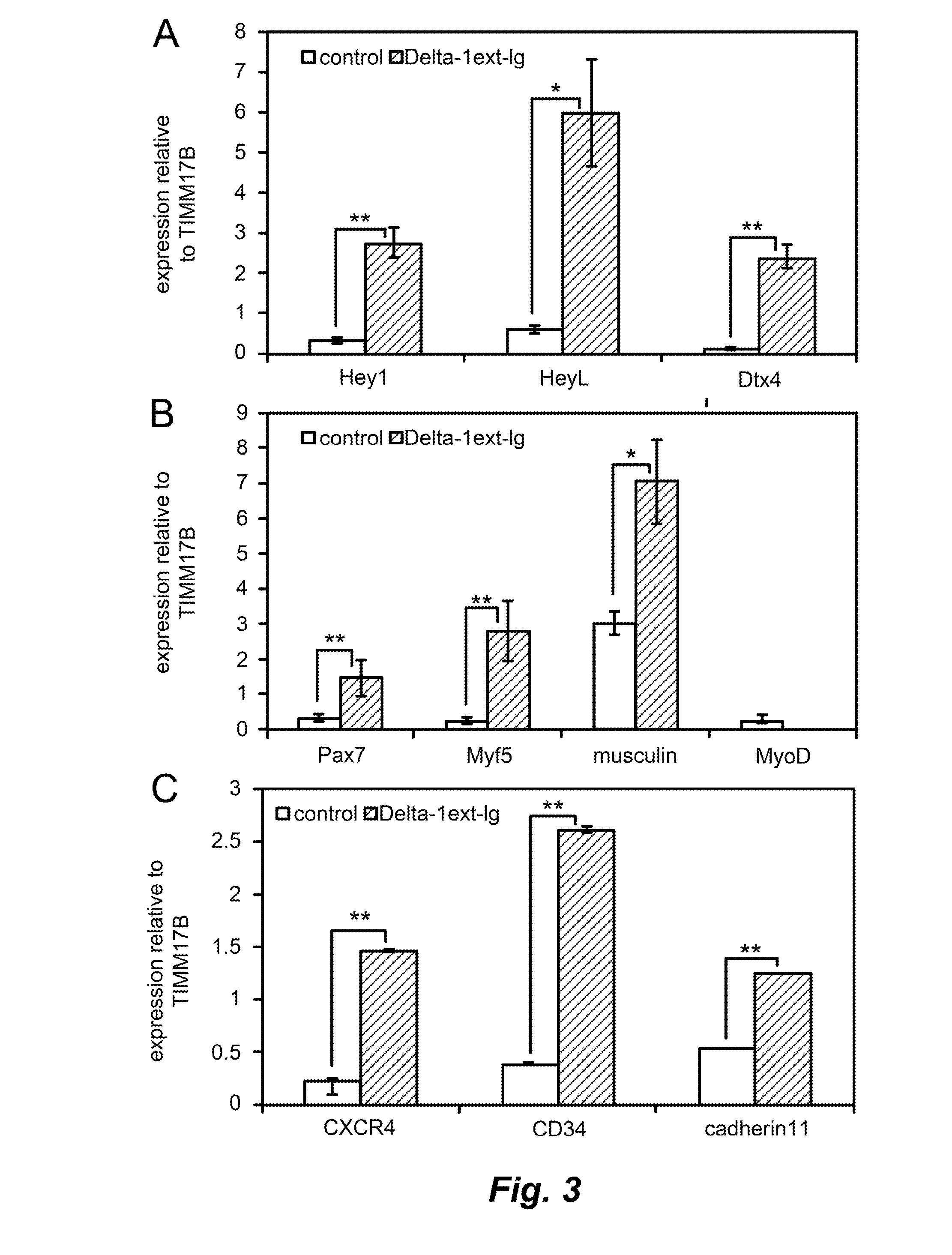
The present invention provides a method for expanding myogenic precursor cells while preserving their engraftment potential. This is achieved by activating Notch signaling in the cells through contact with an immobilized Notch ligand. The method results in a statistically significant increase in the expression of marker genes, such as Hey1, NP—001002953, NM—012258, NM—036390.3, and NM—001040708. The method also involves detectable activation of Notch signaling in a statistically significant percentage of myogenic precursor cells. The immobilized Notch ligand can be an extracellular domain of human delta-like-1 or a fusion protein containing the Notch ligand and a fusion domain polypeptide.

Problems solved by technology

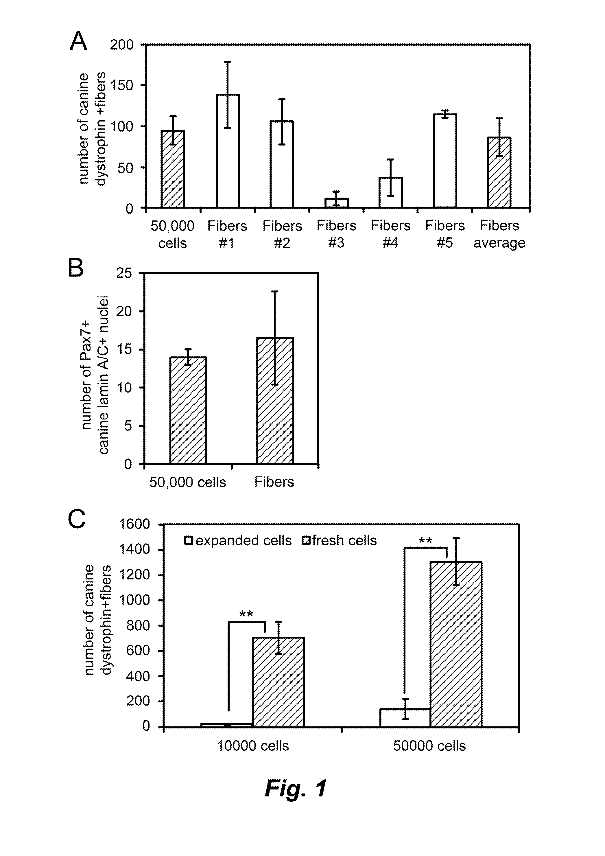
However, it is also possible that time away from the fiber or niche has a negative effect on donor satellite cell engraftment.
However, multiple muscle groups within the body will need to be targeted, and a single donor muscle biopsy is unlikely to provide enough cells to effectively transplant the muscle mass of a patient affected by muscular dystrophy.
Traditional means of expanding satellite cell-derived myoblasts ex vivo results in a dramatic loss of engraftment potential [4, 5].

Method used


Image

  • Ex vivo expansion of myogenic stem cells by notch activation
  • Ex vivo expansion of myogenic stem cells by notch activation
  • Ex vivo expansion of myogenic stem cells by notch activation

Examples


example 1

Ex Vivo Expansion of Myogenic Precursors that are Capable of Muscle Engraftment

[0049]Materials and Methods:

[0050]Donor Cell Isolation.

[0051]The Institutional Animal Care and Use Committee at the Fred Hutchinson Cancer Research Center, which is fully accredited by the Association for Assessment and Accreditation of Laboratory Animal Care, approved this study. Elevated enclosed runs were used for housing, and dogs were maintained in social groups wherever possible. All dogs were enrolled in a veterinary preventative medicine program that included a standard immunization series against canine distemper, parvovirus, adenovirus type 2, parainfluenza virus, coronavirus, and rabies.

[0052]Each donor canine underwent a maximum of 4 skeletal muscle biopsies. For each canine-to-murine transplantation experiment, a 1 cm×1 cm×0.5 cm skeletal muscle biopsy was harvested from the biceps femoris muscle of the donor canine. The muscle biopsy was trimmed and cut into smaller pieces along the length o...

example 2

Upregulation of Wnt Signaling Pathway Components by Notch Activation

[0127]A survey by RT-qPCR of Wnt receptor expression in proliferating myoblasts and in myogenic precursor cells expanded on either Delta-1ext-IgG or human IgG (as described in Example 1) demonstrated that Fzd2, Fzd4, Fzd7, Ror2, and Ryk were expressed in canine muscle derived cells (FIG. 7A). Activation of Notch signaling in the same cells increased expression of Fzd4, a mediator of non-canonical Wnt signaling, and Dkk2, an extracellular antagonist of canonical Wnt signaling (FIG. 7B). Wnt3a has been shown to stimulate proliferation of Pax7+ cells in vitro, yet Brack and colleagues demonstrated that treating muscle after injury with Wnt3a activated canonical Wnt signaling, and stimulated differentiation at the expense of myogenic progenitor proliferation (Brack et al., 2007 Science 317:807; Brack et al., 2008 Cell Stem Cell 2:50; see also Otto et al., 2008 J. Cell Sci. 121:2939). On the other hand, Wnt7a, acting thr...


PUM

PropertyMeasurementUnit
densityaaaaaaaaaa
temperatureaaaaaaaaaa

Abstract

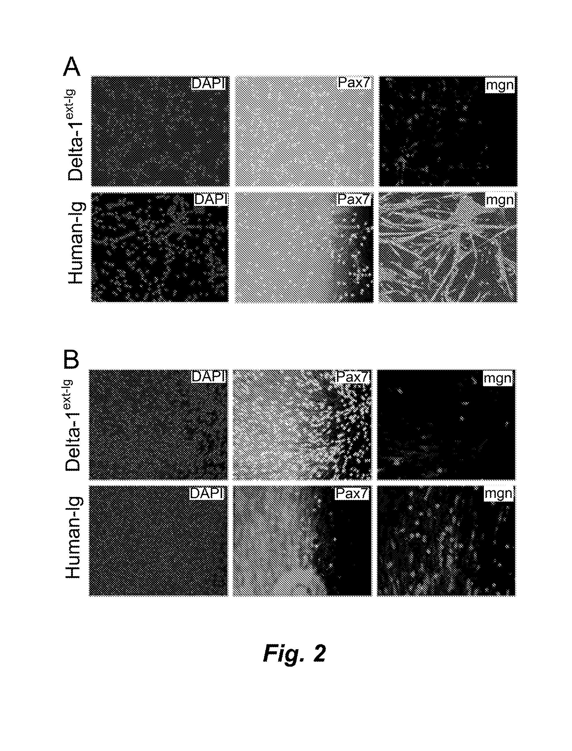
Activating Notch signaling in cultured canine muscle derived cells inhibited myogenic differentiation, and increased the number of myogenic progenitor cells that were similar to quiescent or newly activated satellite cells. Importantly, cells expanded in the presence of Notch activation maintained engraftment potential, indicating the potential for therapeutic benefit. Activation of Notch signaling to inhibit myogenic differentiation in cultured human muscle-derived cells is also contemplated, for maintaining engraftment potential using such human cells in transplantation.

Description

CROSS REFERENCE TO RELATED APPLICATION[0001]This application claims the benefit of U.S. provisional patent application Ser. No. 61 / 659,912, filed Jun. 14, 2012, which is incorporated herein by reference in its entirety.STATEMENT OF GOVERNMENT INTEREST[0002]This invention was made with government support under Grant No. P01-NS046788-07 awarded by the National Institute of Neurological Disorders and Stroke, and Grant No. U01-HL100395 awarded by the National Heart, Lung, and Blood Institute. The government has certain rights in this invention.STATEMENT REGARDING SEQUENCE LISTING[0003]The Sequence Listing associated with this application is provided in text format in lieu of a paper copy, and is hereby incorporated by reference into the specification. The name of the text file containing the Sequence Listing is 360056—415WO_SEQUENCE_LISTING_.txt. The text file is 172 KB, was created on Jun. 14, 2013, and is being submitted electronically via EFS-Web.BACKGROUND[0004]1. Technical Field[00...

Claims


Application Information

Patent Timeline
Patent Type & Authority Applications(United States)
IPC IPC(8): C12N5/077A61K35/34
CPCC12N5/0658C12N2501/42C12N2501/415A61K35/34C07K14/47C07K2319/61C12Q1/68
Inventor PARKER, MAURA H.TAPSCOTT, STEPHEN J.
Owner FRED HUTCHINSON CANCER RES CENT
Learn More