Multiplexed method for diagnosing classical hodgkin lymphoma

a multi-method, classical hodgkin lymphoma technology, applied in the field of diagnostics of classical hodgkin lymphoma, can solve the problems of consuming a dozen, difficult to assess, and difficult to diagnose, and achieve the effects of enhancing information, improving patient care, and ensuring accuracy

Inactive Publication Date: 2015-08-13
GE HEALTHCARE BIO SCI CORP
View PDF1 Cites 9 Cited by
  • Summary
  • Abstract
  • Description
  • Claims
  • Application Information

AI Technical Summary

Benefits of technology

[0009]The invention includes embodiments that provide innovative classical Hodgkin lymphoma diagnostics which enable qualitative and quantitative assessment of biomarkers and allow for visualization of biomarker co-localization at the cellular level on a single slide. This provides greater precision and true concordance between biomarker signals, thus enhancing the information provided by pathologists to oncologists, resulting in better patient care. The method is suited for the diagnosis of classical Hodgkin lymphoma, in which often only a small amount of tissue is available for testing. Similarly, the method could be adapted to suit the diagnosis of other diseases where only a small or rare tissue sample is obtainable or only rare atypical cells are present in the biopsy.

Problems solved by technology

The diagnosis of classical Hodgkin lymphoma may be very difficult because cells resembling RS / H cells may be associated with other diseases, e.g., reactive lymphadenopathy or non-Hodgkin lymphomas.
This is difficult to assess in serial slides and could consume a dozen slides and hamper a diagnosis of Hodgkin lymphoma.
Some of the key challenges of this technique are that serial immunostains are used and hence it can be difficult or impossible to locate the same Hodgkin cell on adjacent slides.
As a consequence, about a third of classical Hodgkin lymphoma cases are considered difficult to diagnose.

Method used


Image

  • Multiplexed method for diagnosing classical hodgkin lymphoma
  • Multiplexed method for diagnosing classical hodgkin lymphoma
  • Multiplexed method for diagnosing classical hodgkin lymphoma

Examples


example 1

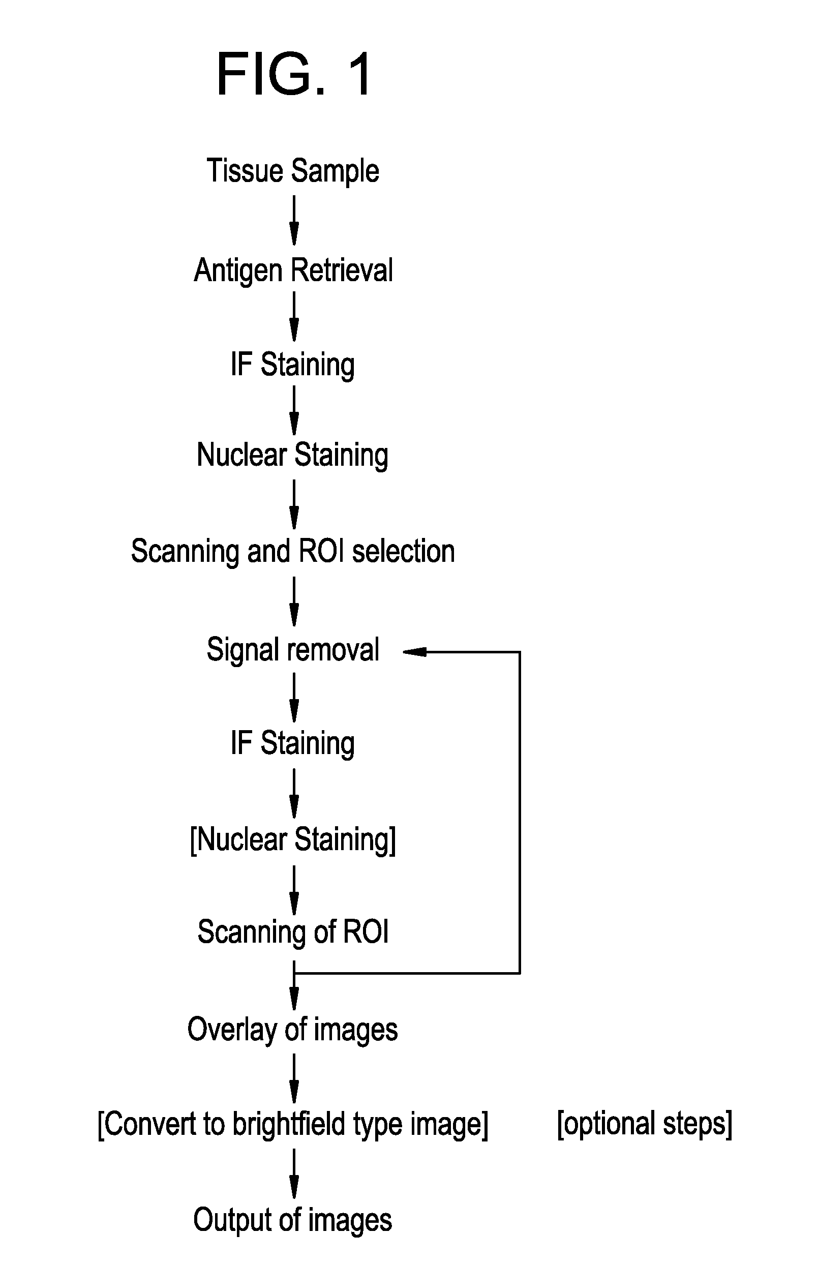
[0253]In one implementation of this invention, lymph node biopsy from patient suspected of having classical Hodgkin lymphoma is obtained from lymph node excision and examined by standard histology methods: the tissue sample is fixed in 10% neutral buffered formalin for 8 hours, and then dehydrated by passage of series of solutions with increasing ethanol concentration (50%, 75%, 80%, 95%, 100%) followed by xylene. The sample is then embedded in paraffin and sections of four micrometer thickness are sectioned using a microtome. Sections are floated onto a waterbath and collected one at a time onto a standard microscope slide. The slides are allowed to dry and baked for 2 hours in a 60° C. oven and then deparaffinized by passage through xylene, then re-hydrated by passage through ethanol followed by a series of water-ethanol mixtures with decreasing ethanol concentration, and finally washed with PBS.

[0254]Next, the slide is subjected to antigen retrieval procedure by heating the slide...

example 2

Multiplexed Analysis of Tissue Samples and

Diagnosis of Classic Hodgkin Lymphoma

Methods:

Specimens

[0260]Tissue microarray slides were run in triplicates. The TMA, noted as AGTA744, consisted of 28 cores: (12) non-Hodgkin cores, (12) Hodgkin Lymphoma cases, (2) tonsils, and (2) reactive lymphoma cases. After initial proof of principle study using the tissue microarrays, a total of (8) whole mount formalin fixed paraffin embedded tissue slides was employed in this study. Of the 8 samples, 4 were from biopsies previously diagnosed as classical Hodgkin lymphoma based on standard brightfield immunohistochemistry analyses. Three of the remaining 4 samples were non-Hodgkin lymphomas with CD30+ cells, while one case had been diagnosed as lymphocyte predominance Hodgkin lymphoma, in which CD30+ cells were present. All specimens were cut at 4 uM onto standard glass slides.

TABLE 1Tissue Microarray MapsAGTA 744marker1234placenta1AGI-4045AGI-4046AGI-4049AGI-4061NHLNHLHLNHL2AGI-4067AGI-4066AGI-4065...

example 3

Multiplexed Analysis of Tissue Samples and Benchmark to the Traditional IHC Methodology

Methods:

Specimens

[0276]A total of 40 whole mount, formalin fixed paraffin embedded tissues were employed to evaluate antibody specificity of the fluorescent stains and benchmark this staining to the traditional IHC methodology. Based on historical characterization from traditional brightfield IHC analyses the tissues were characterized as follows: 19 classical Hodgkin Lymphoma cases, 5 reactive lymph node cases, 7 B-Cell lymphoma cases, 1 nodular lymphocyte predominant lymphoma, 1 plasma cell lymphoma, and 3 T-Cell lymphoma cases, 2 tonsil, and 2 breast carcinomas

Slide Processing & Imaging:

[0277]The slides were baked at 60° C. for 1 hour and then deparaffinized and rehydrated through a series of xylene and alcohol washes. The slides were subjected to a two-step, citrate pH6.0 and Tris pH 8.5 antigen retrieval method via standard pressure cooker methods. The slides were blocked with a generic prote...


PUM

PropertyMeasurementUnit
sizeaaaaaaaaaa
wavelengthsaaaaaaaaaa

Abstract

A method of analyzing a biological sample suspected of having classic Hodgkin lymphoma, comprising (1) detecting, in a single sample, for the expression of at least two biomarkers selected from CD30, CD15, CD20, CD45, CD3, Pax-5, CD79A, BOB1 and OCT-2; and (b) analyzing the sample based on the presence, absence and / or expression level of the at least two biomarkers. Also provided is a method wherein all the nine biomarkers are analyzed on a single sample. Further provided are method for diagnosing classical Hodgkin lymphoma, as well as system and kit that comprise the means for executing the novel methods.

Description

FIELD OF THE INVENTION[0001]The present invention is directed to the detection of biomarkers on a biological sample. More specifically, the present invention is directed to the diagnosis of classical Hodgkin lymphoma using a multiplexed assay method which enables the fluorescent detection of multiple biomarkers on the same section of a biological sample. Also provided are a kit and a system for performing the novel method.BACKGROUND OF THE INVENTION[0002]Classical Hodgkin lymphoma, also known as Hodgkin's lymphoma and previously known as Hodgkin's disease, is a type of lymphoma, which is a cancer originating from lymphocytes. Hodgkin lymphoma is characterized by the orderly spread of disease from one lymph node group to another and by the development of systemic symptoms with advanced disease. When Hodgkin cells are examined microscopically, the presence of multinucleate Reed-Sternberg cells (RS cells, or RSC) and mononuclear variants, Hodgkin cells (H cells), is the characteristic ...

Claims


Application Information

Patent Timeline
Patent Type & Authority Applications(United States)
IPC IPC(8): G01N33/574
CPCG01N33/57407G01N2333/70578G01N2333/70596G01N2458/00G01N2333/7051G01N2333/47G01N2333/70589G01N33/57426
Inventor WEISS, LAWRENCE M.BECK, RODNEY A.KAANUMALLE, LAKSHMI SIREESHASEPPO, ANTTI E.HENDERSON, DAVID LAVANCORWIN, ALEX DAVIDKENNY, KEVIN BERNARDMCCULLOCH, COLIN CRAIGGINTY, FIONABORDWELL, ALEXANDERR G.LAZARE, MICHAEL S.HOLLMAN-HEWGLEY, DENISE A.
Owner GE HEALTHCARE BIO SCI CORP
Why Eureka
Try Eureka