Identifying taxane sensitivity in prostate cancer patients
a prostate cancer patient and taxane technology, applied in the field of identifying taxane sensitivity in prostate cancer patients, can solve the problem that prostate cancer patients may not benefit from taxane drug treatmen
- Summary
- Abstract
- Description
- Claims
- Application Information
AI Technical Summary
Benefits of technology
Problems solved by technology
Method used
Image
Examples
example 2
Cell Capturing Device
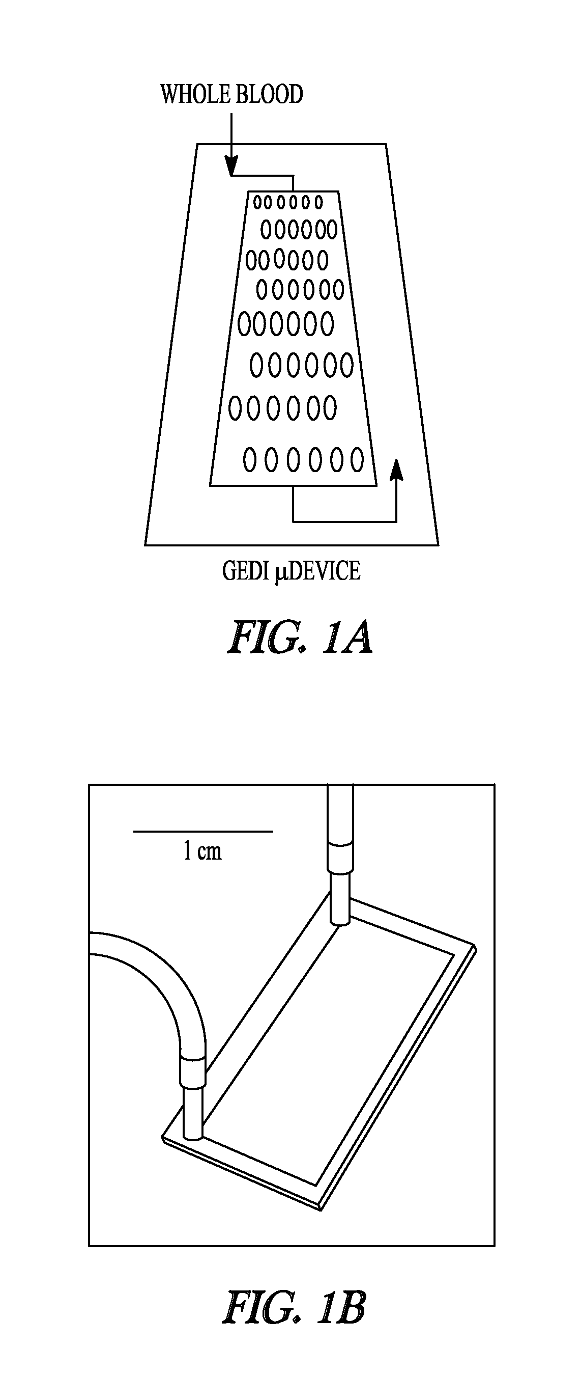
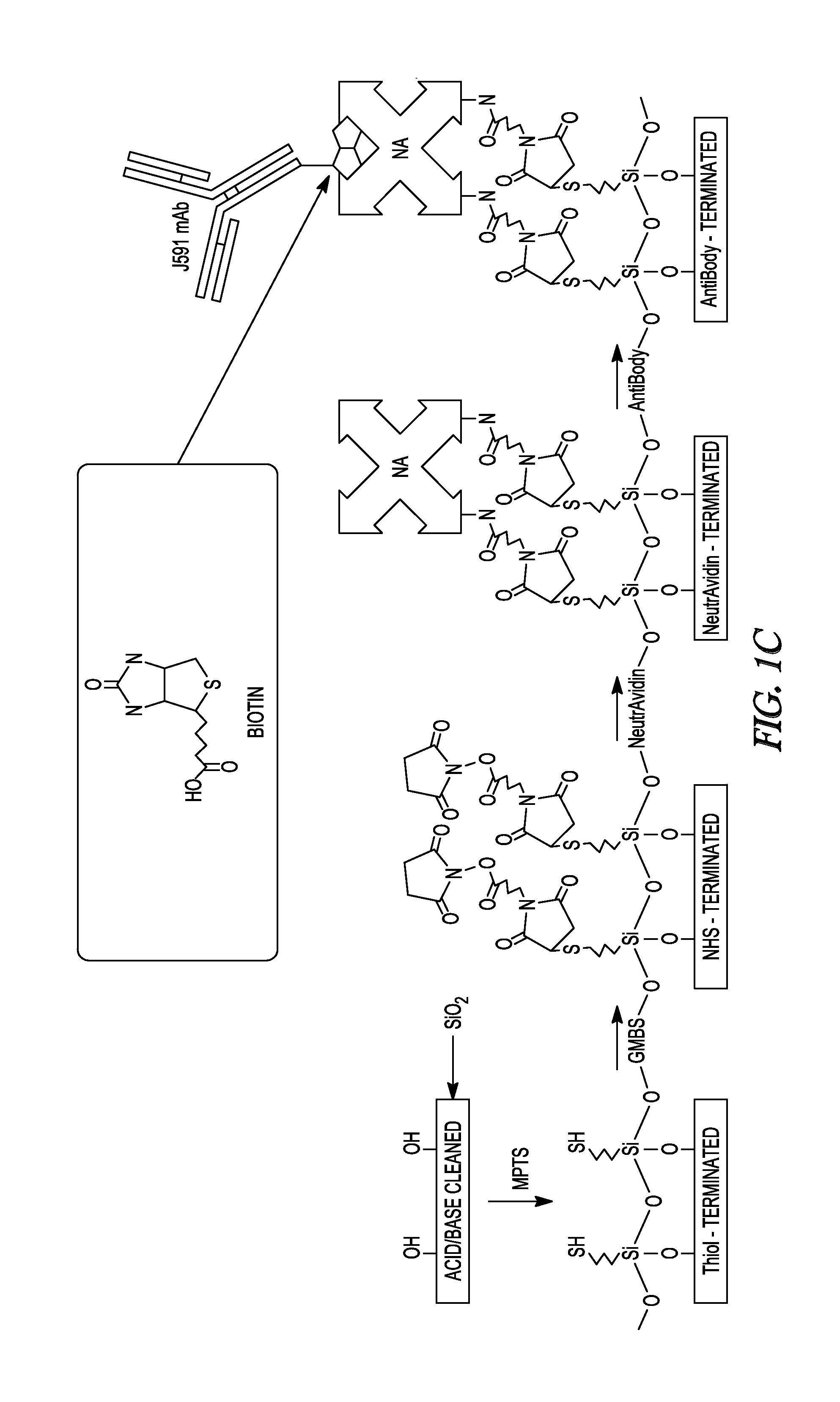
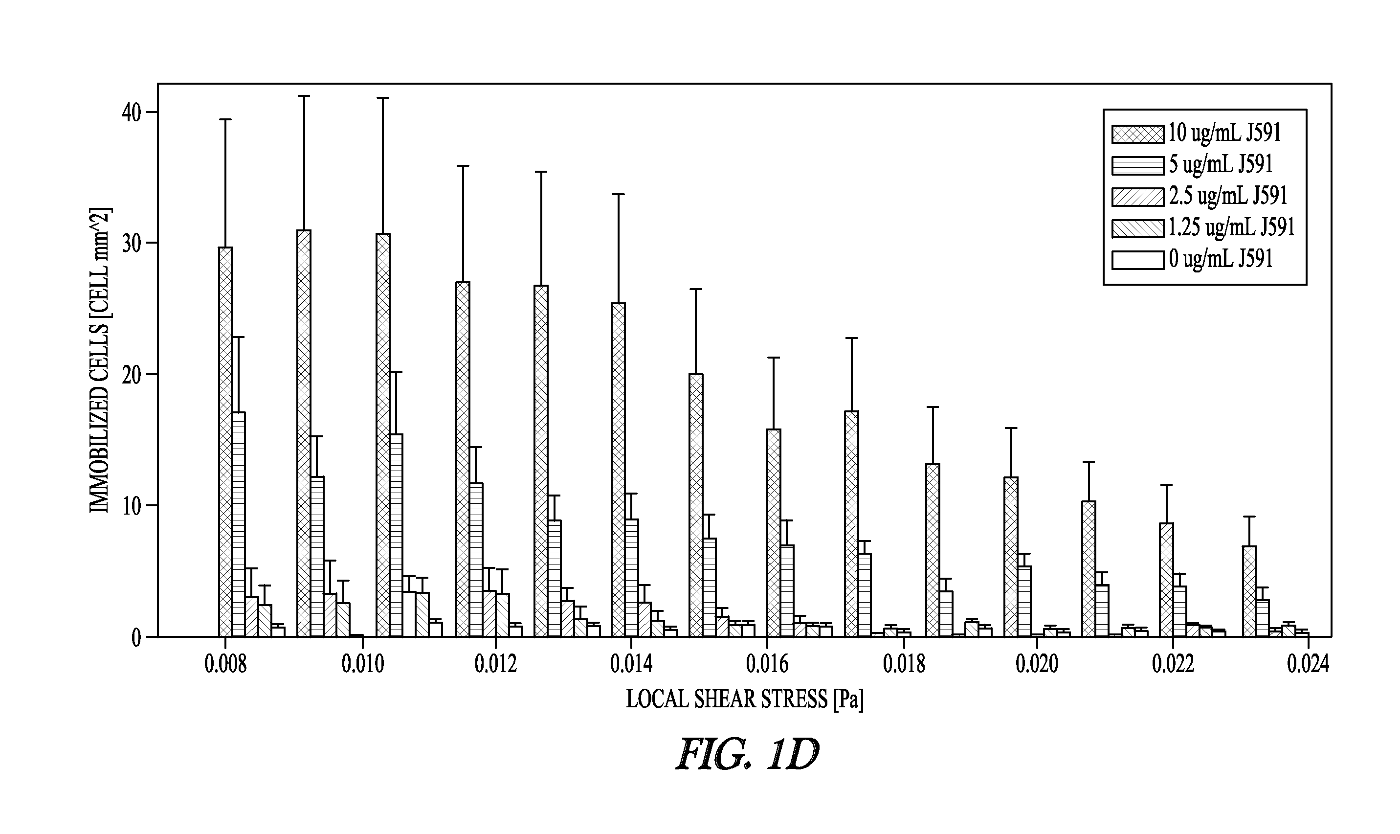
[0147]The cell capture rates of PSMA-positive cancer cells were determined to evaluate the performance of the device depicted in FIG. 1A. Cell capture was determined as a function of varying mAb J591 concentrations (1.5-20 ug / ml) using shear stress magnitudes representative of those experienced by the functionalized surfaces of the device (0.08-0.24 Pa). These experiments revealed a dose-dependent increase in cell capture up to mAb concentration of 10 ug / ml, which was used for all subsequent experiments (FIG. 1D).
[0148]Although the J591 antibody is specific for PSMA-expressing cells, non-specific leukocyte adhesion has been a major problem for all blood-based immunocapture techniques. To minimize leukocyte adhesion, a parametric study was conducted to characterize collision rate (CpR; collision per row) as a function of cell size and obstacle offset. Collision rates from a subset of these offsets exhibit a sharp cutoff according to cell size, as shown in FIG. 2B...
example 3
Cell Capture, Imaging, and Enumeration of Circulating Tumor Cells from Metastatic Prostate Cancer Patients
[0151]This Example describes use of the GEDI device to capture and characterize circulating tumor cells (CTCs) from the blood of patients with metastatic castration-resistant prostate cancer.
[0152]One ml of peripheral blood was passed through the device, captured cells were fixed and immunostained for PSMA, CD45, EpCAM and DAPI, and the captured cells were analyzed by confocal microscopy. Circulating tumor cells were defined as intact, nucleated, PSMA+ / CD45− cells. Different cell lines were used as controls for antibody staining for PSMA, CD45, and EpCAM, as follows: the C4-2 prostate cancer cells are PSMA+ / EpCAM+ / CD45− while the PC3 prostate cancer cells are PSMA− / EpCAM− / CD45−. The U937 leukemic cell line was used as a positive control for the leukocyte marker CD45. DAPI was used to stain the DNA.
[0153]Representative examples of circulating tumor cells and leucocytes are shown ...
example 4
Markers for Circulating Tumor Cells
[0156]This Example provides a comparison of cell capture by the GEDI device and the CellSearch® device.
[0157]The results presented in FIG. 3 illustrate both that the EpCAM expression level of captured PSMA+ / DAPI+ / CD45− cells is variable and that the correlation between GEDI PSMA+ capture and CellSearch® EpCAM+ capture is only weak. The two capture methodologies both correlate with the disease state, but the population captured by the GEDI device is different from that captured by the currently available CellSearch® device, presumably owing to different expression levels of EpCAM and PSMA in the circulating tumor cell population. The data described herein shows detection of a significantly higher circulating tumor cell population using the GEDI microfluidic device compared with CellSearch®. Thus the GEDI microdevice has enhanced sensitivity for detecting circulating tumor cells in samples obtained from prostate cancer patients. Because circulating t...
PUM
| Property | Measurement | Unit |
|---|---|---|
| length | aaaaa | aaaaa |
| time | aaaaa | aaaaa |
| time | aaaaa | aaaaa |
Abstract
Description
Claims
Application Information
 - R&D
- Intellectual Property
- Life Sciences
- Materials
- Tech Scout
- Unparalleled Data Quality
- Higher Quality Content
- 60% Fewer Hallucinations
Browse by: Latest US Patents, China's latest patents, Technical Efficacy Thesaurus, Application Domain, Technology Topic, Popular Technical Reports.
© 2025 PatSnap. All rights reserved.Legal|Privacy policy|Modern Slavery Act Transparency Statement|Sitemap|About US| Contact US: help@patsnap.com


