Recruitment of mensenchymal cells using controlled release systems
a technology of controlled release and mensenchymal cells, which is applied in the direction of peptide/protein ingredients, peptide sources, drug compositions, etc., can solve the problems of life-threatening and debilitating, and achieve the effect of altering the chemokine release kinetics and profil
- Summary
- Abstract
- Description
- Claims
- Application Information
AI Technical Summary
Benefits of technology
Problems solved by technology
Method used
Image
Examples
example i
MSC Migration to an Arthritic Injury
[0250]This example describes a mouse “sports injury” model that could be used to induce articular joint damage analogous to many human joint conditions including, but not limited to, injury-induced arthritis, rheumatoid arthritis and / or osteoarthritis.
[0251]Under brief anesthesia, mice are injected intra-articularly with mono-iodoacetate (MIA) (Sigma-Aldrich, USA) dissolved in 25 μl of saline, or with only 25 μl of saline (controls). The syringe is inserted through the patellar ligament into the intra-articular joint space of the left knee. Animals are randomly assigned to each group.
[0252]Subsequently, each mouse is injected with alginate PDGF microparticles and luciferin-MSCs. See, FIG. 4A. The MIA injection is shown to be responsible for the observed articular cartilage destruction because the injection of a blank microparticle population is shown not cause any damage. See, FIG. 4B. The lucifern loaded MSCs are then subjected to fluorescence t...
example ii
In Vitro Degradation of Alginate Microprticles
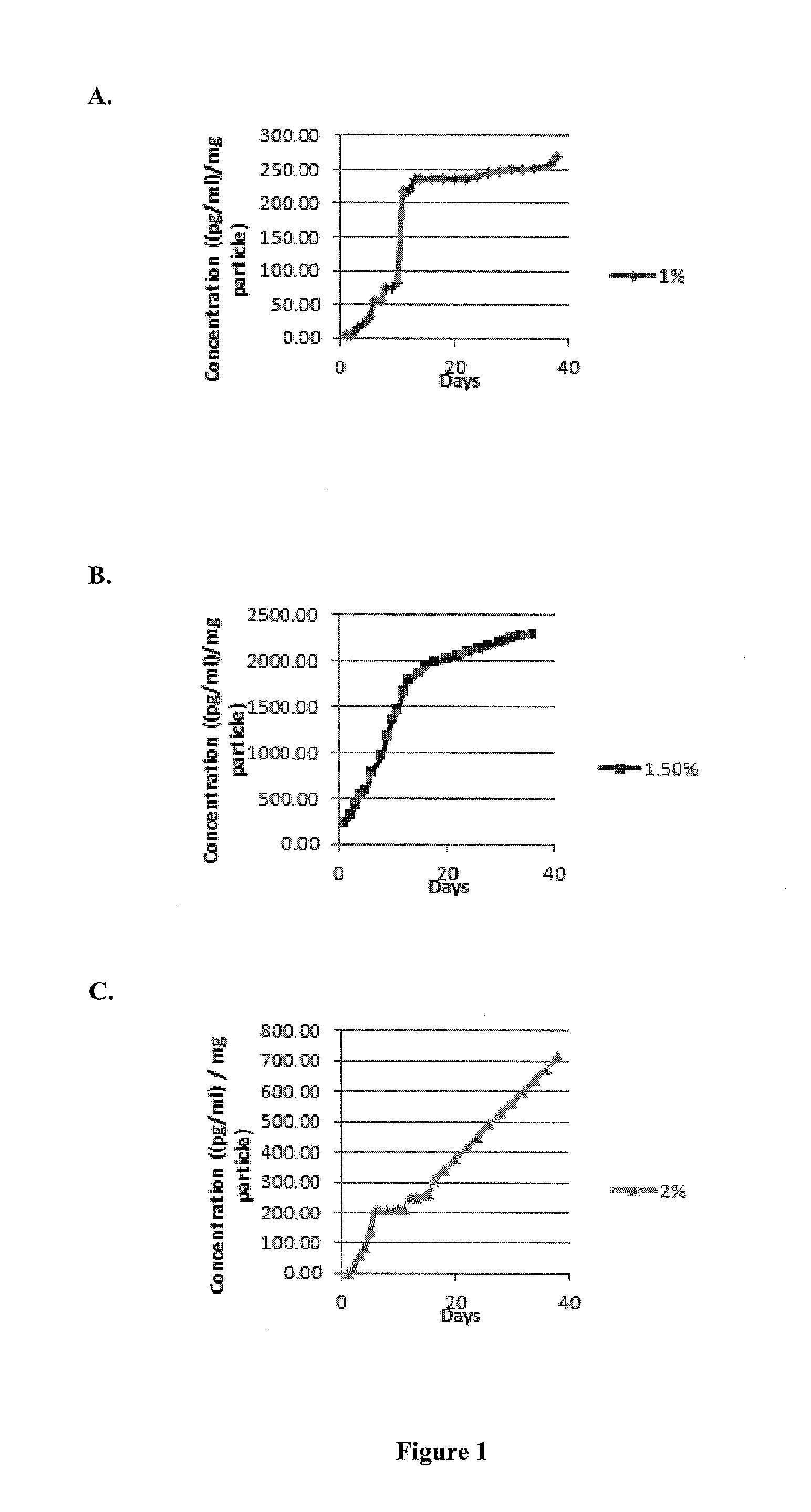
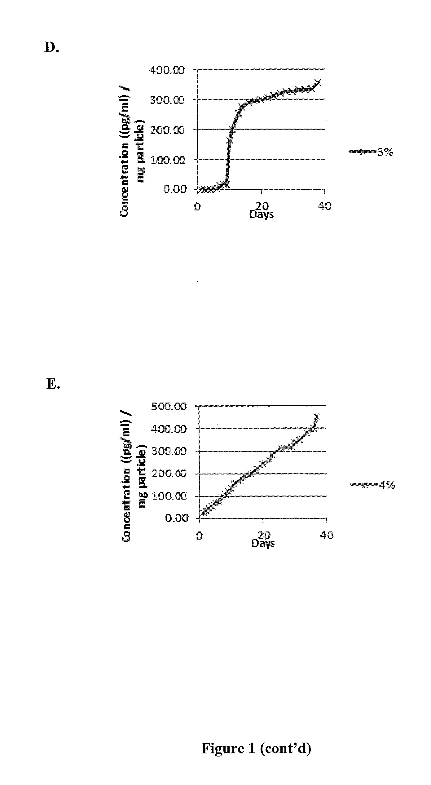
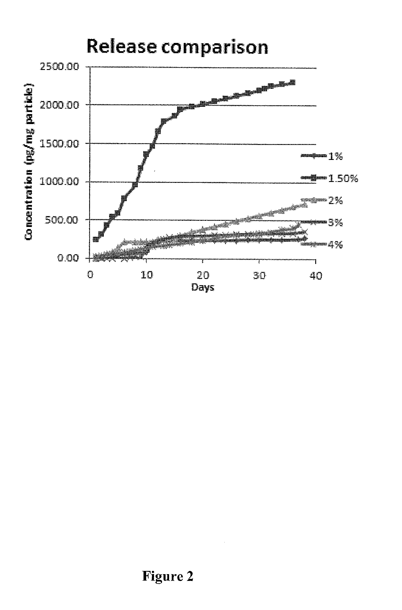
[0254]This example demonstrates that the alginate microparticles contemplated by this invention degrade in a manner that correlates with the in vitro and in vivo chemokine release patterns.
[0255]Photomicrographs taken immediately after placement into the in vitro release system showed no degradation of the microparticles. See, FIGS. 6A and 6B. Photomicrographs demonstrated a physical erosion pattern after thirty (30) days that correlated with the observed PDGF release profiles. See, FIGS. 6C and 6D.
example iii
Cytotoxicity of Alginate Microparticles
[0256]This example demonstrates that the alginate microparticles contemplated by this invention are not cytotoxic to MSCs as determined in an in vitro co-incubation assay.
[0257]Alginate microparticles and MSCs were mixed together in a physiologically balanced buffer solution. Photomicrographs demonstrated a lack of cytotoxicity of the microparticles on the MSCs by demonstrating that MSCs observed in a bright field image co-localized with calcein stained MSCs. See, FIGS. 7A and 7B, respectively. The lack of non-viability was confirmed by the lack of red stain subsequent to ethidium heterodimer-1. See, FIG. 7C. The colocalization of bright field MSCs images with the calcein / ethidium hetereodimer-1 stain images is demonstrated by an overlay of the FIG. 7A-C photomicrographs. See, FIG. 7D.
PUM
| Property | Measurement | Unit |
|---|---|---|
| Fraction | aaaaa | aaaaa |
| Fraction | aaaaa | aaaaa |
| Fraction | aaaaa | aaaaa |
Abstract
Description
Claims
Application Information
 - R&D
- Intellectual Property
- Life Sciences
- Materials
- Tech Scout
- Unparalleled Data Quality
- Higher Quality Content
- 60% Fewer Hallucinations
Browse by: Latest US Patents, China's latest patents, Technical Efficacy Thesaurus, Application Domain, Technology Topic, Popular Technical Reports.
© 2025 PatSnap. All rights reserved.Legal|Privacy policy|Modern Slavery Act Transparency Statement|Sitemap|About US| Contact US: help@patsnap.com


