
Isothermal amplification under low salt condition
a technology of low salt and isothermal amplification, applied in the direction of enzymology, biochemistry apparatus and processes, transferases, etc., can solve the problems of incomplete amplification of dna sequences, undesirable chimeric products, poor performance of these systems
- Summary
- Abstract
- Description
- Claims
- Application Information
AI Technical Summary
Benefits of technology
Problems solved by technology
Method used
Image

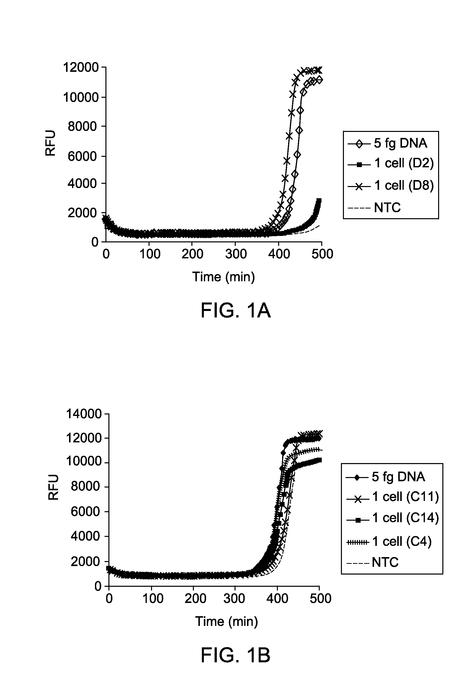
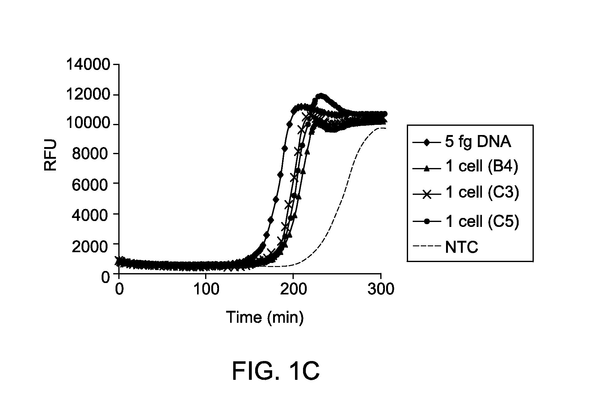
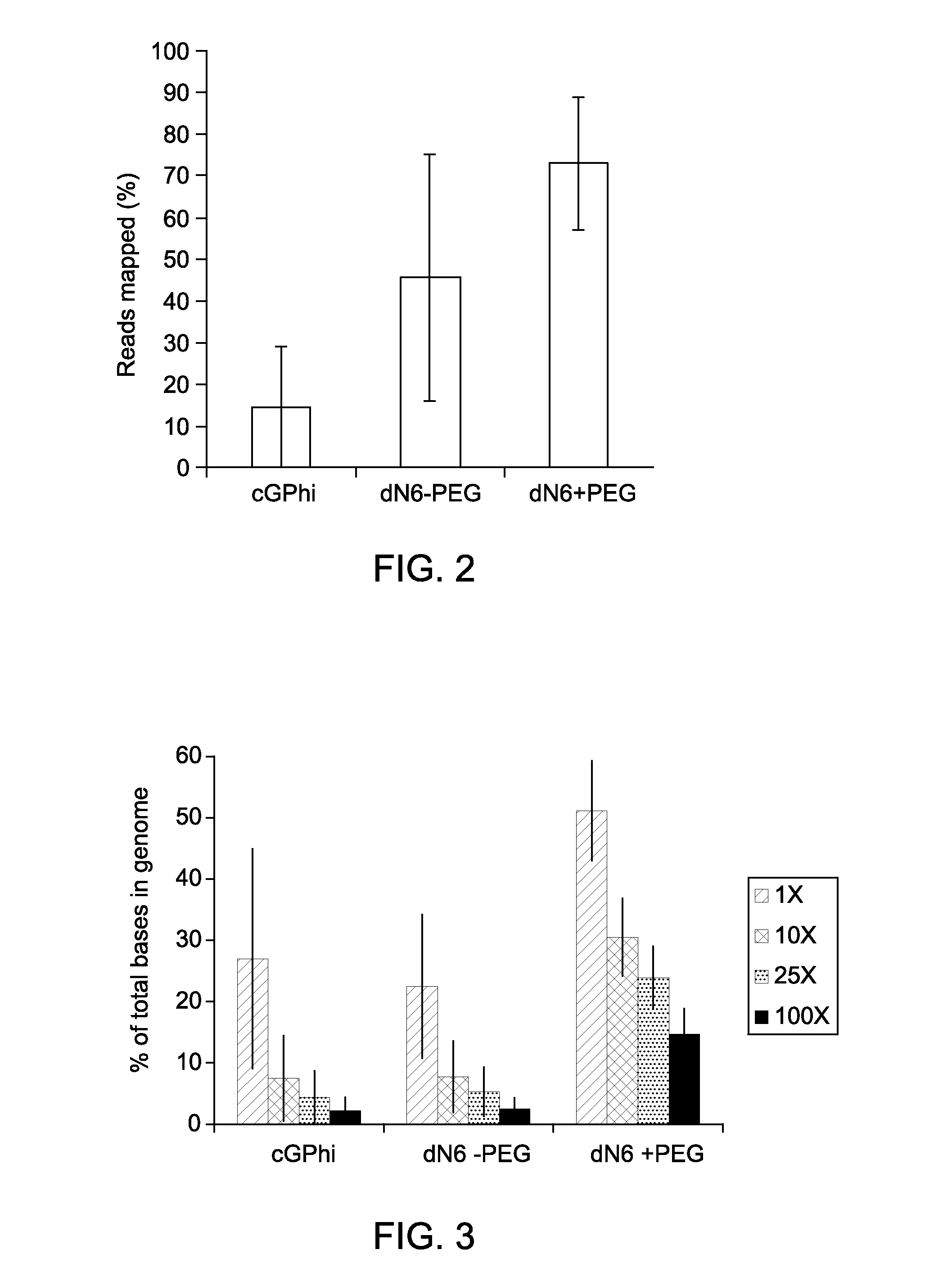
Examples
example 1
Reaction Kinetics and Sensitivity of MDA Reactions of DNA from Single Cells in Presence of PEG and Low Salt Condition
[0063]Cultures of E. coli MG1655 were grown to log phase in LB media, harvested by centrifugation, and washed three times using TEN buffer (10 mM Tris, pH 7.5, 100 mM NaCl, and 0.1 mM EDTA). After washing, cells were resuspended in buffer TEN+30% glycerol and serial dilutions were made. Cells were then stained with 10 μM FM1-43FX dye (F-35355, from Invitrogen, Life Technologies) for 10 minutes at room temperature, added stained cells into each of the wells of a transparent-bottom 384-well plate, and counted using an inverted fluorescent microscope (Nikon Eclipse TE2000-U). Following identification of wells containing single cells, lysis was initiated by addition of 2 μl of 0.2 M KOH, 50 mM DTT, 0.015% Tween-20 and freezing at −80° C. overnight. The following morning, plates were thawed and lysate was further incubated at 65° C. for 10 minutes, cooled, and neutralized ...
PUM
| Property | Measurement | Unit |
|---|---|---|
| Temperature | aaaaa | aaaaa |
| Temperature | aaaaa | aaaaa |
| Temperature | aaaaa | aaaaa |
Abstract
Description
Claims
Application Information