Assessment of risk of aneuploidy

Active Publication Date: 2015-11-26
HANDYSIDE ALAN
View PDF1 Cites 1 Cited by
  • Summary
  • Abstract
  • Description
  • Claims
  • Application Information

AI Technical Summary

Benefits of technology

[0178]FIG. 1—chromosome segregation in female meiosis I and II (1) Female meiosis is initiated in the fetal ovary before birth during the early development of the female germ cells (primary oocytes), which will eventually form mature eggs or oocytes in the adult female. (2) To reduce the number of chromosomes from the normal (euploid) 23 pairs of homologous chromosomes (one of each pair inherited from the father (left hand side, including representative allele A) and one from the mother (right hand side, including representative allele B), or 46 in total, to 23 single chromosomes, there is one round of DNA replication in which each chromosome is duplicated into two sister chromatids followed by two specialised meiotic divisions, meiosis I and II. The two sister chromatids have identical DNA sequences and therefore SNP genotypes at this stage, including the regions flanking the centromere as represented here in the boxes

Problems solved by technology

However, biopsying both polar bodies from each oocyte is labour intensive for clinics and multiple arrays are required to test each oocyte.
However amplification bias makes it difficult to accurately quantify the products, thereby limiting possible

Method used


Image

  • Assessment of risk of aneuploidy
  • Assessment of risk of aneuploidy
  • Assessment of risk of aneuploidy

Examples


example 1

Meiotic Errors that can Lead to Aneuploidy in a Fertilized Egg

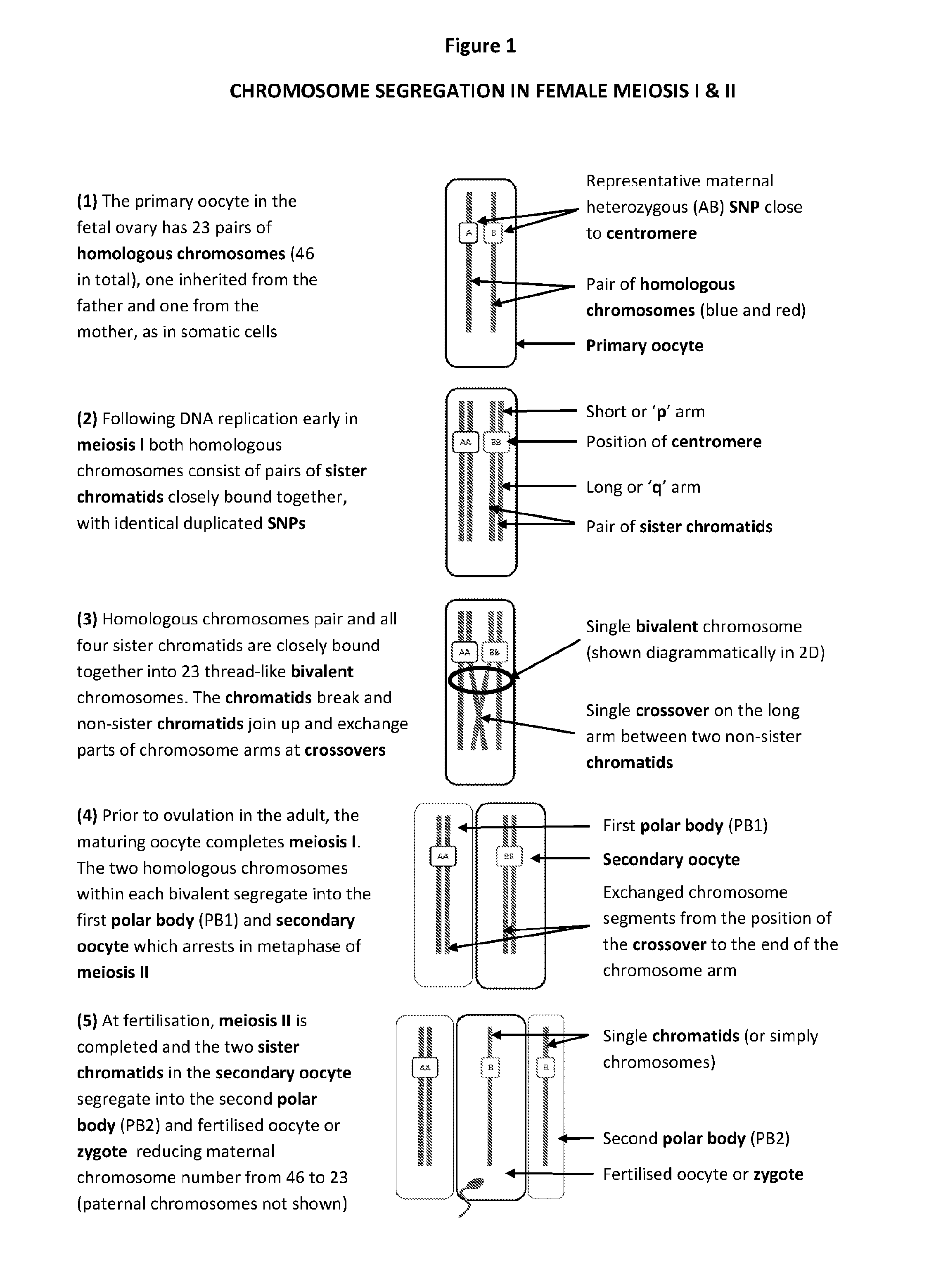
[0203]As illustrated in FIG. 1, normally in meiosis I the two homologues of each chromosome, now consisting of pairs of sister chromatids, pair up and join together, homologous non-sister chromatids undergo one or more recombinations or crossovers.

[0204]Subsequently the homologous chromosomes of the condensed bivalent chromosome are to-oriented' to the same spindle poles, so that the homologous chromosomes separate away from each other into PB1 and the secondary oocyte. In meiosis II, the two sister chromatids separate into PB2 and fertilised oocyte (zygote or egg) following fertilisation.

[0205]It can thus be seen that in the in the normal (euploid) oocyte all centromeric regions of each chromosome are homozygous in PB1, since this part of the sister chromatids will generally not be significantly affected by recombination or crossovers.

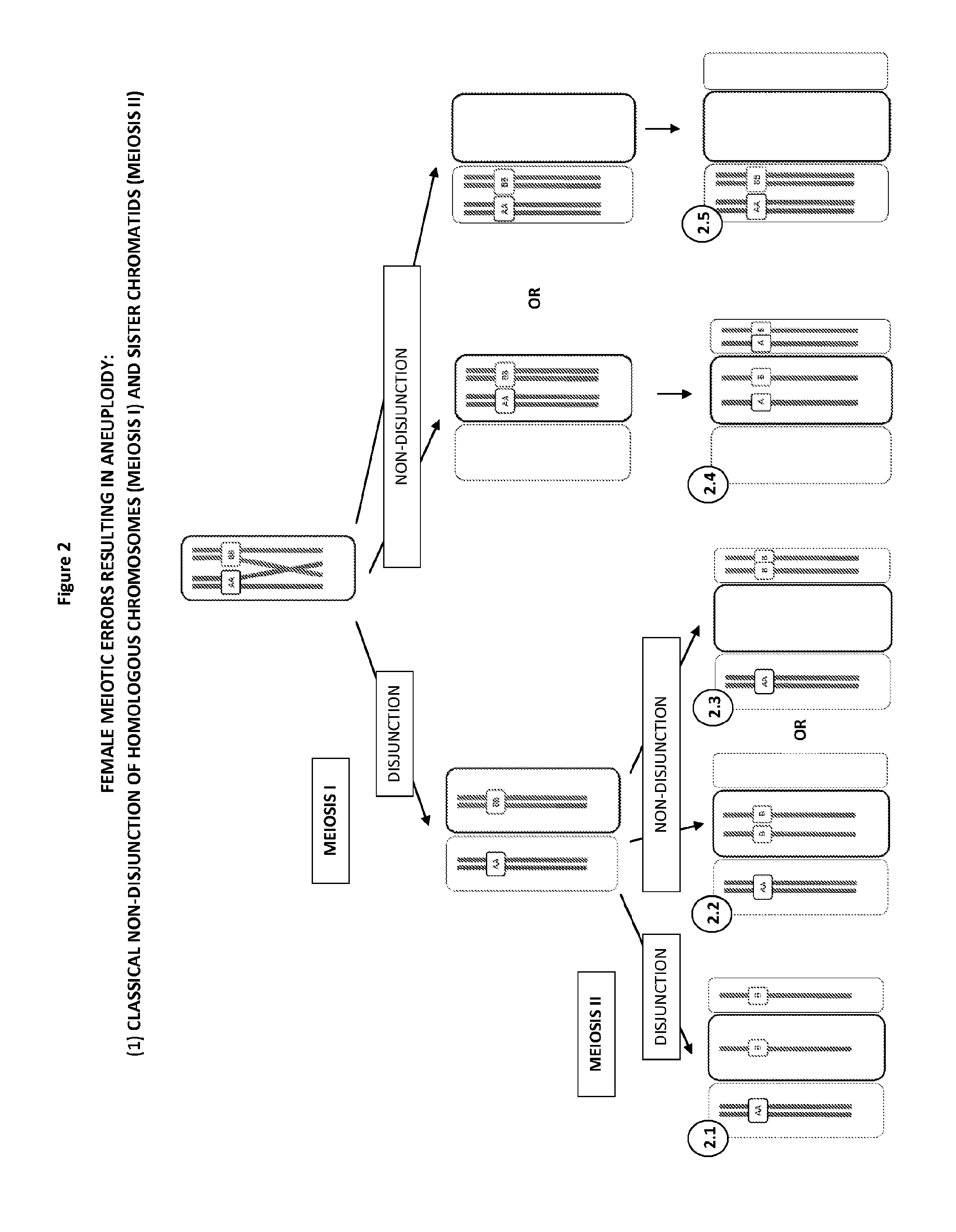
[0206]As illustrated in FIG. 2, a classical textbook mechanism causing errors in meiosi...

example 2

Assessment of CH in PB1 from Number of Different Oocytes

[0213]In this example the status of defined SNPs were assessed in PBs (“PB1 genotype”) and corresponding secondary oocytes (“Egg genotype”).

[0214]Methods

[0215]PBs and oocytes were lysed and the whole genome amplified (WGA) by multiple displacement amplification according to manufacturer's instructions (Repli-g, Qiagen). WGA products were then genotyped on a SNP genotyping bead array again according to the manufacturer's protocol (Infinium Human CytoSNP-12, Illumina). The genotype data was exported as a text file and imported into Microsoft Excel and a macro was used to identify SNPs flanking the centromeres of each chromosome and display the results. The macro also calculated the percentage of heterozygous SNPs.

[0216]The maternal genotype was ascertained using the same bead array but genomic DNA isolated from a blood sample by standard methods was used. This data was also imported into Excel and used to identify all of the hete...

example 3

Comparison of Array CGH for Quantitative Detection of Aneuploidy in PB1 and PB2 with SNP Genotypinq, Maternal Haplotyping and Total Heterozygosity and / or CH Analysis in PB1 alone, or PB1 and PB2

[0235]Ten mature MII arrested oocytes were collected from a patient having aneuploidy testing by array CGH of PB1 and PB2. PB1 was biopsied from each oocyte prior to intracytoplasmic sperm microinjection and, following fertilisation and resumption of meiosis, PB2 was also biopsied. Both polar bodies were lysed, DNA amplified by WGA and aliquots of the products used for array CGH. The array CGH results indicated that all of the embryos had one or more copy number abnormalities in PB1 and / or PB2 except one, presumed euploid embryo (Embryo # 1), which was therefore selected for transfer (Table 2). With the patients consent, the remaining 9 presumed aneuploid embryos were lysed and the DNA amplified by WGA. Genomic DNA from both parents, WGA products from all polar bodies and the corresponding em...


PUM

PropertyMeasurementUnit
Fractionaaaaaaaaaa
Fractionaaaaaaaaaa
Fractionaaaaaaaaaa

Abstract

The present disclosure relates generally to methods and materials for use in detecting abnormalities of the number of whole chromosomes or chromosome regions (aneuploidy). It has particular utility for assessing the risk of aneuploidy of eggs (i.e., oocytes), fertilised eggs or embryos developed therefrom in the context of in vitro fertilisation.

Description

TECHNICAL FIELD[0001]The present disclosure relates generally to methods and materials for use in detecting abnormalities of the number of whole chromosomes or chromosome regions (aneuploidy). It has particular utility for assessing the risk of aneuploidy of eggs (i.e., oocytes), fertilised eggs or embryos developed therefrom in the context of in vitro fertilisation.BACKGROUND ART[0002]In normal female meiosis the precursor cells of the ova multiply and then reduce the number of chromosomes to one half set in each gamete in two specialised meiotic divisions.[0003]Meiosis is initiated in the fetal ovary before birth during the early development of the female germ cells (primary oocytes), which will eventually form mature eggs (or oocytes, the terms are used interchangeably) in the adult female.[0004]To reduce the number of chromosomes from the normal (euploid) 23 pairs of homologous chromosomes (one of each pair inherited from the father and one from the mother, so 46 in total) to 23...

Claims


Application Information

Patent Timeline
IPC IPC(8): C12Q1/68
CPCC12Q2600/156C12Q1/6883C12Q1/6827C12Q2539/115
Inventor HANDYSIDE, ALAN
Owner HANDYSIDE ALAN
Why Eureka
Try Eureka