

Neuronal imaging and treatment
a neuronal imaging and treatment technology, applied in the field of methods and systems of imaging, can solve the problems of poor spatial resolution of mibg spect images, less clinical significance, and difficult acquisition of hydroxyephedrine pet images
- Summary
- Abstract
- Description
- Claims
- Application Information
AI Technical Summary
Benefits of technology
Problems solved by technology
Method used
Image

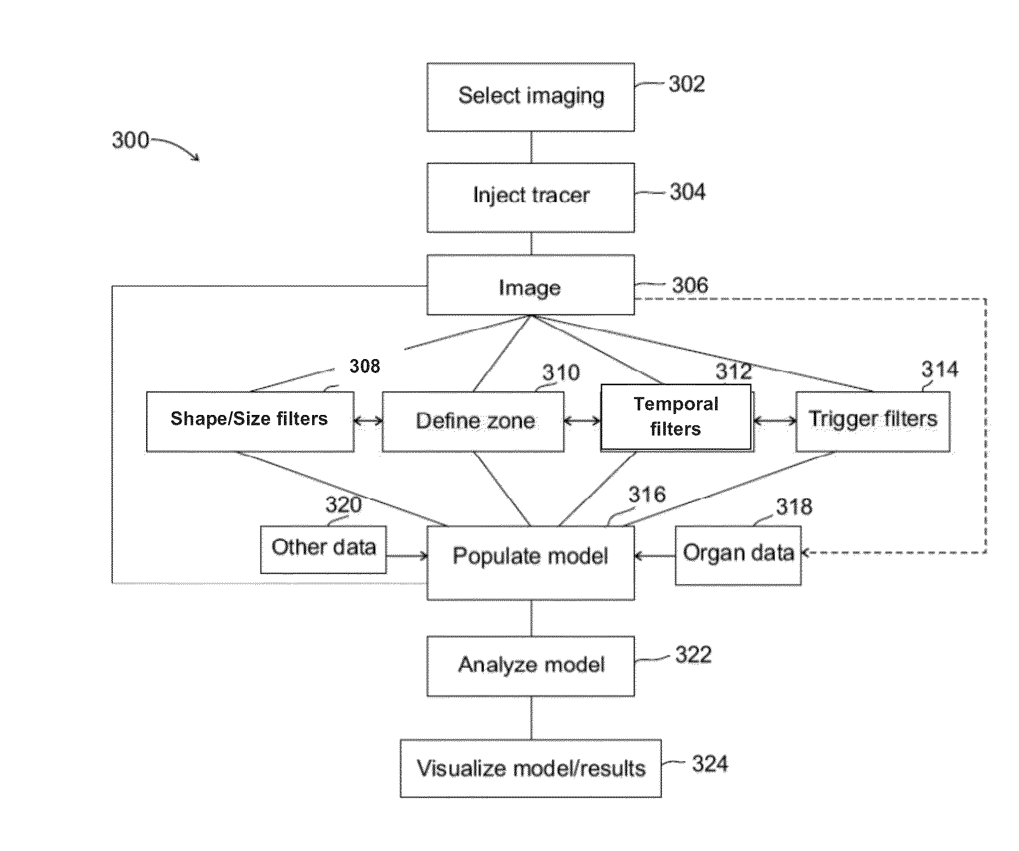
Examples
example 1
Imaging of Adrenergic Synaptic Activity in Heart
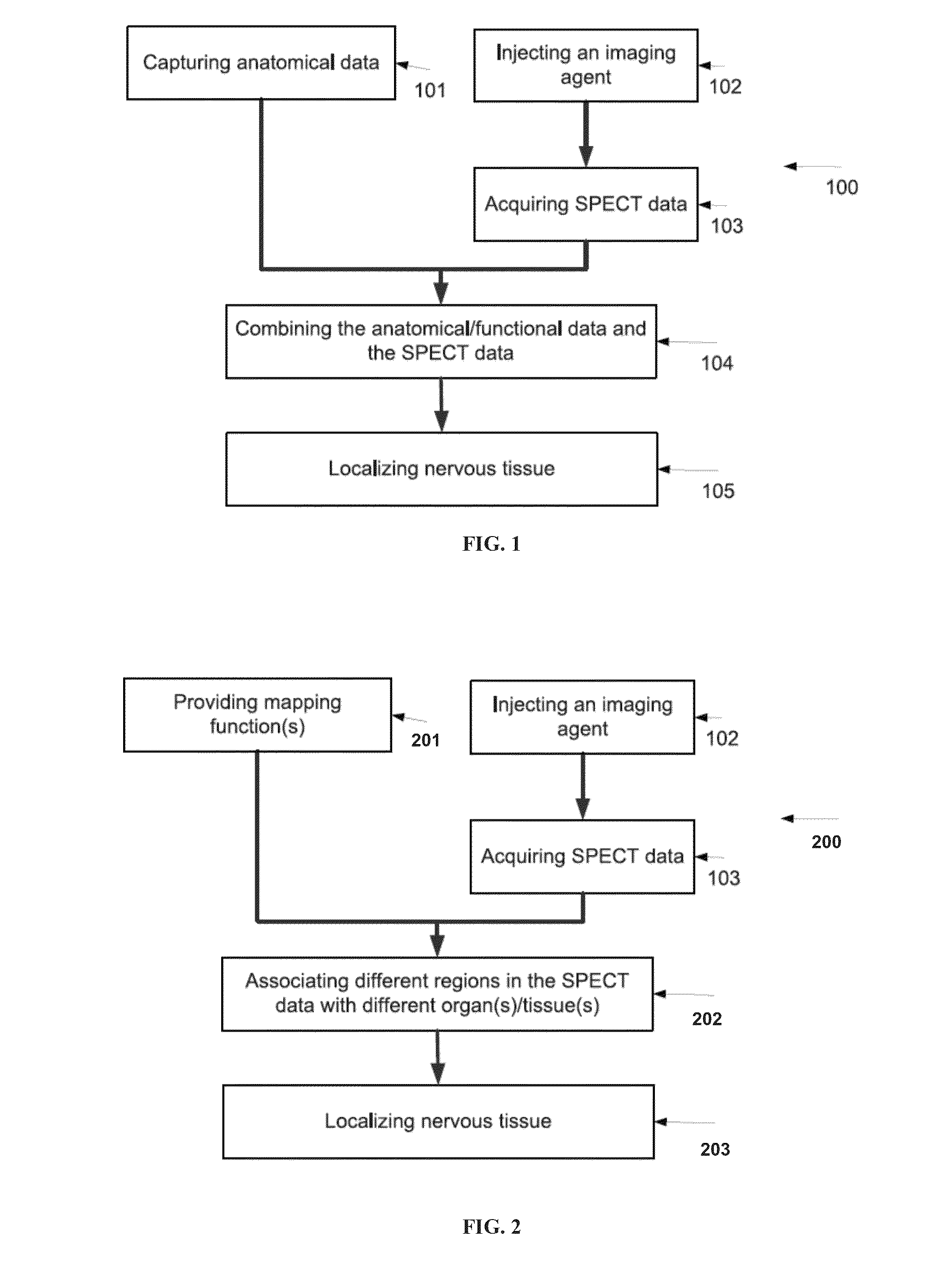
[0684]A human patient was administered I-123-radiolabeled mIBG, a radioactive tracer for adrenergic synapses of the sympathetic nervous system. An image based on mIBG distribution was obtained by SPECT using a D-SPECT® Cardiac Imaging System (Spectrum Dynamics, Israel). The anatomy of the heart was determined by performing x-ray CT imaging and segmenting the obtained x-ray CT images. The x-ray CT data was then combined with the SPECT data so as to construct an image showing mIBG distribution in the heart. Images showing levels of adrenergic synapse activity in the left atrium and right ventricle are presented in FIGS. 6A-8B.
[0685]As shown in FIGS. 6A-6D, as well as in FIGS. 7A and 7B, regions with a high level of adrenergic synapses (red) were observed in certain regions of the left atrium, whereas most of the left atrium was characterized by a low level of adrenergic synapses (green). FIG. 6A shows with a high level of adrenergic syna...
example 2
Imaging of Cardiac Ganglionated Plexuses
[0688]In order to image cardiac ganglionated plexuses, a 60 year old male subject, having a body mass index (BMI) of 22 (height 176 cm; weight 69 kg) and a medical history of ventricular arrhythmias, chest discomfort, low ejection fraction (LVEF (left ventricular ejection fraction)=35%), and non-ischemic cardiomyopathy, was administered I-123-radiolabeled mIBG, and images based on mIBG distribution were obtained by SPECT using a D-SPECT® Cardiac Imaging System (Spectrum Dynamics, Israel). The anatomy of the heart and its surroundings was determined by performing x-ray CT imaging and segmenting the obtained x-ray CT images. The x-ray CT data was then combined with the SPECT data so as to construct an image showing mIBG distribution in the region of the heart, as described in FIG. 1.
[0689]FIGS. 9A-9C and 10A-10C show the location of three identified (relatively large) synaptic centers adjacent to the heart of the subject. FIG. 10C further shows ...
example 3
Imaging, Verification and Ablation of Cardiac Ganglionated Plexuses
[0692]An electrophysiological study was performed in order to verify the identification of cardiac GP (ganglionated plexus) locations by imaging as described in Example 2, and to evaluate the utility of such imaging in ablation therapy.
[0693]Using the procedures described in Example 2, four synaptic centers representing ganglionic plexuses (GPs) were identified adjacent to the heart of a 47 year old male subject having a medical history of paroxysmal atrial fibrillation, catheter ablation for PVI (pulmonary vein isolation) and CVI (cavo-tricuspid isthmus), and normal LV (left ventricle) contraction ((LVEF (left ventricular ejection fraction)=60%, LVDd (left ventricular end-diastolic dimension)=47 mm, and LAD (left atrial dimension)=36 mm).
[0694]After imaging and identification of the four GPs, verification was performed by measuring the reaction of individual target sites to high frequency stimulation (HFS), as deter...
PUM
| Property | Measurement | Unit |
|---|---|---|
| Diameter | aaaaa | aaaaa |
| Time | aaaaa | aaaaa |
| Concentration | aaaaa | aaaaa |
Abstract
Description
Claims
Application Information