Robot Assisted Volume Removal During Surgery

a robotic and volume technology, applied in the field of robotic assisted volume removal during surgery, can solve the problems of repetitive motion disorders, serious complications, difficult process of removal/trimming of lamina, etc., and achieve the effect of effectively removing volume inside a patient, shortening surgery time, and freeing surgeons from repetitive and laborious tasks

Active Publication Date: 2016-06-02
KB MEDICAL SA
View PDF6 Cites 79 Cited by
  • Summary
  • Abstract
  • Description
  • Claims
  • Application Information

AI Technical Summary

Benefits of technology

[0004]Described herein is a device and method used to effectively remove volume inside a patient in various types of surgeries, such as spinal surgeries (e.g. laminotomy), neurosurgeries (various types of craniotomy), ENT surgeries (e.g. tumor removal), and orthopedic surgeries (bone removal). The disclosed technology provides robotic assistance linked with a navigation system and medical imaging to shorten surgery time, make the surgery safer and free surgeons from doing repetitive and laborious tasks. The disclosed technology is also compatible with robotic surgical system described in U.S. patent application Ser. No. 14 / 266,769, filed Apr. 30, 2014, and entitled “Apparatus, Systems, and Methods for Precise Guidance of Surgical Tool” which is hereby incorporated by reference in its entirety, thereby, for example, allowing the same robotic surgical system to assist with different aspects of spinal surgery.

Problems solved by technology

Removal / trimming of the lamina is a difficult process.
The surgeon must be particularly careful not to damage spinal dura mater (tissue protecting spinal cord) which would lead to serious complications.
Furthermore, removing bony tissue requires repetitive movements which are tiring and may lead to repetitive motion disorders for surgeons performing these surgeries daily.

Method used


Image

  • Robot Assisted Volume Removal During Surgery
  • Robot Assisted Volume Removal During Surgery
  • Robot Assisted Volume Removal During Surgery

Examples


Embodiment Construction

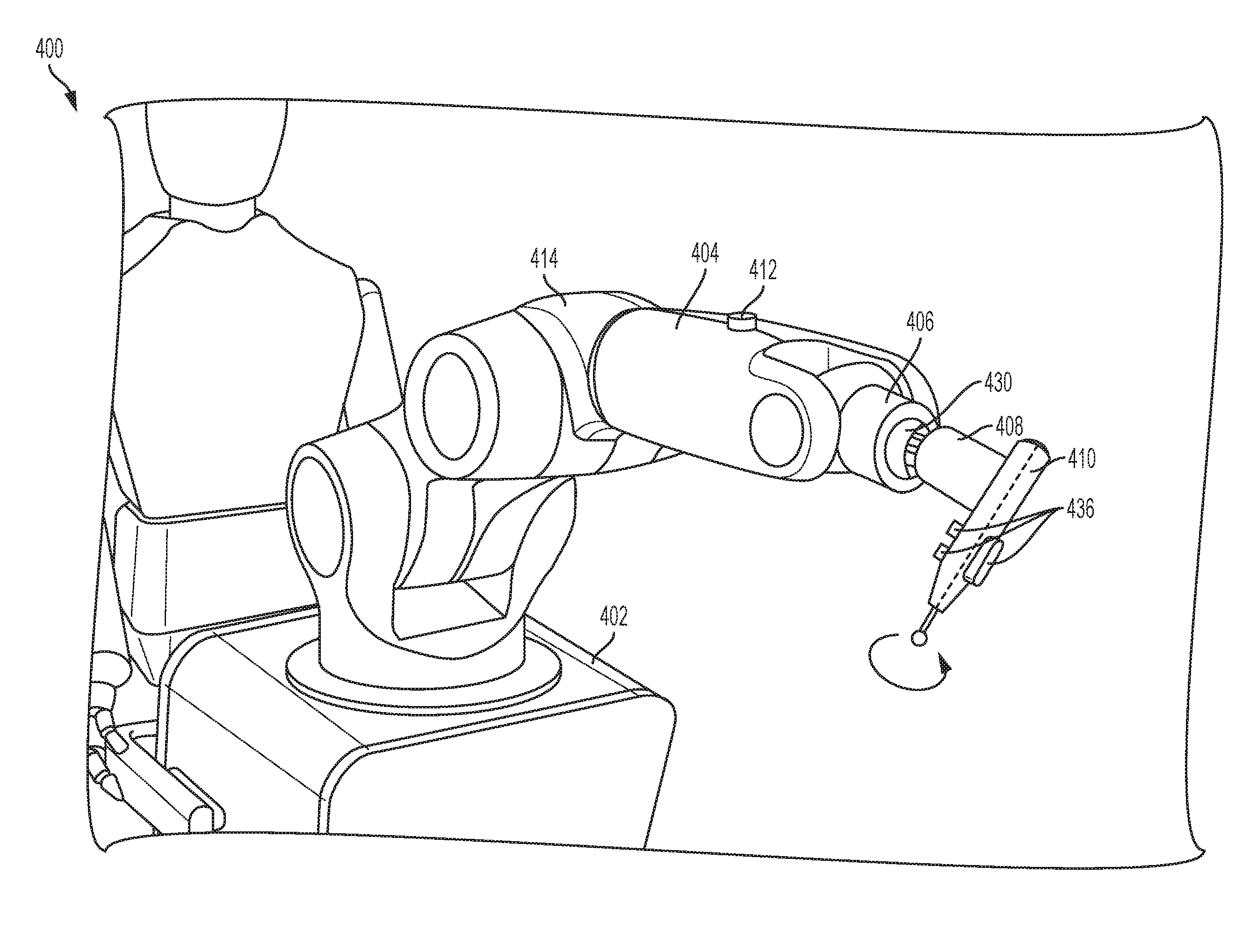
[0073]The disclosed technology includes the robotic surgical system used to remove a target volume from a patient. Initially, an incision is made and the vertebra is exposed. In some implementations, the frame of a navigation system is attached to the patient in the place selected by the surgeon. Intra-operative medical images of the target anatomy may be obtained. Alternatively, images are acquired pre-operatively. Once the images are obtained, the images must be matched to the actual patient position by a process called registration. For intra-operative images, an automatic algorithm may be used to register the actual patient position with the intra-operative images. Alternatively, point-to-point registration or surface matching may be used. The disclosed technology provides an effective and quick way for the surgeon to define volume to be removed and thereafter remove the volume. In another example, the robotic surgical system may be used to place a screw in a vertebra by assisti...


PUM


Abstract

Described herein is a device and method used to effectively remove volume inside a patient in various types of surgeries, such as spinal surgeries (e.g. laminotomy), neurosurgeries (various types of craniotomy), ENT surgeries (e.g. tumor removal), and orthopedic surgeries (bone removal). Robotic assistance linked with a navigation system and medical imaging it can shorten surgery time, make the surgery safer and free surgeon from doing repetitive and laborious tasks. In certain embodiments, the disclosed technology includes a surgical instrument holder for use with a robotic surgical system. In certain embodiments, the surgical instrument holder is attached to or is part of an end effector of a robotic arm, and provides a rigid structure that allows for precise removal of a target volume in a patient.

Description

PRIORITY APPLICATION[0001]This application claims priority to U.S. Provisional Patent Application No. 62 / 086,677, filed Dec. 2, 2014, titled “Robot Assisted Volume Removal During Surgery”, the content of which is expressly incorporated by reference herein in its entirety.BACKGROUND OF THE INVENTION[0002]Spinal stenosis (narrowing of the spinal canal) is the single most common diagnosis that leads to spinal surgery. A laminotomy may be performed to alleviate pain associated with spinal stenosis by partially removing / trimming the vertebra lamina shown in FIG. 1, thereby decompressing the corresponding spinal cord and / or spinal nerve root. During this type of surgery, after obtaining access to the vertebrae, a surgeon manually removes portions of the lamina. The surgical instruments used for this task include surgical pliers and a high-speed burr as shown in FIGS. 2A and 2B, respectively.[0003]Removal / trimming of the lamina is a difficult process. The surgeon must be particularly caref...

Claims


Application Information

Patent Timeline
Patent Type & Authority Applications(United States)
IPC IPC(8): A61B34/30
CPCA61B34/30A61B2017/00017A61B2576/00A61B2017/00477A61B17/1671A61B2090/064A61B17/3403A61B90/50A61B2090/508A61B2090/066A61B2034/2059A61B17/1757A61B2017/00207A61B2017/00123A61B2034/2068A61B34/25A61B2034/107A61B2017/00203A61B34/76
Inventor KOSTRZEWSKI, SZYMONPATEL, CHETANMASON, ALEXANDER
Owner KB MEDICAL SA
Why Eureka
Try Eureka