Personalized protease assay to measure protease activity in neoplasms

a protease and protease technology, applied in the field of personalized protease assay to measure protease activity in neoplasms, can solve the problems of unable to remediate the poor outcome, extra trauma and expense, and difficult to deliver markers, drugs, nucleic acids, etc., to inhibit or prevent the cellular uptake of portion b, and the effect of preventing the cellular uptake of portion

Active Publication Date: 2016-06-09
RGT UNIV OF CALIFORNIA
View PDF1 Cites 15 Cited by
  • Summary
  • Abstract
  • Description
  • Claims
  • Application Information

AI Technical Summary

Benefits of technology

[0017]In some embodiments, the present invention provides an ex vivo method for detecting the presence of one or more protease activities in a neoplasia sample comprising a) combining ex vivo said neoplasia sample from a subject with a molecule of the structure A-X-B-C, wherein B is a peptide portion of about 5 to about 20 basic amino acid residues, which is suitable for cellular uptake, A is a peptide portion of about 2 to about 20 acidic amino acid residues, which when linked with portion B is effective to inhibit or prevent cellular uptake of portion B, and X is a cleavable linker of about 2 to about 100 atoms joining A with B, where X is cleavable under physiological conditions, and C is a detectable moiety; and b) detecting cleavage of A-X-B-C by detecting a change in said detectable moiety C, wherein said change in C is indicative of cleavage and said cleavage is indicative of the presence of one or more protease activities in said neoplasia.
[0018]In some embodiments, the present invention provides an ex vivo method of determining a treatment regimen based on the protease profile of a neoplasia sample, comprising a) combining ex vivo said neoplasia sample from a subject with a molecule of the structure A-X-B-C, wherein B is a peptide portion of about 5 to about 20 basic amino acid residues, which is suitable for cellular uptake, A is a peptide portion of about 2 to about 20 acidic amino acid residues, which when linked with portion B is effective to inhibit or prevent cellular uptake of portion B, and X is a cleavable linker of about 2 to about 100 atoms joining A with B, where X is cleavable under physiological conditions and C is a detectable moiety; and b) detecting cleavage of A-X-B-C by detecting a change in detectable moiety C, wherein said change in C is indicative of cleavage and said cleavage is indicative of the presence of one or more protease activities and wherein the presence and / or absence of one or more protease activities allows for determining a medical treatment regimen.
[0037]In some embodiments, the method comprises an in vivo method of determining a treatment regimen based on the protease profile of a neoplasia, comprising a) providing to a subject a molecule of the structure A-X-B-C, wherein B is a peptide portion of about 5 to about 20 basic amino acid residues, which is suitable for cellular uptake, A is a peptide portion of about 2 to about 20 acidic amino acid residues, which when linked with portion B is effective to inhibit or prevent cellular uptake of portion B, and X is a cleavable linker of about 2 to about 100 atoms joining A with B, where X is cleavable under physiological conditions and C is a detectable moiety; and b) detecting cleavage of A-X-B-C by detecting a change in detectable moiety C, wherein said change in C is indicative of cleavage and said cleavage is indicative of the presence of one or more protease activities and wherein the presence and / or absence of one or more protease activities allows for determining a medical treatment regimen.

Problems solved by technology

This barrier function of the cell membrane makes difficult the delivery of markers, drugs, nucleic acids, and other exogenous material into cells.
As in most solid tumors, salvage surgery (i.e., re-excision of the positive margin) or adjuvant chemotherapy and / or radiation not only cause extra trauma and expense but also often fail to remediate the poor outcome (Hague R., et al., BMC Ear Nose Throat Disord.
The reason for this observation is likely multifactorial and related in part to the difficulty in identifying the residual cancer during repeat surgery.
In our experience, such other sites of MMP activity are unlikely to confuse any experienced clinician, just as the enormous 18F signal in normal brain, heart, and bladder during [18F]-FDG PET scans does not prevent the usefulness of such imaging in locating tumors and metastases with high glucose utilization.

Method used


Image

  • Personalized protease assay to measure protease activity in neoplasms
  • Personalized protease assay to measure protease activity in neoplasms
  • Personalized protease assay to measure protease activity in neoplasms

Examples


example 1

Methods for Measuring Ex Vivo Cleavage of FRET-Based Enzymatically Cleavable Peptide Probes by Tumor Extract

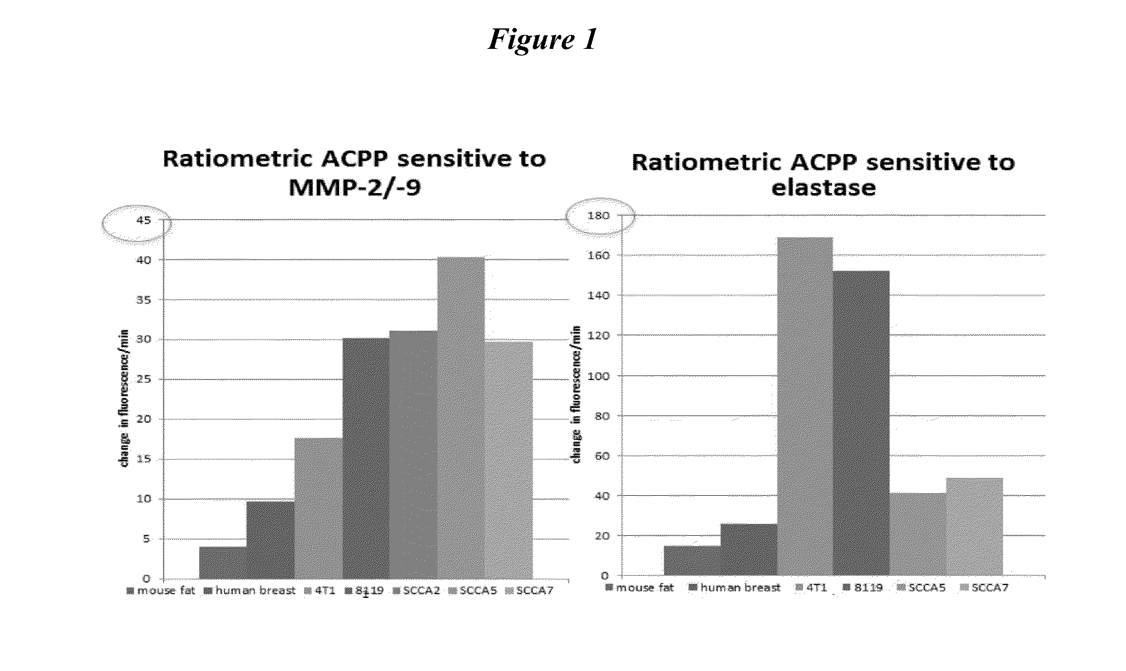
[0256]The rationale is to identify enzymatically positive tumors in a patient population that may benefit from the use of enzymatically cleavable peptide probes for early tumor detection and intraoperative margin evaluation. Experiments have been performed using probes that detected either MMPs (PLGLAG (SEQ ID NO: 1) and PLGC(met)AG (SEQ ID NO: 2) or elastases (RLQLK(acetyl)L (SEQ ID NO: 26). A panel of probes will be expanded to include other tumor expressed proteases.

[0257]Xenograft Tumor Extracts:

[0258]Animals models of a variety of cancers cancer were generated as previously described. Tumors were grown to 0.5 cm-1 cm and then surgically excised. Following dissection, tissue was gently homogenized in PBS, while kept cooled on ice to minimize the release of intracellular and intraorganellar proteases. Homogenates were microcentrifuged at 14,000×g for 1 minute, and supernata...

example 2

Generation of Panel of ACPP for Profiling Tumor Protease Activity

[0269]Multiple ACPPs with varied protease cleavage sequence will be used to establish each specific tumors protease profile. Currently the panel consist of ACPPs that are selective for MMPs, elastases, plasmin, thrombin. MMP 2,9 cleavable sequence PLGLAG (SEQ ID NO: 1), PLGC(met)AG (SEQ ID NO: 2). Other MMP selective substrates could include RS-(Cit)-G-(homoF)-YLY (SEQ ID NO: 4), CRPAHLRDSG (SEQ ID NO: 5), SLAYYTA (SEQ ID NO: 6), NISDLTAG (SEQ ID NO: 7), PPSSLRVT (SEQ ID NO: 8), SGESLSNLTA (SEQ ID NO: 9), RIGFLR (SEQ ID NO: 10). Elastase cleavable sequence RLQLA(acetyl)L (SEQ ID NO: 11). Plasmin selective substrate RLQLKL (SEQ ID NO: 12). Thrombin selective substrates DPRSFL (SEQ ID NO: 13), PPRSFL (SEQ ID NO: 14), Norleucine-TPRSFL (SEQ ID NO: 15). Chymase selective substrate GVAY|SGA (SEQ ID NO: 16). Urokinase-type plasminogen activator (uPA) and tissue plasminogen activator (tPA) selective substrate YGRAAA (SEQ ID N...

example 3

Personalized Protease Assay

[0270]A personalized protease (PePA) assay will be provided which will find use in a variety of situations. A PePA assay will provide an assay for use in understanding the heterogeneity in levels of tumor specific enzymes between patients for a given cancer diagnosis. A PePA assay will also provide an assay for use in correlating tumor specific enzyme activity with known histologic grade / stage and patient prognosis. A PePA assay will provide an assay for use in identifying which patients may benefit from the use of individual enzymatically activatable probes for staging, medical and surgical management in a variety of cancers.

Example 4

Abstract

Objective:

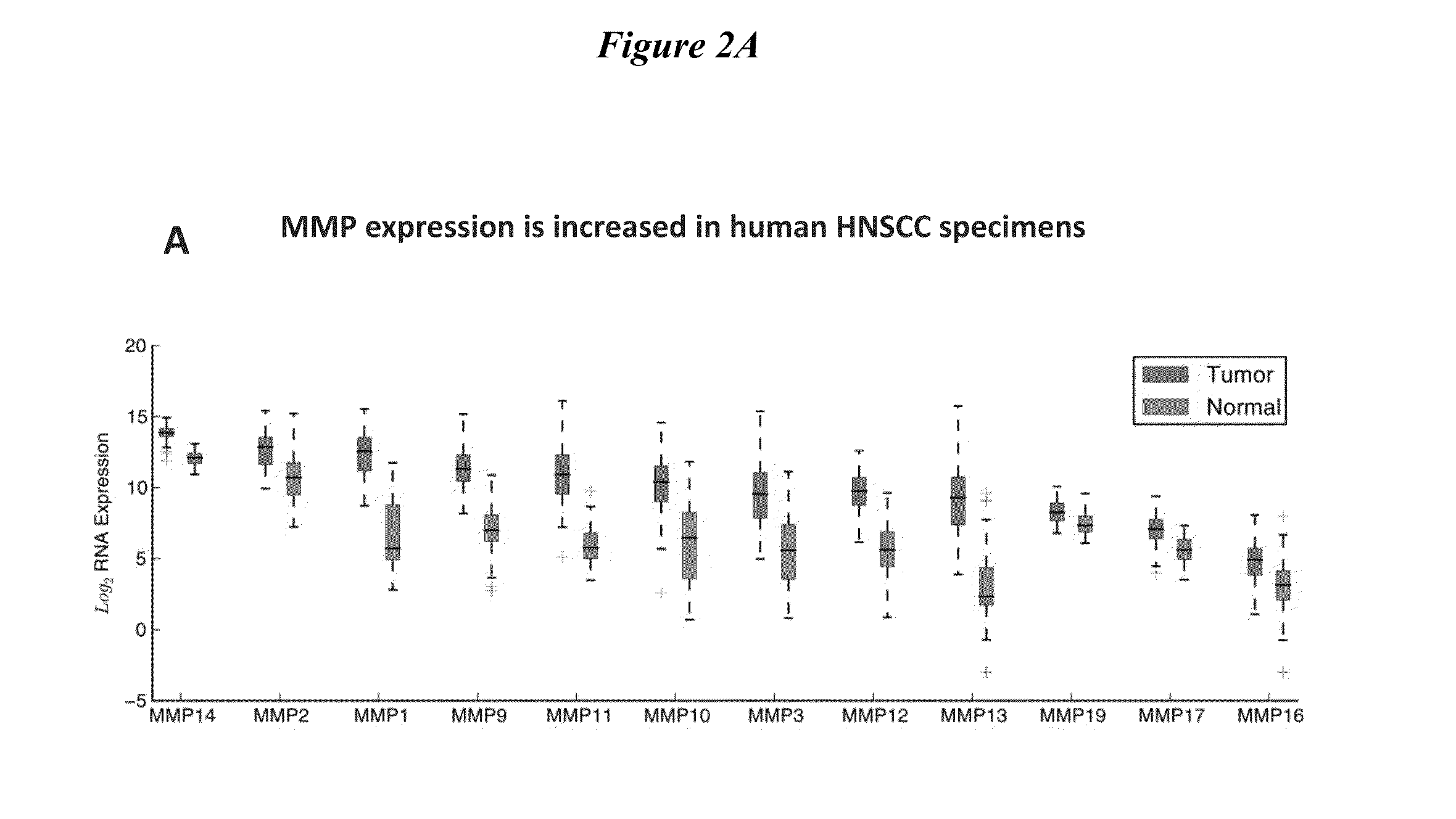
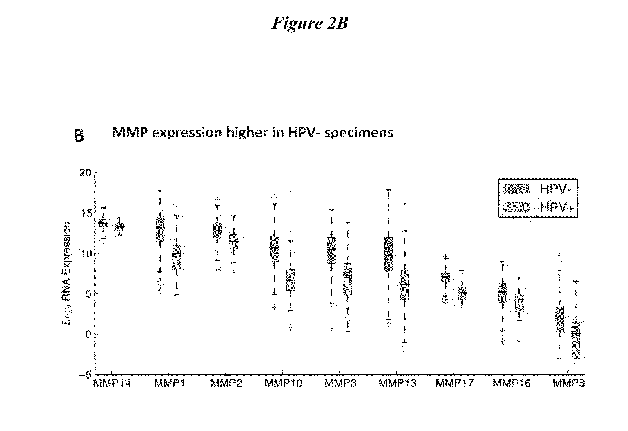
[0271]1. Obtain matrix-metalloproteinase(MMP) expression profiles for head and neck squamous cell carcinoma(HNSCC) specimens from The Cancer Genomic Atlas (TCGA)[0272]2. Demonstrate HNSCC imaging using MMP-cleavable, fluorescently-labeled ratiometric activatable cell-penetrating peptide (RACPP).

Study Design:...


PUM

PropertyMeasurementUnit
waiting timeaaaaaaaaaa
pHaaaaaaaaaa
timeaaaaaaaaaa

Abstract

Disclosed herein, the invention pertains to methods and compositions that find use in diagnostic, prognostic and characterization of neoplasia samples based on the ability of a neoplasia sample to cleave a MTS molecule of the present invention. In some embodiments, a MTS molecule disclosed herein has the formula (A-X-B-C), wherein A is a peptide with a sequence comprising 5 to 9 consecutive acidic amino acids, wherein the amino acids are selected from: aspartates and glutamates; B is a peptide with a sequence comprising 5 to 20 consecutive basic amino acids; X is a linker; and C is a detectable moiety.

Description

CROSS REFERENCE TO RELATED APPLICATIONS[0001]This application claims priority to U.S. Provisional Application No. 62 / 059,081, filed on Oct. 2, 2014, and which is incorporated by reference herein in its entirety for all purposes.STATEMENT OF FEDERALLY-SPONSORED RESEARCH[0002]This work was supported in part by grants from the Howard Hughes Medical Institute, the Department of Defense (W81XWH-09-1-0699), National Cancer Institute (CA158448-01 and P50 CA 097007-DRP), NIBIB (K08 EB008122-01), and Burroughs Wellcome Fund. The government has certain rights in this invention.FIELD OF THE INVENTION[0003]This invention pertains to methods and composition that find use in diagnostic, prognostic and characterization of neoplasia samples based on the ability of a neoplasia sample to cleave a MTS molecule of the present invention.BACKGROUND OF THE INVENTIONIntroduction[0004]Cell membranes delimit the outer boundaries of cells, and regulate transport into and out of the cell interior. Made primari...

Claims


Application Information

Patent Timeline
Patent Type & Authority Applications(United States)
IPC IPC(8): C12Q1/37
CPCC12Q1/37
Inventor WHITNEY, MIKENGUYEN, QUYEN T.TSIEN, ROGER Y.
Owner RGT UNIV OF CALIFORNIA
Why Eureka
Try Eureka