Device for processing tomographic data for visualizing the course of a therapy

a technology of tomographic data and visualizing the course of therapy, which is applied in the field of devices for processing and visualizing data relating to the three-dimensional thoracic dimension of the lungs, and achieves the effect of improving the shape of the contour

Active Publication Date: 2016-06-09
DRAGERWERK AG
View PDF0 Cites 14 Cited by
  • Summary
  • Abstract
  • Description
  • Claims
  • Application Information

AI Technical Summary

Benefits of technology

[0047]A comparison with a predetermined, anatomically typical, stored comparison shape is performed for determining the characteristic contour in a special embodiment variant. The anatomically typical comparison shape is preferably stored in the form of a transverse visualization of the lungs and may have preferably been obtained by means of an e

Problems solved by technology

Due to the position of the EIT electrodes around the thorax, it is not possible therefore to generate frontal views or lateral view

Method used


Image

  • Device for processing tomographic data for visualizing the course of a therapy
  • Device for processing tomographic data for visualizing the course of a therapy

Examples


Embodiment Construction

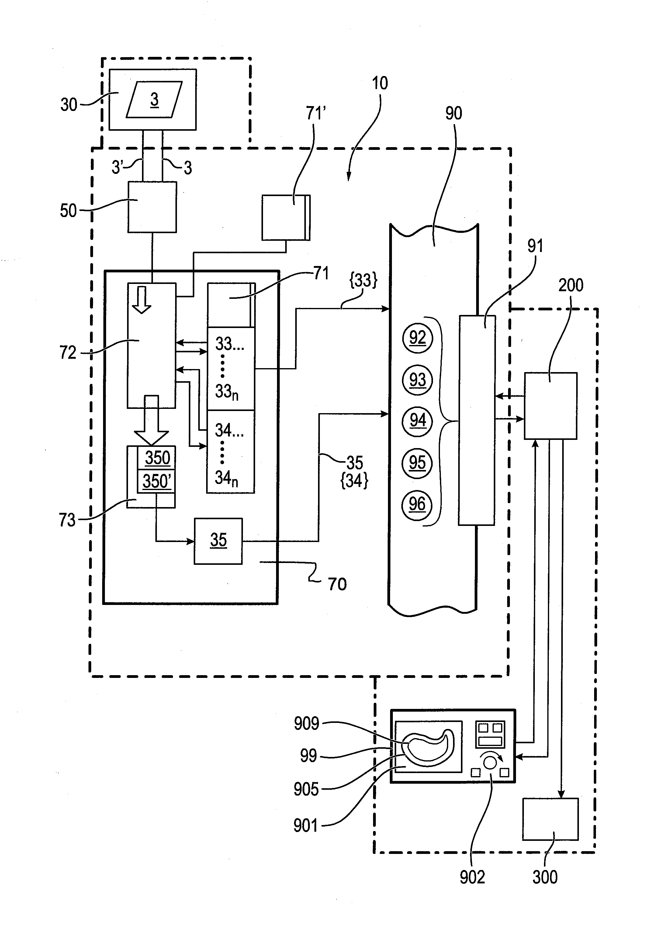
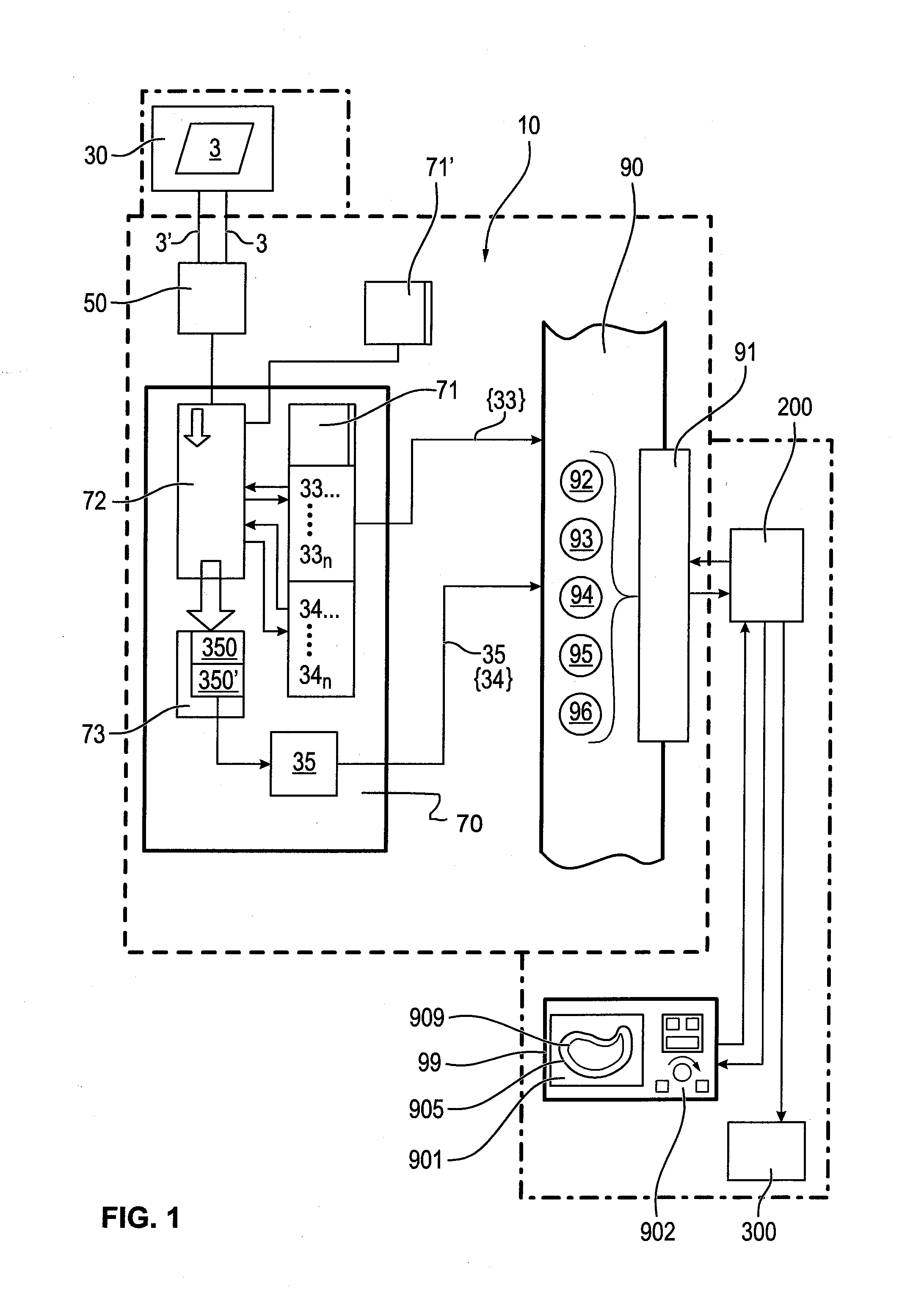
[0055]Referring to the drawings, FIG. 1 shows a device 10 composed of functional elements for processing EIT data 3 in a schematic form.

[0056]This device 10 comprises as the basic components a data input unit 50, a computing and control unit 70 and a data output unit 90. The connections between the elements and units of the device 10 are shown only schematically in this embodiment according to this FIG. 1; for example, the essential data connections and data inputs and data outputs are shown, but no supply lines are shown, and not all connection lines between the elements and units with one another are shown. Furthermore, a display unit 99 connected to the data output unit 90 is shown in this FIG. 1. The display unit 99 comprises visualization means 901, such as display elements, display screens, displays for visualizing graphics, curves, diagrams or images or even numerical value displays for reproducing numerical values. Furthermore, the display unit 99 comprises input elements an...


PUM


Abstract

A device (10) for processing data (3), which were obtained from a medical device suitable for imaging the lungs or the thorax, particularly an electrical impedance tomography device (30), provides improved visualization of a three-dimensional thoracic dimension (350) of the lungs. A characteristic contour (34, 350) is determined continuously by continuous reference to a previously determined outer contour (905) of the lungs as a comparison variable and is outputted, provided and visualized as an output signal (35).

Description

CROSS REFERENCE TO RELATED APPLICATIONS[0001]This application claims the benefit of priority under 35 U.S.C. §119 of German Patent Application 10 2014 018107.3 filed Dec. 9, 2014, the entire contents of which are incorporated herein by reference.FIELD OF THE INVENTION[0002]The present invention pertains to a device for processing and visualizing data relating to a three-dimensional thoracic dimension of the lungs, wherein the data were obtained from a medical device suitable for generating data for imaging, especially from an electrical impedance tomography device. The three-dimensional thoracic dimension of the lungs corresponds to a position and extension of the lungs of a patient within the patient's thorax.BACKGROUND OF THE INVENTION[0003]Devices for electrical impedance tomography (EIT) are known from the state of the art. These devices are designed and intended for generating an image, a plurality of images or a continuous sequence of images from signals obtained by means of e...

Claims


Application Information

Patent Timeline
IPC IPC(8): G06T7/00A61B8/00A61B5/055A61B5/053A61B6/03
CPCG06T7/0085A61B5/0536A61B6/032G06T2207/10088A61B8/00G06T2207/30061G06T2207/10081A61B5/055G06T7/0016G06T2207/10072G06T2207/30076G06T7/62G06T7/13
Inventor GARBER, YVO
Owner DRAGERWERK AG
Why Eureka
Try Eureka