Multi-protein biomarker assay for brain injury detection and outcome

Active Publication Date: 2016-06-23
THE JOHN HOPKINS UNIV SCHOOL OF MEDICINE
View PDF0 Cites 12 Cited by
  • Summary
  • Abstract
  • Description
  • Claims
  • Application Information

AI Technical Summary

Benefits of technology

[0004]The present invention is based, at least in part, on the discovery that the biomarkers GFAP (glial fibrillary acidic protein), S100B, MT3 (metallothionein 3), NSE (neuron specific enolase) and ICAM5 (intracellular adhesion molecule 5) (and/or any of their post-translational modified forms or their isoforms) in combination can discriminate which patient

Problems solved by technology

Objective diagnoses of TBI and especially mTBI remain challenging.
At present, there is no reliable diagnostic test to distinguish between patients with TBI and those without TBI.
Thirdly, BDNF expression is predominantly in brain and spinal cord tissue, limit

Method used


Image

  • Multi-protein biomarker assay for brain injury detection and outcome
  • Multi-protein biomarker assay for brain injury detection and outcome
  • Multi-protein biomarker assay for brain injury detection and outcome

Examples


Example

Example 1

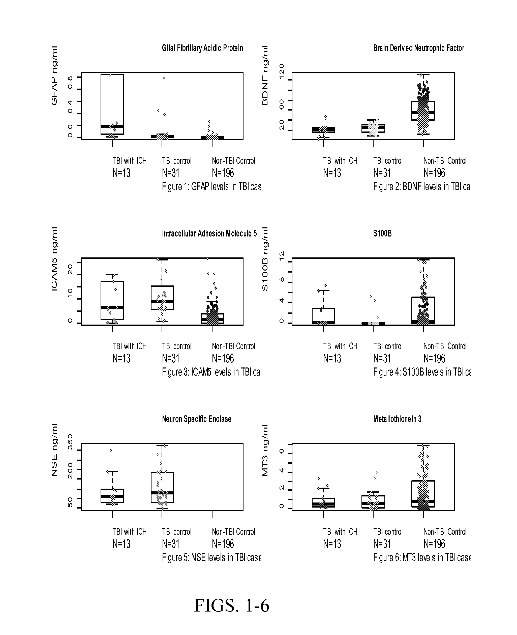
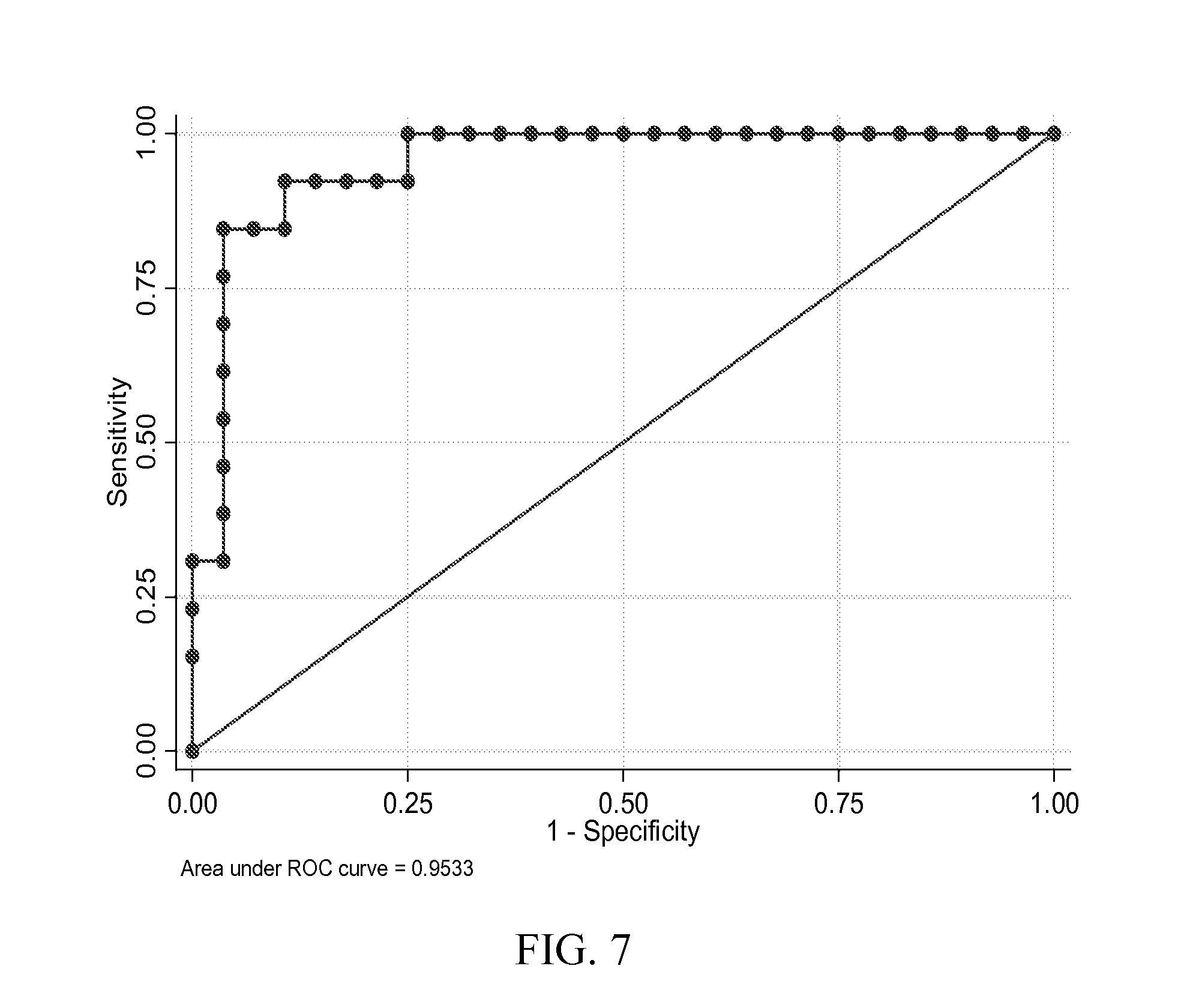
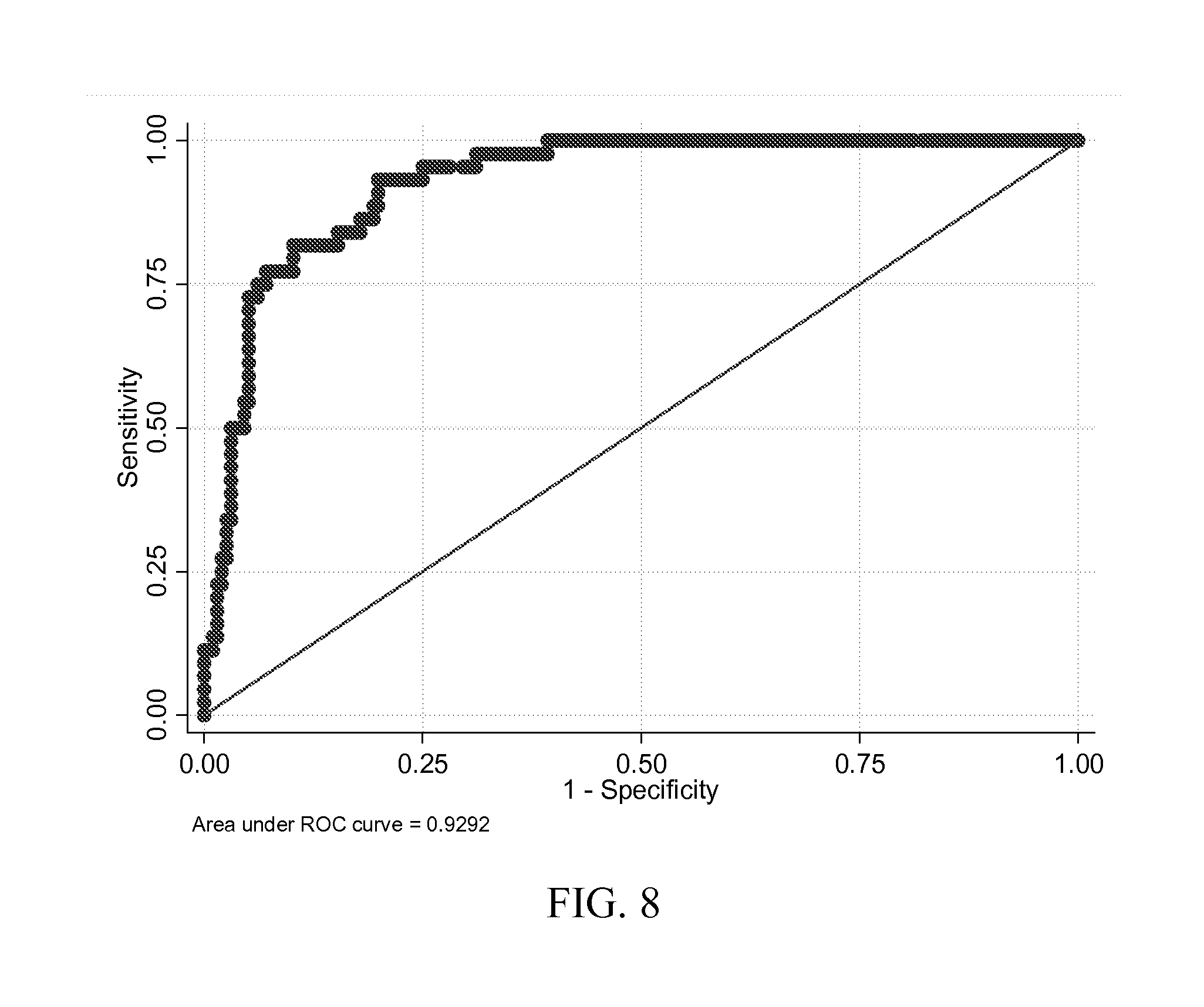
Use of Protein Biomarker Panels for Acute Traumatic Brain Injury (TBI) at Time of Presentation

[0139]Combinations of protein biomarkers and their variants (including protein splice variants (isoforms), polymorphisms, and degraded and other post-translational modifications including, but not limited to, citrullination, phosphorylation, acetylation, methylation, etc.) used as a screening test prior to obtaining a head computerized tomography (CT) scan for diagnosing acute intracranial pathology will allow a safe decrease in avoidable head CT scans.

Materials and Methods

[0140]Study Design:

[0141]Case control.

[0142]Study Population. TBI Cases:

[0143]Cases were consecutive adult patients (18 years or older) presenting to the Johns Hopkins Hospital Emergency Department (ED) with blunt trauma to the head that occurred within 24 hours of arrival, who had a head CT scan that demonstrated acute traumatic intracranial hemorrhage, and who additionally, had excess serum specimen available i...

Example

Example 2

Brain Derived Neurotrophic Factor (BDNF) and its Variants at Time of Presentation Differentiates Between Patient with and without Traumatic Brain Injury (TBI)

[0164]Brain Derived Neurotrophic Factor (BDNF) and its variants (including its splice variants (isoforms), polymorphisms, pro-hormone, active form, pro-hormone inactive cleavage fragment and degraded and other post-translational modifications including, but not limited to, citrullination, phosphorylation, acetylation, methylation and etc., as well as BDNF receptors) at time of presentation differentiates between patient with and without traumatic brain injury (TBI). Furthermore, BDNF, its variants or receptors may be used therapeutically in the treatment of TBI.

Materials and Methods

[0165]Study Design.

[0166]Case control study.

[0167]Study Population. TBI Cases:

[0168]Adult patients (18 years or older) presenting to the Johns Hopkins Hospital Emergency Department (ED) with blunt trauma to the head that occurred within 24 h...

Example

Example 3

Combination of Serum Glial Fibrillary Acidic Protein (GFAP) and Brain Derived Neurotrophic Factor (BDNF) Concentrations Obtained at Presentation Yields Improved Prediction of Short and Long-Term Post-Concussive Symptoms and Disability

Materials and Methods

[0177]Study Design.

[0178]Prospective cohort study.

[0179]Study Population: TBI Cases:

[0180]Cases were consecutive adult patients (18 years or older) presenting to the Johns Hopkins Hospital Emergency Department (ED) with blunt trauma to the head that occurred within 24 hours of arrival, who had a head computerized tomography CT scan that demonstrated acute traumatic intracranial hemorrhage, and who additionally, had excess serum specimen available in the clinical chemistry laboratory. Patients were excluded if they had a prior surgical procedure on the brain or a brain tumor.

[0181]TBI Controls:

[0182]TBI Controls were consecutive adult patients (18 years or older) who presented to the ED within 5 days of cases, for blunt head...


PUM

PropertyMeasurementUnit
Areaaaaaaaaaaa
Threshold limitaaaaaaaaaa

Abstract

The present invention relates to the field of brain injuries. More specifically, the present invention provides methods and compositions useful in the diagnosis/prognosis/assessment of brain injuries. In a specific embodiment, a method for identifying which patients with traumatic brain injury (TBI) require a head computerized tomography (CT) scan for diagnosing acute intracranial pathology comprises the steps of (a) obtaining or collecting a sample from the patient; (b) measuring the levels of one or more biomarkers in the blood sample obtained from the patient, wherein the biomarkers comprise glial fibrillary acidic protein (GFAP), S100B, metallothionein 3 (MT3), neuron specific enolase (NSE) and intracellular adhesion molecule 5 (ICAM5); and (c) identifying the patient as requiring or not requiring a head CT scan based on the measured levels of one or more of biomarkers comprising GFAP, S100B, MT3, NSE and ICAM5.

Description

CROSS-REFERENCE TO RELATED APPLICATIONS[0001]This application claims the benefit of U.S. Provisional Application No. 61 / 847,213, filed Jul. 17, 2013, which is incorporated herein by reference in its entirety.FIELD OF THE INVENTION[0002]The present invention relates to the field of brain injuries. More specifically, the present invention provides methods and compositions useful in the diagnosis / prognosis / assessment of brain injuries.BACKGROUND OF THE INVENTION[0003]Clinical tools such as physical exam, and central nervous system (CNS) imaging (computerized tomography (CT) scan or magnetic resonance imaging (MRI)) are subjective, not widely available, not sensitive or specific enough and too costly to identify all patients with CNS injury and therefore has a high false negative rate. This can include individuals on life support or cardiopulmonary bypass, trauma, loss of oxygen, etc. regardless of the initial injury or disease. There is a great clinical need to identify patients with C...

Claims


Application Information

Patent Timeline
IPC IPC(8): G01N33/68
CPCC12Q1/6883C12Q2600/158G01N33/6893G01N2800/2871G01N2800/28G01N2333/70525G01N2333/825G01N2333/988G01N2333/4727
Inventor EVERETT, ALLEN D.VAN EYK, JENNIFER E.KORLEY, FREDERICK
Owner THE JOHN HOPKINS UNIV SCHOOL OF MEDICINE
Why Eureka
Try Eureka