Microbial analysis

a microorganism and analysis method technology, applied in the field of microorganism analysis, can solve the problems of not widely applicable, inability to reliably distinguish between different strains of the same species, and inability to determine the identity of other microbial organisms, etc., and achieve the effect of faster processing technique and higher analysis throughpu

Active Publication Date: 2016-08-18
KRATOS ANALYTICAL
View PDF2 Cites 7 Cited by
  • Summary
  • Abstract
  • Description
  • Claims
  • Application Information

AI Technical Summary

Benefits of technology

[0019]At its most general, the present invention provides a method of microbial analysis that can differentiate microbial strains based on the constituent lipid analysis. The methods use MALDI mass spectrometry. In some cases, the method analyses microbial phospholipids. Optionally the method also analyses microbial proteins. Thus, “protein fingerprinting” and “lipid fingerprinting” can be performed on the same microbes. Indeed, the same extraction process can be used to obtain extracted lipids and extracted proteins. This facilitates subsequent MALDI-MS analysis, and that analysis can include merging the data from lipid and protein m / z ranges.

Problems solved by technology

However, protein fingerprinting cannot reliably differentiate between different strains of the same species.
However, this method was developed for and tailored to the specific extraction of these glycolipids and the inventors have found that it is not widely applicable.
For example, it cannot be used to determine the identity of other microbial organisms (e.g. yeasts or fungi), which do not contain these glycolipids.
One of the first examples of using MS for the classification of microorganisms was the use of gas-chromatography (GC)-MS.[8] GC-MS is used for cellular fatty acid analysis because of its good chromatographic separation potential.[9] However, lipids need to be broken down into constituent fatty acid molecules to be analysed, and thus the phospholipid composition within the cells cannot be determined.
Recently, MALDI-MS has been reported as a method for bacterial phospholipid analysis.[14][15] However, a lack of reliable sample preparation protocols and instrumentation techniques has prevented wider application of these methods.
Routine bacterial identification by phospholipid analysis using MALDI-MS has not been possible for closely related bacteria, preventing its use in taxonomy.
Moreover, no successful lipid MALDI-MS analysis has been reported for other types of microbe (e.g. yeasts and fungi), which must still be identified by cell morphology using light microscopy, genotyping, and / or protein fingerprinting.
Furthermore, differentiation of microbial strains (within a species) using MALDI-MS based phospholipid profiling has not been reported.

Method used


Image

  • Microbial analysis
  • Microbial analysis
  • Microbial analysis

Examples


example 1

Comparison of Extraction Solvents

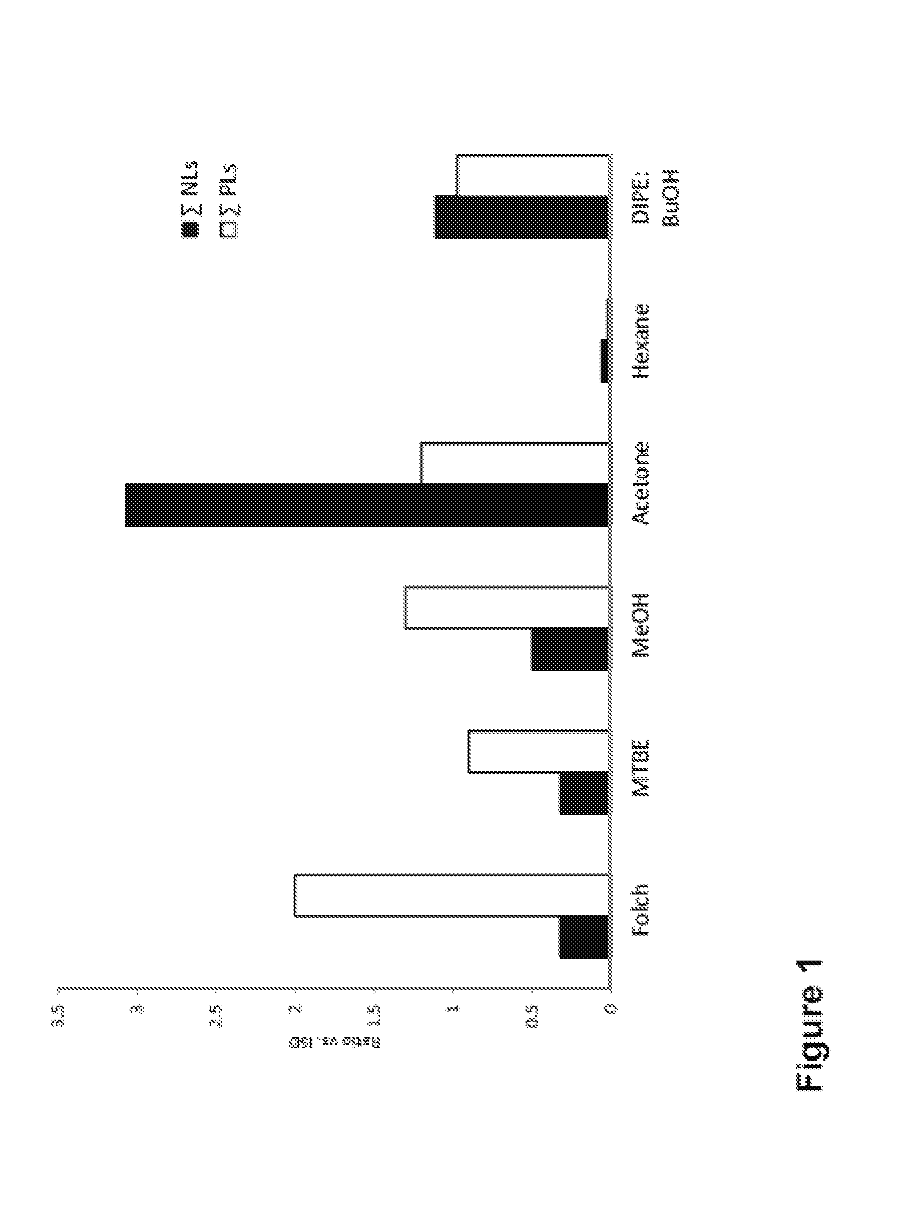
[0259]FIG. 1 shows the extraction efficiency of 6 different organic solvents, for both charged phospholipids (PLs) and neutral lipids (NLs). The extraction efficiency is calculated based on the signal intensity of the corresponding lipid species in a MALDI-MS spectrum.

[0260]It can be seen that Folch, MTBE and MeOH solvents preferentially allows detection of PLs, whereas acetone favours the detection of NLs. An almost equal efficiency for both lipid classes was observed using DIPE / BuOH while the worst results were obtained for Hexane.

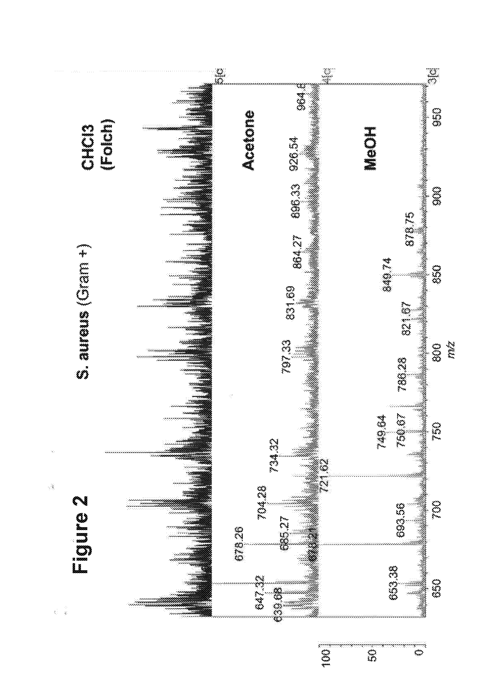
[0261]Based on these preliminary results, Folch, MeOH and acetone were further evaluated for the use in the lipid extraction step and MALDI-MS analysis of the PLs from the different bacteria, yeasts and fungal species.

[0262]These three solvents were applied to a Gram+ bacterium (S. aureus). It can be seen from FIG. 2 that methanol returned a lipid MALDI-MS spectrum with the best signal-to-noise (S / N) ratio.

example 2

Comparison of Matrix Substances

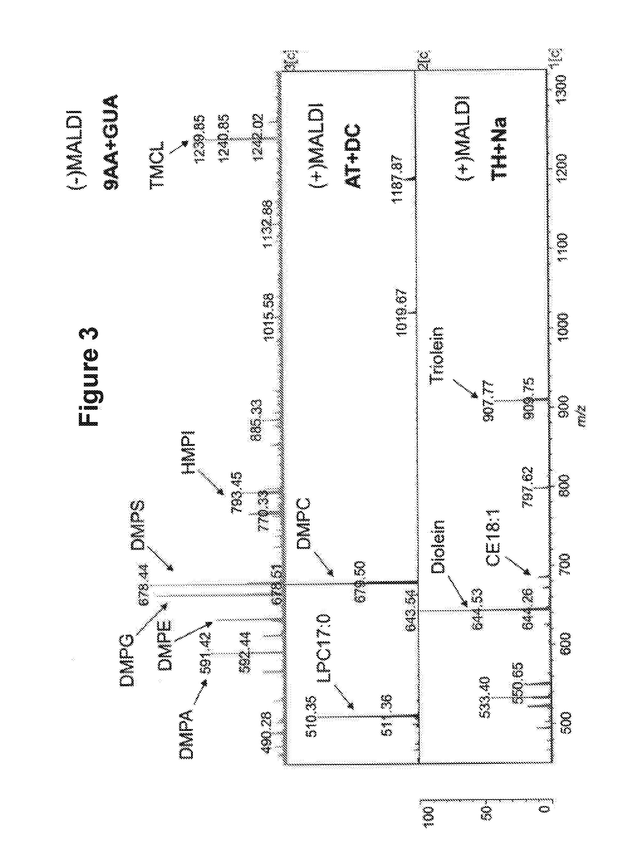
[0263]FIG. 3 shows three different mass spectra of the same lipid sample, containing a mixture of different lipid standards. The spectra were obtained using different matrix substances according to the method described above. It can be seen that in the negative mode, 9AA-GUA allows detection of a large number of peaks. These correspond to anionic phospholipids.

[0264]It can also be seen that both ATT and THAP return clear spectra in the positive mode. The ATT-DC spectrum peaks correspond to cationic phospholipids, and the THAP-Na peaks correspond to neutral lipids. It can be seen that in the positive mode, fewer peaks are detected.

[0265]The inventors assessed the selectivity of the three different matrix substances for ionization of the different major PL-classes present in biological membranes. The results are shown in FIG. 4.

[0266]The data show that in the positive mode, cationic PLs (PC and SM) from an equimolar PL-mixture are more readily ionised an...

example 3

Effect of Culture Medium and Ionization Mode

[0269]FIG. 5 shows the mass spectrum obtained from blood agar in the presence of a methanol lipid-extraction solvent. Several background peaks are seen in the lipid profiling mass range (m / z 700-1500) when measured in the (+)MALDI mode, but not in the (−)MALDI mode.

[0270]This is because blood agar contains some plasma lipids (e.g. derived from blood cells and lipoproteins) which are mostly cationic PL-species (mainly LPC, PC, and SM), and which are therefore preferentially detected in the (+) ionization mode

[0271]This problem can be circumvented in the (+)MALDI mode by using minimal medium (devoid of exogenous lipids) instead of blood agar. The resultant lipid mass spectrum is essentially the same, but with fewer culture medium contaminants. However, the use of minimal medium generally leads to less favourable culture conditions, and results in longer incubation times to obtain a sufficient number of cells for analysis. The (−) mode allows...


PUM

PropertyMeasurementUnit
timeaaaaaaaaaa
timeaaaaaaaaaa
vol %aaaaaaaaaa

Abstract

The present invention is concerned with a method of identifying microbial strains (e.g. from a cell culture), the method comprising; i) a lipid extraction step, comprising extraction of phospholipids from the microbe, suitably with an extraction composition comprising more than 50 vol % MeOH; ii) a sample preparation step, comprising preparation of a MALDI sample incorporating the extracted lipids; iii) a data gathering step, comprising performing MALDI-based mass spectrometry on the MALDI sample, and iv) a microbe identification step, comprising analysis of the mass spectrometry data to characterise or identify the microbial strain. Suitably the method also includes extracting proteins from the microbes and analysing the extracted proteins using MALDI-based mass spectrometry so as to obtain not only lipid m / z data but also protein m / z data.

Description

FIELD[0001]The present invention relates to a method of analysing microbes. The analysis is based on lipid profiling, and can be used in microbial identification.BACKGROUND[0002]It is desirable to swiftly and simply, reliably identify microbes. Reliable identification allows the pathogenicity and / or other characteristics of a microbial sample to be identified. Determining the biomolecule make-up of a microbe can assist in identification and hence diagnosis.[0003]Mass spectrometry can be used to analyse biomolecules, typically using soft ionization techniques. Matrix-Assisted Laser Desorption / lonization (MALDI)-MS is one such technique, and has been widely used in the analysis of large biomolecules including proteins. Protein fingerprinting has enabled the application of MALDI-MS for microbial identification and diagnostics.[0004]However, protein fingerprinting cannot reliably differentiate between different strains of the same species. Serological testing or PCR analysis is needed t...

Claims


Application Information

Patent Timeline
Patent Type & Authority Applications(United States)
IPC IPC(8): C12Q1/04H01J49/04
CPCC12Q1/04H01J49/0418G01N33/92G01N33/6851G01N33/68
Inventor STUBIGER, GERALDBELGACEM, OMAR
Owner KRATOS ANALYTICAL
Why Eureka
Try Eureka