Methods and systems for disrupting calcified walls of biological conduits and calcified lesions therein

a biological conduit and calcification technology, applied in the field of methods and systems for disrupting the calcification walls of biological conduits and calcified lesions therein, can solve the problems of systematic disruption of any calcification within the intimal and/or medial layers of the subject artery, softening of calcification within the occlusion or lesion, and improving compliance of the vessel, as well as the lesion itself.

Active Publication Date: 2016-08-25
CARDIOVASCULAR SYST INC
View PDF1 Cites 6 Cited by
  • Summary
  • Abstract
  • Description
  • Claims
  • Application Information

AI Technical Summary

Benefits of technology

[0017]The result achieved by use of at least one eccentric head, smooth or abrading, that, during high-speed rotation within the exemplary lumen, has for the first time been found to produce a combination of a low-frequency orbital motion comprising a force that is exerted against the lumen wall, with concomitant deflection of same, and a high-frequency pulsatile frequency, also with concomitant exertion of force against the lumen wall and deflection of same. These force-driven circumferential deflections produce a series of shockwaves within the layers of the exemplary artery's wall layers, resulting in systematic disruptions of any calcification within the intimal and / or medial layers of the subject artery. In addition, any calcification within the occlusion or lesion is also softened and disrupted. Thus, as this process proceeds, the compliance of the vessel, as well as the lesion itself, improves.

Problems solved by technology

In certain embodiments, however, the eccentric head that is used is not an abrading head and may therefore be a smooth head lacking in abrasive coating.
These force-driven circumferential deflections produce a series of shockwaves within the layers of the exemplary artery's wall layers, resulting in systematic disruptions of any calcification within the intimal and / or medial layers of the subject artery.
In addition, any calcification within the occlusion or lesion is also softened and disrupted.

Method used


Image

  • Methods and systems for disrupting calcified walls of biological conduits and calcified lesions therein
  • Methods and systems for disrupting calcified walls of biological conduits and calcified lesions therein
  • Methods and systems for disrupting calcified walls of biological conduits and calcified lesions therein

Examples


working example 1

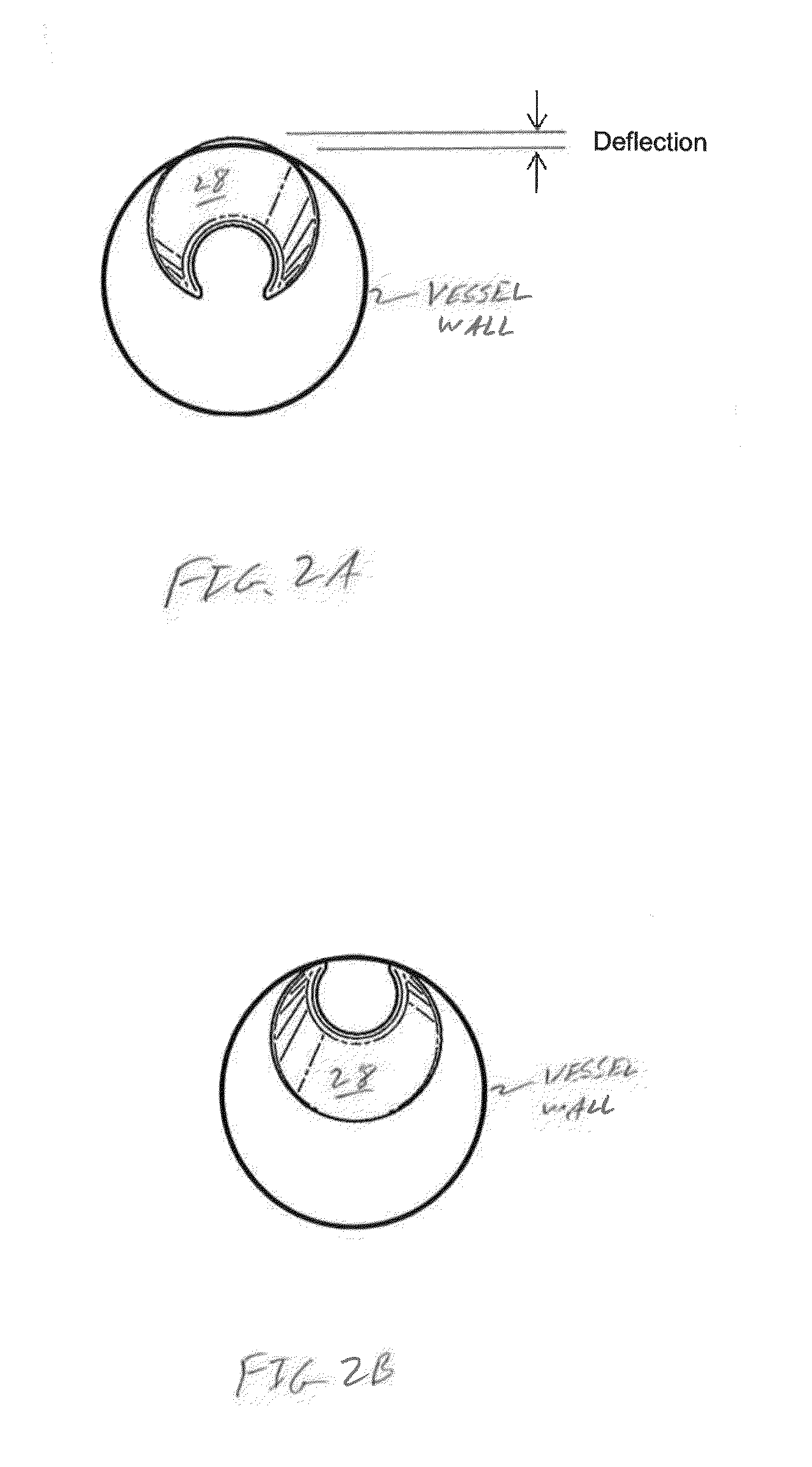
[0048]The following working example investigates the motion and contact forces of an eccentric abrading during high-speed orbital atherectomy. A high-speed camera and image processing technique were utilized to visualize and quantify the crown motion and its interaction with the wall of a transparent arterial phantom made of tissue-mimicking polyvinyl chloride (PVC). Forces were measured simultaneously by a piezoelectric force dynamometer with sufficient sensitivity and bandwidth for such rapid dynamic measurements.

[0049]Materials and Methods

[0050]The experimental setup consisted of three modules—the atherectomy device, an arterial phantom, and the measurement system—in the following sections.

[0051]Atherectomy Device

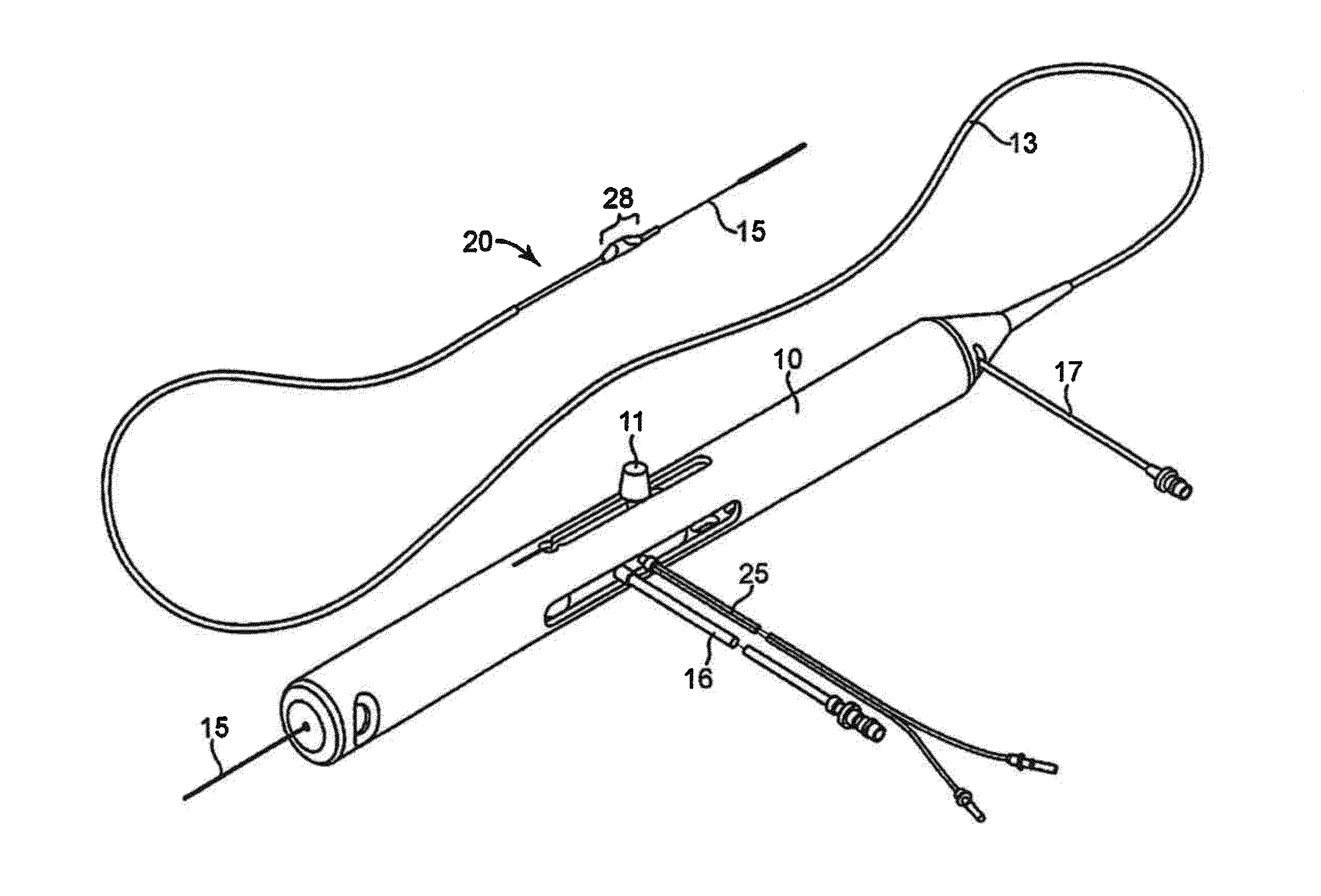
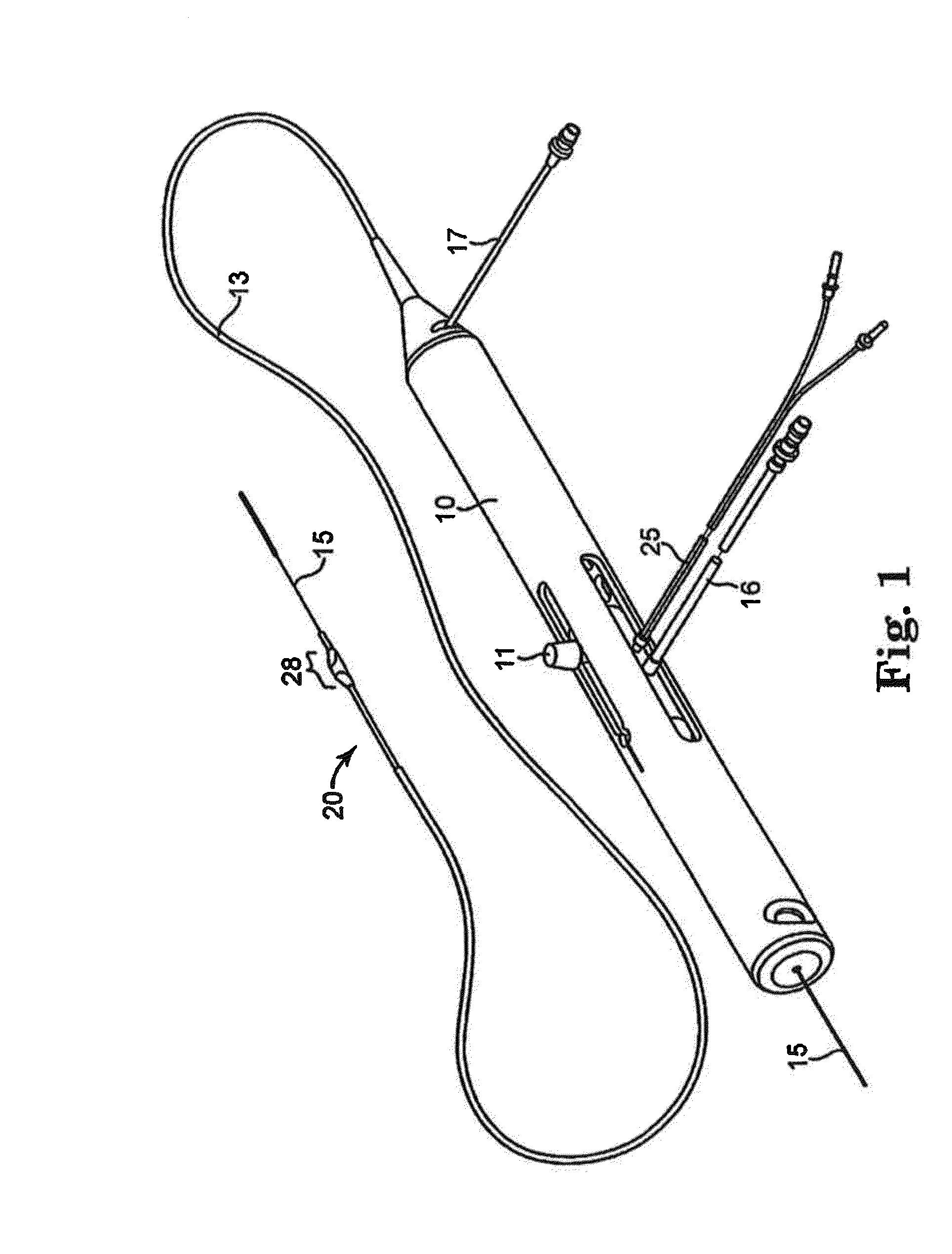
[0052]The orbital atherectomy device in Working Example 1 is the Diamondback 360® manufactured by Cardiovascular Systems Inc. (St. Paul, Minn.), assignee of the present disclosure and is similar to the device illustrated in FIG. 1. This device consists of three units: (1...


PUM


Abstract

The present system is directed in various embodiments to rotational atherectomy systems and methods generally. More specifically, a method for methodically softening and otherwise disrupting calcification located within atherosclerotic plaque, lesion or occlusion and/or within the wall of a biological conduit or lumen. For example, calcification within the occlusion and/or intimal and/or medial layer walls of a blood vessel, e.g., an artery, may be methodically softened or otherwise disrupted with various embodiments of the present invention. The softening and/or disruption of the calcification in the walls of the exemplary artery is accomplished in conjunction with abrading removal of any occlusion located on the interior surface of the exemplary artery and, therefore, located within the artery's lumen. This result is achieved by use of at least one eccentric head that, during high-speed rotation within the exemplary lumen, has been found to produce a combination of a low-frequency orbital motion comprising a force that is exerted against the lumen wall, with concomitant deflection of same, and/or a high-frequency pulsatile frequency, also with concomitant exertion of force against the lumen wall and deflection of same. These force-driven deflections produce a series of shockwaves within the layers of the exemplary artery's wall layers, resulting in systematic disruptions of any calcification within the intimal and/or medial layers of the subject artery. In addition, any calcification within the occlusion is also softened and disrupted. Thus, as this process proceeds, the compliance of the vessel, as well as the lesion itself, is increased.

Description

CROSS-REFERENCE TO RELATED APPLICATIONS[0001]This application claims priority to App. Ser. No. 62 / 118,696, entitled “Secondary Mechanism Of Action Of Orbital Atherectomy,” filed Feb. 20, 2015; Appl. Ser. No. 62 / 134,408, entitled “Secondary Mechanism Of Action of Orbital Atherectomy,” filed Mar. 17, 2015; and App. Ser. No. 62 / 190,411, entitled “Experimental Investigation Of The Abrasive Crown Dynamics In Orbital Atherectomy,” filed Jul. 9, 2015, the entire contents of each of which are hereby incorporated by reference.INVENTORS[0002]Robert E. Kohler, a citizen of the United States, resident in Lake Elmo, Minn.[0003]Brad J. Martinsen, a citizen of the United States, resident in St. Louis Park, Minn.;[0004]Michael X. Yang, a citizen of the United States, resident in Maple Grove, Minn.;[0005]Yihao Zheng, a citizen of the People's Republic of China, resident in Ann Arbor, Mich.;[0006]Albert Shih, a citizen of the United States, resident in Ann Arbor, Mich.; and[0007]Rohit Deokar, a citiz...

Claims


Application Information

Patent Timeline
Patent Type & Authority Applications(United States)
IPC IPC(8): A61B17/32A61B17/3207
CPCA61B17/3207A61B17/320068A61B17/22012A61B17/320758A61B2017/22011A61B2017/22051A61B2017/320004A61B2017/320766
Inventor KOHLER, ROBERT E.MARTINSEN, BRAD J.YANG, MICHAEL X.ZHENG, YIHAOSHIH, ALBERTDEOKAR, ROHIT
Owner CARDIOVASCULAR SYST INC
Why Eureka
Try Eureka