Methods and compositions for preserving retinal ganglion cells

a technology of retinal ganglion cells and compositions, applied in drug compositions, heterocyclic compound active ingredients, enzyme inhibitor ingredients, etc., can solve the problems of retinal ganglion cell loss, glaucoma and optic nerve injury, pan-caspase inhibitor z-vad, etc., to prevent retinal ganglion cell loss, reduce or prevent the loss of retinal ganglion cell viability, and reduce the loss of visual function

Active Publication Date: 2016-12-22
MASSACHUSETTS EYE & EAR INFARY
View PDF0 Cites 7 Cited by
  • Summary
  • Abstract
  • Description
  • Claims
  • Application Information

AI Technical Summary

Benefits of technology

[0010]The invention is based, in part, on the discovery that a necrosis inhibitor, e.g., RIP kinase inhibitor, e.g., a necrostatin, e.g., necrostatin-1, can be used to reduce or prevent the loss of retinal ganglion cell viability, especially when the necrosis inhibitor is combined with an apoptotic inhibitor (e.g., a pan-caspase inhibitor, e.g., Z-VAD and / or IDN-6556). It was previously understood that retinal ganglion cell death associated with glaucoma and optic nerve injury was primarily caused by apoptosis. However, studies have shown that the administration of Z-VAD, an apoptosis inhibitor (i.e., a pan-caspase inhibitor), fails to thoroughly prevent retinal ganglion cell loss in glaucoma and optic nerve injury. The studies described hereinbelow indicate that, in the presence of an apoptosis inhibitor (e.g., a pan-caspase inhibitor), retinal ganglion cells die by necrosis, including necroptosis (or programmed necrosis). These studies show that programmed necrosis is a critical mechanism for ocular disorders wherein a symptom of the disorder is the loss of retinal ganglion cell viability in the presence of a pan-caspase inhibitor. As a result, it is possible to reduce the loss of visual function associated with an ocular disorder, in particular while the ocular disorder is being treated, by reducing the loss of retinal ganglion cell viability.
[0012]In another aspect, provided herein is a method of preserving the viability of retinal ganglion cells within the retina of a subject with an ocular condition. A symptom of the ocular condition may be the loss of retinal ganglion cells in the retina of the eye with the condition. The ocular condition may be glaucoma, optic nerve injury, optic neuritis, optic neuropathies, central retinal artery occlusion, central retinal vein occlusion and diabetic retinopathy. The method comprises administering to the eye of the subject an effective amount of a necrosis inhibitor and an apoptosis inhibitor thereby preserving the viability of the retinal ganglion cells with the retina of the subject with the condition. After the administration of the necrosis inhibitor and the apoptosis inhibitor, the retinal ganglion cell is capable of supporting axonal regeneration.
[0014]In another aspect, provided herein is a method of preserving the visual function of an eye of a subject affected with an ocular condition wherein a symptom of the ocular condition is loss of retinal ganglion cell viability in the retina of the eye. The method comprises (a) reducing the production or activity of a RIP-1 kinase and / or a RIP-3 kinase in the eye thereby to preserve the viability of the retinal ganglion cells disposed within the retina of the eye; and (b), after treatment, measuring visual function (e.g., visual acuity) of the eye. In certain embodiments, the reduction in the production or activity of the RIP-1 kinase and / or the RIP-3 kinase can be achieved by administering an effective amount of RIPK inhibitor, e.g., a necrostatin. After administration of the RIPK inhibitor the visual function of the eye may be preserved or improved relative to the visual function of the eye prior to administration of the RIPK inhibitor.

Problems solved by technology

However, studies have shown that the administration of Z-VAD, an apoptosis inhibitor (i.e., a pan-caspase inhibitor), fails to thoroughly prevent retinal ganglion cell loss in glaucoma and optic nerve injury.
A symptom of the ocular condition may be the loss of retinal ganglion cells in the retina of the eye with the condition.

Method used


Image

  • Methods and compositions for preserving retinal ganglion cells
  • Methods and compositions for preserving retinal ganglion cells
  • Methods and compositions for preserving retinal ganglion cells

Examples


example 1

Efficacy of a Necrosis Inhibitor and a Pan-Caspase Inhibitor in the Treatment of Glaucoma and Optic Nerve Injury

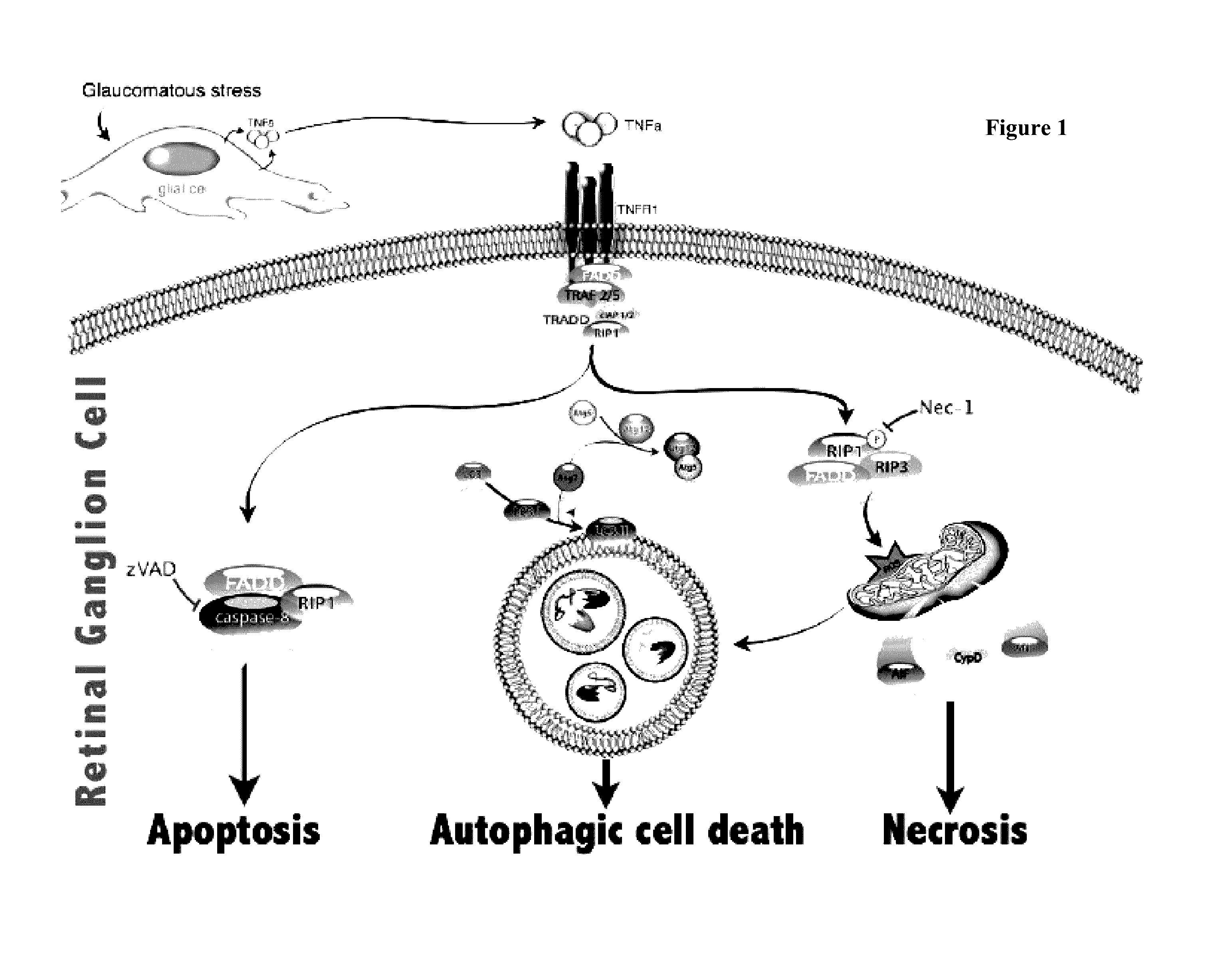
[0336]Glaucoma is a group of ocular disorders, characterized by optic nerve injury. In most cases of glaucoma, the optic nerve injury is caused by elevated intraocular pressure. Furthermore, higher intraocular pressures are generally associated with greater nerve damage. In this example, mouse models of optic nerve (ON) injury was used to assess the role of RIP-mediated programmed necrosis and apoptosis in this ocular disorder. Given that ON injury is a hallmark of glaucoma, this model also provided an assessment of RIP-mediated programmed necrosis in RGC loss in glaucoma.

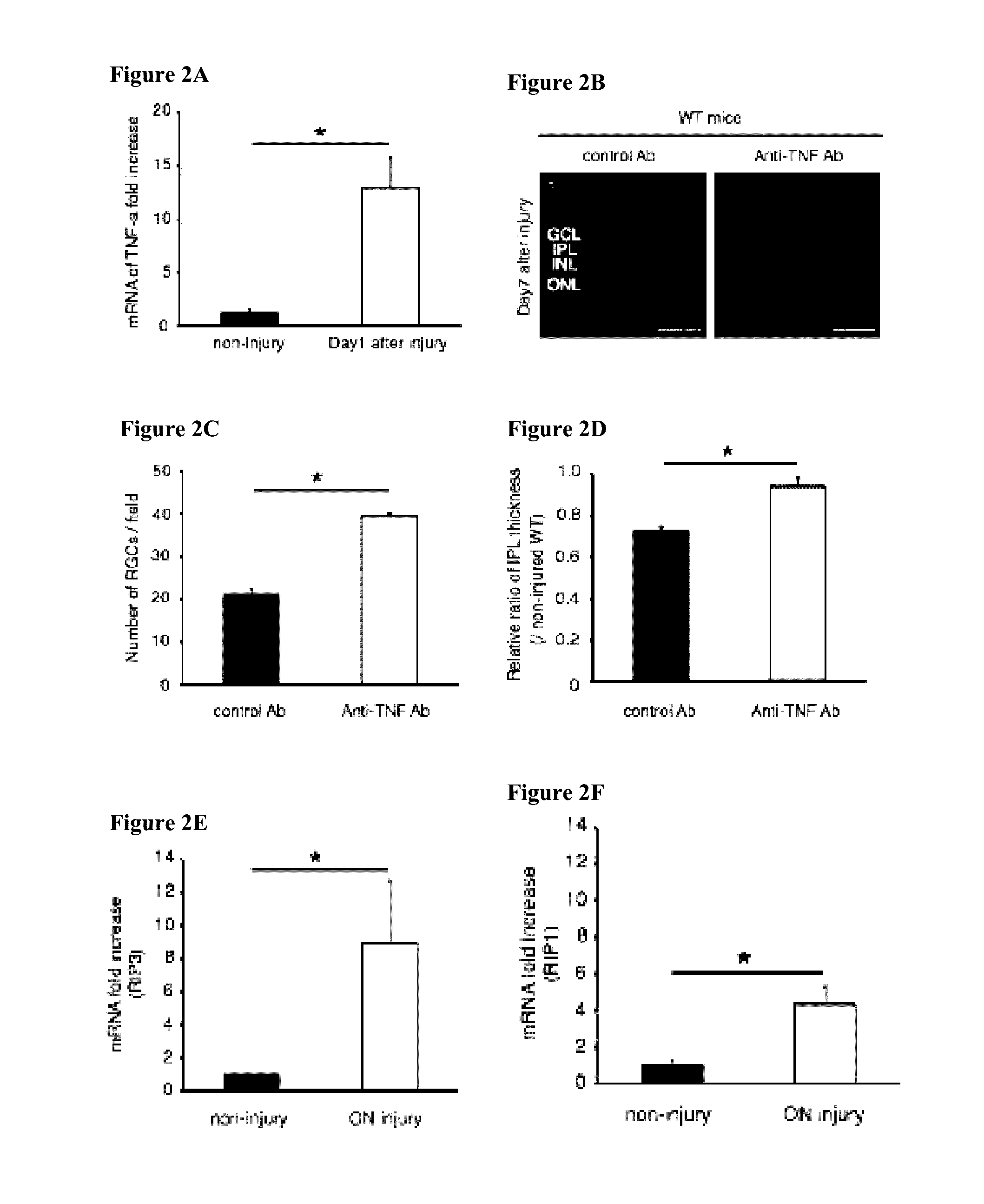
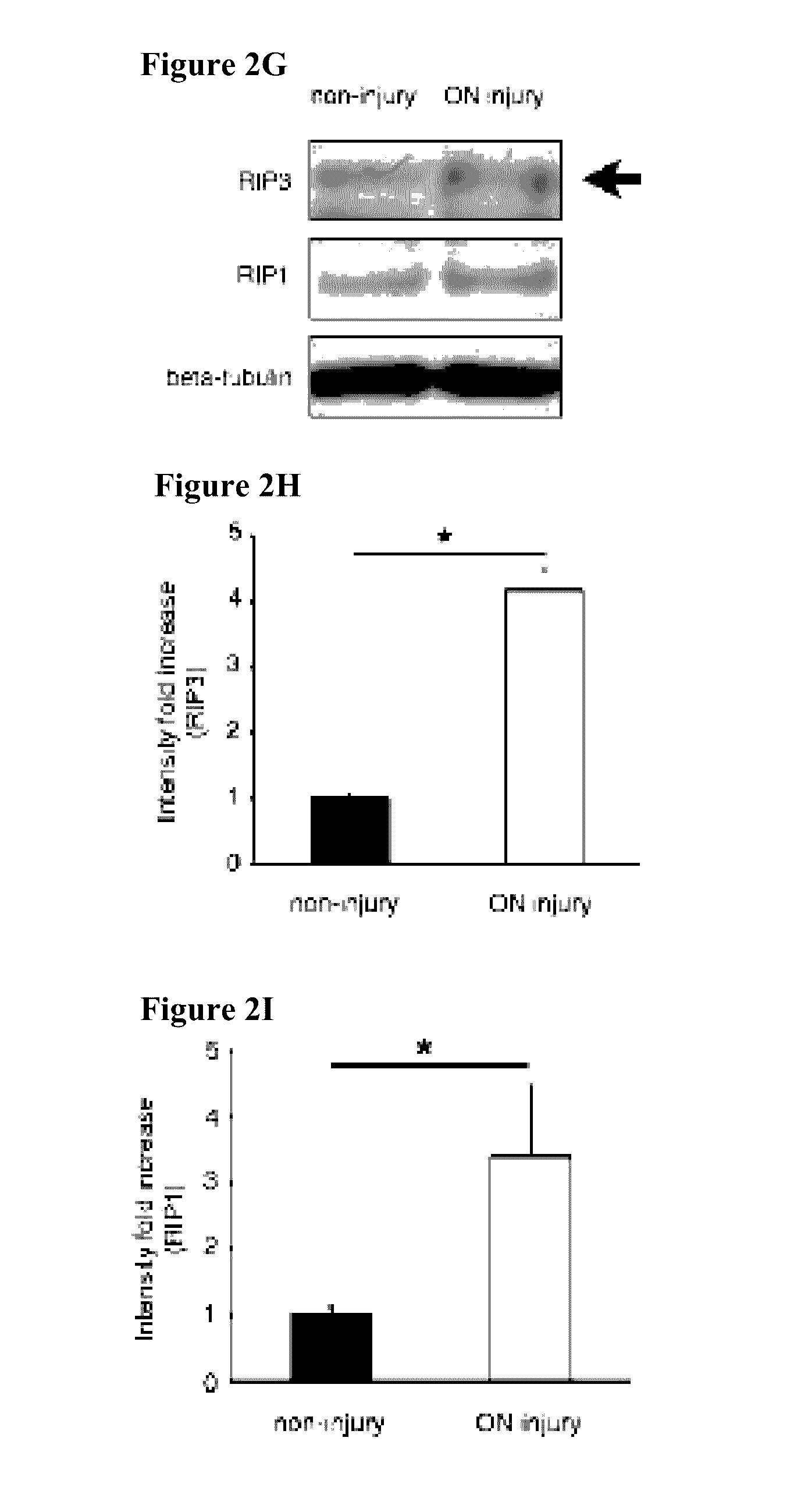
A. Neutralization of TNF-α Prevents Optic Nerve Injury-Induced RGC Death

[0337]Recent evidence has demonstrated the role of TNF-α as a mediator of retinal ganglion cell (RGC) death in glaucoma (Nakazawa et al., (2006) J NEUROSCI 26:12633-12641)). Furthermore, in the glaucomatous eye, death receptors of TNF...

example 2

Efficacy of a Necrosis Inhibitor and a Pan-Caspase Inhibitor in Promoting RGC Survival and Axon Regeneration

[0365]Like most pathways in the mature central nervous system, the optic nerve cannot regenerate if injured, leaving victims of traumatic nerve injury or degenerative diseases such as glaucoma with life-long visual losses. This situation can be, at least, partially reversed by enhancing the intrinsic growth state of retinal ganglion cells (RGCs). In this example, the efficacy of necrosis inhibitor and a pan-caspase inhibitor in promoting RGC survival and axon regeneration is investigated using a mouse optic nerve crush model.

A. A Necrosis Inhibitor in Combination with a Caspase Inhibitor Promotes RGC Survival in a Optic Nerve Crush Model

[0366]Mice were subjected to optic nerve crush surgery. Specifically, animals were anesthetized with an intraperitoneal injection of ketamine (60-80 mg / kg: Phoenix Pharmaceutical, St. Joseph, Mo.) and xylazine (10-15 mg / kg: Bayer, Shawnee Missi...


PUM

PropertyMeasurementUnit
volumeaaaaaaaaaa
diameteraaaaaaaaaa
distanceaaaaaaaaaa

Abstract

Provided are methods and compositions for maintaining the viability of retinal ganglion cells in a subject with an ocular disorder including, for example, glaucoma and optic nerve injury. The viability of the retinal ganglion cells can be preserved by administering a necrosis inhibitor either alone or in combination with an apoptosis inhibitor to a subject having an eye with the ocular condition. The compositions, when administered, maintain the viability of the cells and / or promote axon regeneration, thereby minimizing the loss of vision or visual function associated with the ocular disorder.

Description

CROSS-REFERENCE TO RELATED APPLICATIONS[0001]This application is a continuation of U.S. patent application Ser. No. 13 / 882,932, filed Sep. 30, 2013, which is the national stage filing under 35 U.S.C. §371 of International Patent Application No. PCT / US2011 / 057327, filed Oct. 21, 2011, which claims the benefit of and priority to U.S. Provisional Application No. 61 / 409,055, filed Nov. 1, 2010; U.S. Provisional Application No. 61 / 414,862, filed Nov. 17, 2010; and U.S. Provisional Application No. 61 / 472,144, filed Apr. 5, 2011; the disclosures of each application are hereby incorporated by reference in their entirety.GOVERNMENT FUNDING[0002]The work described in this application was sponsored, in part, by the National Eye Institute under Grant No. EY14104. The United States Government has certain rights in the invention.FIELD OF THE INVENTION[0003]The field of the invention relates generally to methods and compositions for preserving the viability of retinal ganglion cells (RGCs), for ex...

Claims


Application Information

Patent Timeline
Patent Type & Authority Applications(United States)
IPC IPC(8): A61K38/06A61K31/4178A61K9/00
CPCA61K38/06A61K31/4178A61K9/0048A61K31/195A61K31/40A61K31/403A61K31/4166A61K38/005A61P27/02A61P27/06A61K2300/00A61K31/401
Inventor VAVVAS, DEMETRIOS G.MILLER, JOAN W.KAYAMA, MAKI
Owner MASSACHUSETTS EYE & EAR INFARY
Why Eureka
Try Eureka