
Hyperspectral imaging for prediction of skin injury after exposure to thermal energy or ionizing radiation

a technology of ionizing radiation and hyperspectral imaging, which is applied in the direction of spectroscopy, instruments, applications, etc., can solve the problems of increased infection risk, prolonged hospitalization, early differentiation of superficial versus deep dermal burns, etc., and achieves rapid identification and effective, efficient and non-invasive detection.

Inactive Publication Date: 2017-05-18
UNIV OF MASSACHUSETTS
View PDF4 Cites 4 Cited by
  • Summary
  • Abstract
  • Description
  • Claims
  • Application Information

AI Technical Summary

Benefits of technology

The patent is about a new way to detect and measure changes in tissue caused by heat or radiation. It uses special methods that can show changes in skin color and blood flow, which can help doctors quickly identify and treat people who have been exposed to these kinds of injuries. The invention is non-invasive and can help predict the extent of harm to normal tissue.

Problems solved by technology

However, early differentiation of superficial dermal versus deep dermal burns presents a diagnostic challenge.
However, deep dermal burns result in prolonged hospitalizations, increased risk of infection, excessive scarring and thus are best managed by excision and skin grafting.
Differentiation between dermal burns that spontaneously heal versus those that require excision remains clinically challenging, with only 50-75% accuracy in early clinical assessments.
Basic clinical overestimation of burn depth results in unnecessary surgery, while underestimation results in prolonged hospital stays, increased morbidity and mortality, delayed surgery, and inferior functional and aesthetic outcomes.
Over or under-estimation of burn injury results in inappropriate fluid resuscitation of the patient and potentially life threatening complications.
Due to iatrogenic injury and sampling error potential, however, it has little application in actual clinical practice.
In addition, exposure to ionizing radiation can also have profound biological consequences to the skin.
The latter posses a significant clinical problem since the development of acute skin reactions during radiotherapy usually signal a treatment break.
It has been estimated that a treatment break of a more than a week during breast cancer radiotherapy can have significant negative impact on recurrence rate and overall survival.

Method used


Image

  • Hyperspectral imaging for prediction of skin injury after exposure to thermal energy or ionizing radiation
  • Hyperspectral imaging for prediction of skin injury after exposure to thermal energy or ionizing radiation
  • Hyperspectral imaging for prediction of skin injury after exposure to thermal energy or ionizing radiation

Examples


example 1

Hyperspectral Imaging as an Early Biomarker for Radiation Exposure and Microcirculatory Damage

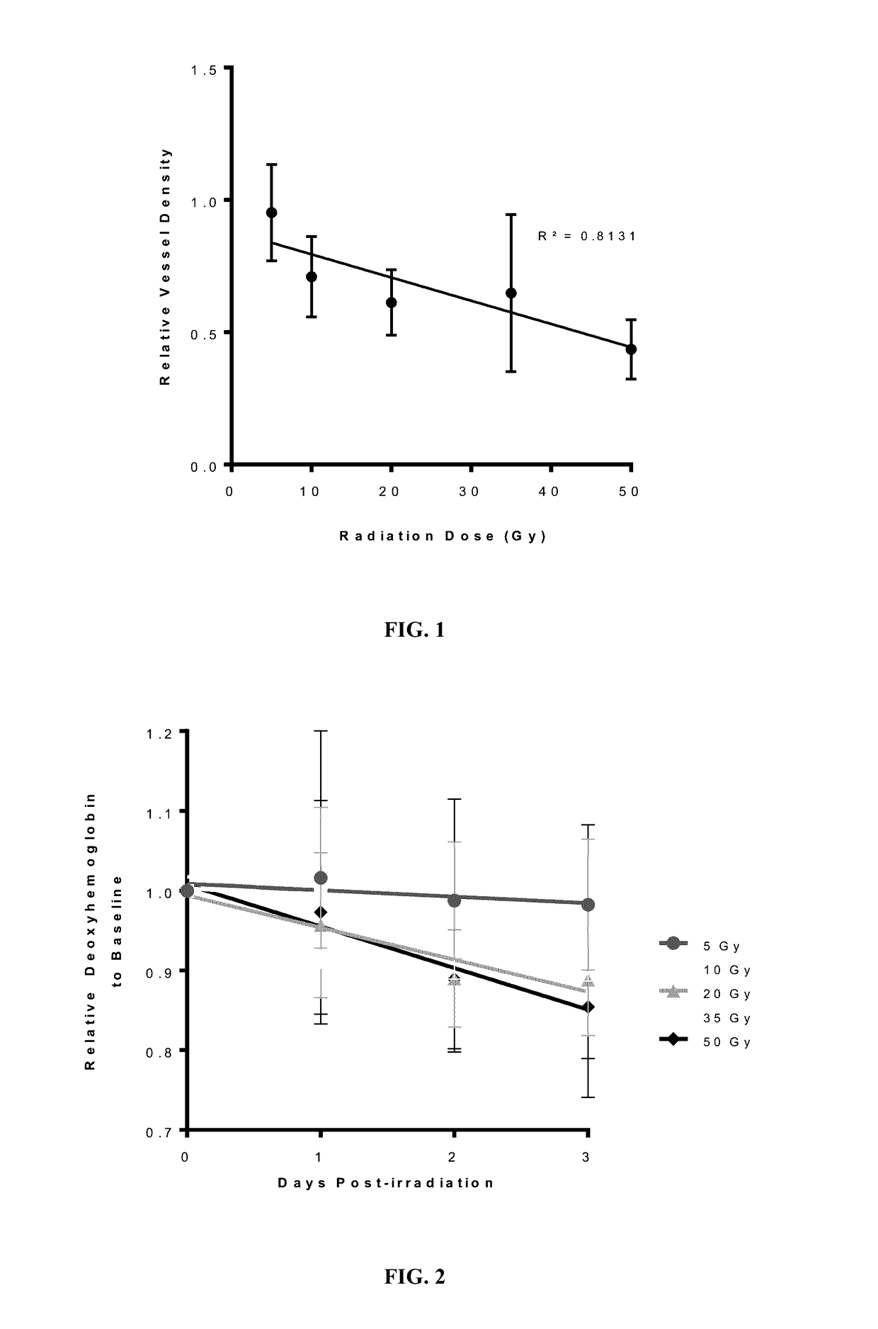
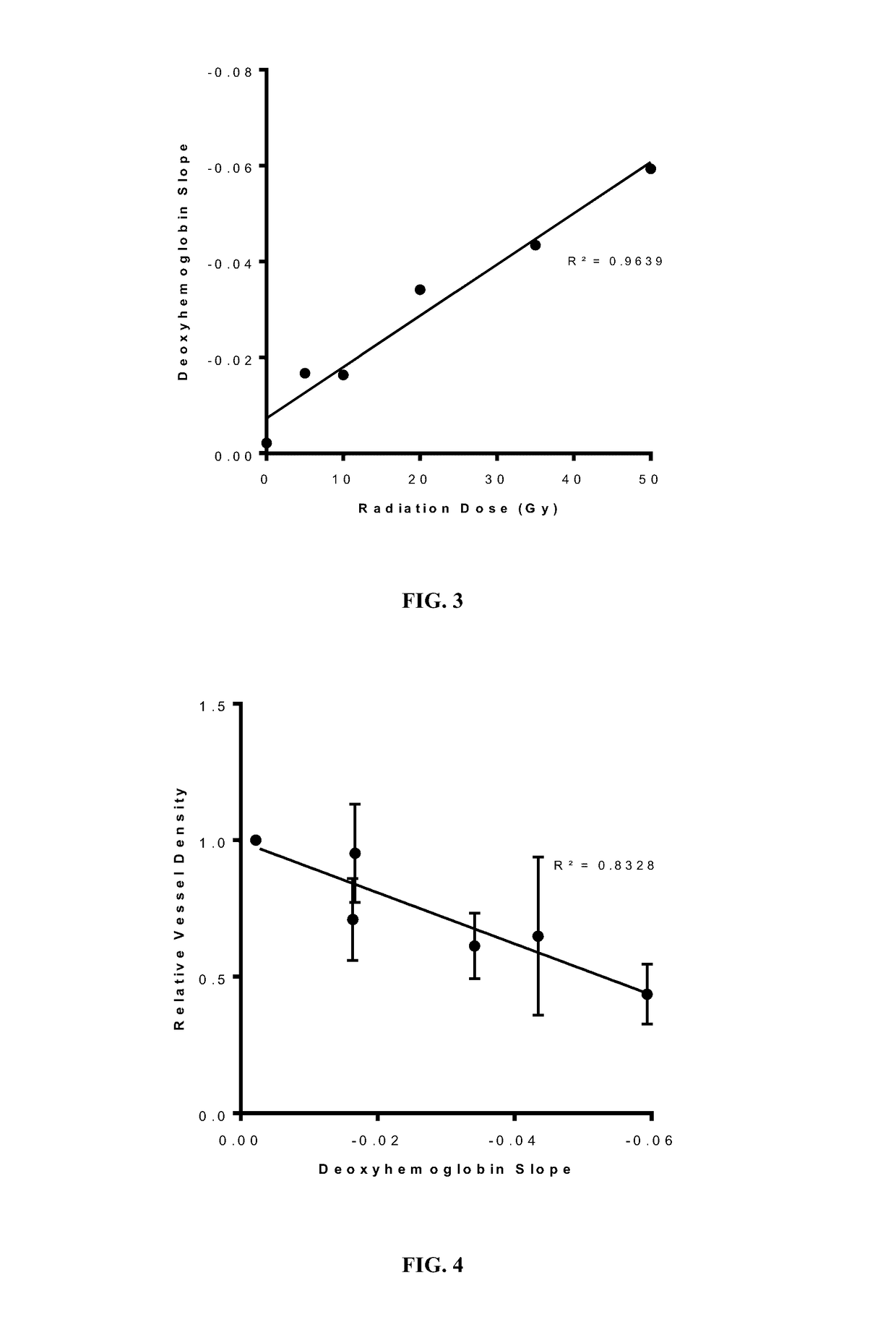
[0073]The study demonstrates that early measurement of cutaneous deoxygenated hemoglobin levels after radiation exposure is a useful biomarker for dose reconstruction and also for chronic microvascular injury. Changes in deoxygenated hemoglobin can also be correlated to acute skin reactions before any are visible.

Animals and Irradiation Procedure

[0074]All handling of and procedures performed with animals was done in accordance with a protocol (UMass IACUC Protocol #A2354) approved by our Institutional Animal Care and Use Committee. Prior to this experiment, a model of acute radiation-induced skin injury was established. (Chin, et al. 2012 J. Biomedical Optics 17(2):026010.) The radiation source utilized was a Strontium-90 beta-emitter with an active diameter of 9 mm, which created an 8 mm diameter skin injury. This beta-emitter source delivers less than 10% of the total dose deeper than 3.6...

example 2

Hyperspectral Imaging as an Early Prediction of Maximal Burn Depth After Thermal Injury

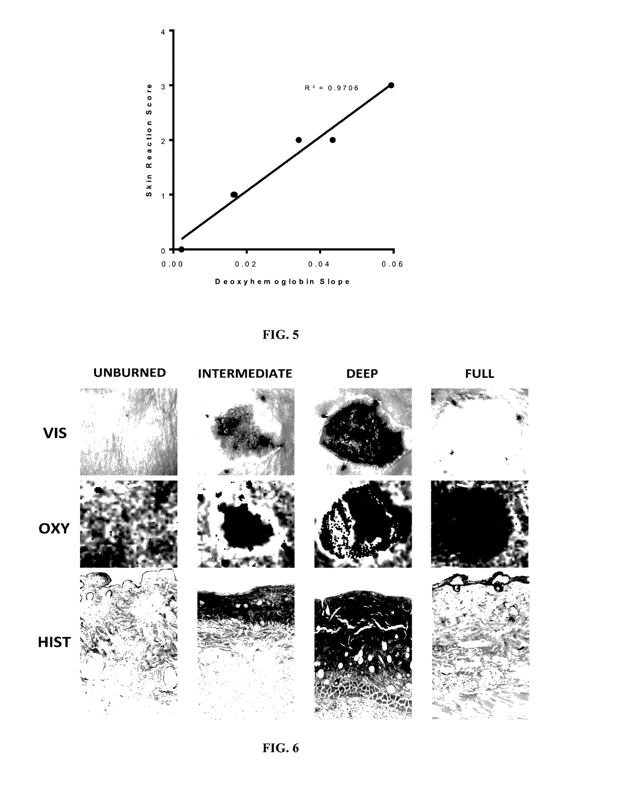
[0092]This example was to characterize dermal perfusion and oxygenation in three sequential depths of burn over a dynamic, three-day period after burn injury, and to assess whether vsHSI could differentiate depths of injury, based on any of the parameters it quantifies: oxyHb, deoxyHb, tHb, or StO2.

Animals and Thermal Burn Procedure

[0093]All handling of and procedures performed with animals was done in accordance with a protocol (UMass IACUC Protocol #A2454) approved by our Institutional Animal Care and Use Committee.

[0094]One hundred and seven hairless immune-competent, adult male mice (SKH-1 Elite, Charles River Laboratories, Wilmington, Mass.) were used. Anesthesia for thermal burn procedure and imaging was performed with a mixture of ketamine (55 mg / kg) and xylazine (5 mg / kg). Tattoo marks were placed on the dorsum of the mice to act as fiducial marks. After anesthesia, the dorsal skin was dis...


PUM


Abstract

The invention provides hyperspectral imaging-based methods that enable effective, efficient and non-invasive detection and characterization of thermal and ionizing radiation exposure in tissue. The methods allow for complete visualization and quantification of oxygenation and perfusion changes in thermal burn or ionizing radiation impacted skin and enables rapid identification of individuals exposed to such exposures and allows early prediction of extent of injury in normal tissue after exposure.

Description

PRIORITY CLAIMS AND RELATED PATENT APPLICATIONS[0001]This application claims the benefit of priority from U.S. Provisional Application Ser. No. 62 / 007,891, filed on Jun. 4, 2014, the entire content of which is incorporated herein by reference in its entirety.TECHNICAL FIELDS OF THE INVENTION[0002]The invention generally relates to methods for detecting and analyzing exposure of tissue to thermal burn or ionizing radiation. More particularly, the invention relates to methods for characterization, evaluation and prediction of injuries from thermal or radiation exposures in tissue and their effect thereof using hyperspectral imaging-based techniques.BACKGROUND OF THE INVENTION[0003]Exposure to thermal burn or ionizing radiation can have profound biological consequences to skin and underlying subcutaneous tissue. Skin structures, especially the dermal plexus, demonstrate changes in response to both thermal and ionizing radiation energy. After exposure to thermal energy, depth of burn in...

Claims


Application Information

Patent Timeline
Patent Type & Authority Applications(United States)
IPC IPC(8): A61B5/00A61B5/1455
CPCA61B5/7275A61B5/0075A61B2576/00A61B5/445A61B5/443A61B5/14551G16H30/40G16H50/20
Inventor CHIN, MICHAEL S.
Owner UNIV OF MASSACHUSETTS
Learn More