
Exosome isolation

a technology of exosomes and isolation, applied in the field of exosomes, can solve the problems of a plethora of peptides that cannot survive the exposed circulatory environment, study exosomes, and a high degree of labileness

Inactive Publication Date: 2017-10-19
EXERKINE
View PDF1 Cites 29 Cited by
  • Summary
  • Abstract
  • Description
  • Claims
  • Application Information

AI Technical Summary

Benefits of technology

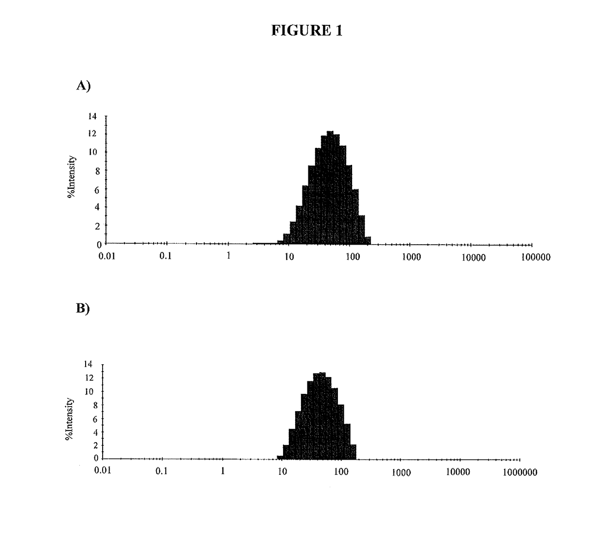
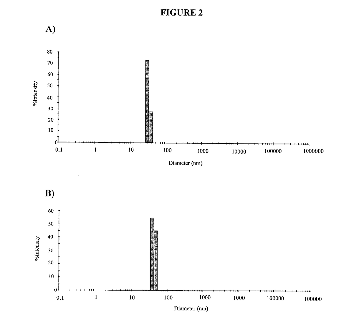
The present invention describes a novel method for isolating exosomes from biological samples, involving several steps including centrifugation, microfiltration, and ultracentrifugation. The isolated exosomes are then used to deliver exogenous cargo to mammals. The method is efficient and effective in isolating pure exosomes without contamination by particles with a diameter of less than 20 nm or greater than 140 nm. The isolated exosomes can also be loaded with various types of cargo and used for in vivo delivery. The invention also provides a kit for conducting the isolated exosome method.

Problems solved by technology

However, there are a plethora of peptides that cannot survive the exposed circulatory environment.
Additionally, both mRNA and miRNA are extremely labile in the extracellular environment and require an encapsulated venue for transfer between tissues and organs.
One of the major limitations in studying exosomes is lack of a standardized / functional exosome isolation protocol.
Many of the isolation protocols that are published lack the capacity to yield a purified exosome population of sufficient quantity and quality for biochemical analyses and various other downstream applications, including the delivery of therapeutic payloads (protein, mRNA, miRNA, etc.).
Additionally, the non-ultracentrifuge / filtration-based methodologies commercially available in the form of kits have severe shortcomings in their ability to isolate a pure exosomal fraction and / or to isolate exosomes of sufficient quality for biological evaluation or therapeutic delivery.

Method used


Image

  • Exosome isolation
  • Exosome isolation
  • Exosome isolation

Examples


example 1

Exosome Isolation From Biological Samples Using a PEG-Based Method

[0073]Exosomes were isolated from various human and other mammalian biological samples as follows.

[0074]Blood samples were collected from healthy human subjects using red top serum collection tubes (e.g. BD, Ref #367812) and blue top plasma collection tubes containing sodium citrate (e.g. BD, Ref #369714) for serum and plasma isolations, respectively. For serum isolation, blood was allowed to clot for 1 hour at room temperature followed by centrifugation at 2,000×g for 15 min at 4° C. For plasma isolation, blood was spun down immediately after collection at 2,000×g for 15 min at 4° C. Plasma and serum was similarly collected from C57B1 / 6J mice and Sprague Dawley rats. Exosomes were then isolated from these samples, as well as from bovine whole milk (Natrel fine-filtered 3.25% milk). From this point onwards, all exosome sources were treated the same.

[0075]Serum, plasma and milk were spun at 2000×g for 15 min at 4° C. T...

example 2

Exosome Isolation From Cell Culture

[0082]Isolation of exosomes from cell culture (such as JawsII cells or Chinese hamster ovary (CHO) cells) was conducted using a similar procedure to that described in Example 1, including centrifugation of the sample, filtration, precipitation in PEG and solubilization in trehalose. In this case, however, the 0.22 μm filtrate was incubated with PEG at 4° C. overnight to enhance the precipitation of exosomes.

[0083]Exosome purity was similar to that achieved from the biological samples of Example 1. About 12-23 μg protein was obtained from about 1 mL of cell culture media.

example 3

Exosome Isolation From Human Biological Samples

[0084]Blood and urine samples were collected from healthy human subjects. For serum isolation, blood was allowed to clot for 1 hour at room temperature followed by spinning at 2,000× g for 15 min at 4° C. Similarly, urine samples were spun at 2,000×g for 15 min at 4° C. to remove any cellular debris. For plasma isolation, blood was spun down immediately after collection at 2,000×g for 15 min at 4° C. and treated with 5 ug of Proteinase K (20 mg / mL stock, Life Technologies) for 20 min at 37° C. From this point onwards, all samples (serum-1 mL, plasma-1 mL, and urine) are treated exactly the same.

[0085]The supernatant from the first centrifugation was spun at 2000×g for 60 min at 4° C. to further remove any contaminating non-adherent cells (optional). The supernatant was then spun at 14,000×g for 60 min at 4° C. (optional). The resultant supernatant was spun at 50,000×g for 60 min at 4° C. The resulting supernatant was then filtered throu...


PUM

PropertyMeasurementUnit
sizeaaaaaaaaaa
diameteraaaaaaaaaa
average molecular weightaaaaaaaaaa

Abstract

Methods of isolating exosomes from a biological sample is provided. In one embodiment, the method may include a series of optional centrifugation steps, and comprises exosome precipitation using a PEG-based solution followed by resuspension in a saccharide-based solution such as trehalose. The method advantageously results in essentially pure exosomes that maintain integrity and stability. The exosomes are useful for the in vivo delivery of cargo, including macromolecules such as protein and nucleic acid.

Description

FIELD OF THE INVENTION[0001]The present invention generally relates to exosomes, and more particularly relates to a method of isolating exosomes from a biological sample.BACKGROUND OF THE INVENTION[0002]In organisms, tissues and cells must continuously correspond with each other to best adapt to their surrounding microenvironment. To date, the transmission of signals between cells and tissues has been described by protein-based signaling systems exemplified by enzymes, hormones, cytokines, and chemokines. However, there are a plethora of peptides that cannot survive the exposed circulatory environment. Additionally, both mRNA and miRNA are extremely labile in the extracellular environment and require an encapsulated venue for transfer between tissues and organs. Thus, these factors are secreted in small double-membrane extracellular vesicles (ECVs) including exosomes, microvesicles, and apoptotic bodies. Depending on their cellular site of origin, these vesicles have distinct struct...

Claims


Application Information

Patent Timeline
Patent Type & Authority Applications(United States)
IPC IPC(8): A61K38/18A61K9/51
CPCA61K38/1866A61K9/5192A61K9/5176A61K9/5184A61K38/22A61P3/00A61K9/16
Inventor TARNOPOLSKY, MARK
Owner EXERKINE
Learn More