Method for the enrichment of circulating tumor DNA

a technology for circulating tumors and dna enrichment, applied in material analysis, instruments, biological material analysis, etc., can solve the problems of high cost of biopsy, limited tissue dna tests, and inability to perform invasive biopsy procedures on patients

Inactive Publication Date: 2018-01-25
BELGIAN VOLITION SPRL
View PDF3 Cites 7 Cited by
  • Summary
  • Abstract
  • Description
  • Claims
  • Application Information

AI Technical Summary

Problems solved by technology

However, tissue DNA tests have limitations as invasive biopsy procedures cannot be performed repeatedly on patients for monitoring purposes.
Biopsy is expensive to perform, uncomfortable for the patient, poses patient risk, and may lead to surgical complications.
Firstly, although all cancers have mutations, the frequency of any particular mutation in a particular cancer disease is usually low.
Secondly, even if the cancer tissue of a patient does contain the mutation, the level or concentration of mutated ctDNA present in the blood of the patient may be low and difficult to detect.
However, raised levels of circulating nucleosomes per se are not used clinically as biomarkers of cancer as nucleosomes are a non-specific product of cell death and raised levels are observed for many conditions involving elevated cell death including acute trauma (Holdenrieder and Stieber, 2009).
There are currently no ctDNA based tests in routine use for clinical oncology purposes due to a number of limitations.
A major methodological limitation is a requirement for high quality DNA.
Current ctDNA sampling methods produce poor quality ctDNA samples due to the nature of the sample.
The main difficulty lies in the presence of large amounts of non-tumor cfDNA in the circulation which complicates any analysis of ctDNA.
Moreover this DNA is from the same subject and hence of similar sequence and will interfere in any method for the quantification or analysis of ctDNA.
A similar problem occurs for the measurement of circulating cell free nucleosomes and / or the epigenetic composition of circulating nucleosomes as biomarkers for cancer because nucleosomes per se are a non-specific indicator of cell death and are released as part of the normal cell turnover process of the body as well as in conditions associated with elevated levels of cell death such as autoimmune diseases, stroke, sepsis, post trauma, burns, myocardial infarction, cerebral stroke, during graft rejection after organ transplantation and after severe exercise.
These non-tumor nucleosomes will interfere in any method for the quantification or epigenetic analysis of nucleosomes of tumor origin.

Method used


Image

  • Method for the enrichment of circulating tumor DNA
  • Method for the enrichment of circulating tumor DNA
  • Method for the enrichment of circulating tumor DNA

Examples


example 1

[0101]An antibody directed to bind specifically to histone isoform H3.1, H3.2 or H3t is biotinylated and immobilized on streptavidin coated magnetic beads (Dynal) by the recommended method of the manufacturer. The beads are washed several times with loading buffer using a magnetic separation system. Serum or plasma taken from a cancer patient is diluted in loading buffer and added to the beads. Nucleosomes containing histone H3 variant are adsorbed to the beads. Other serum / plasma components remain in solution and are removed by means of magnetic separation. The beads are washed with buffer. The nucleosomes containing histone H3 variant are now isolated on the beads. ctDNA associated with the isolated nucleosomes is extracted by the phenol / chloroform method or other standard extraction methods. The extracted DNA may be analysed for genetic or epigenetic features of cancer.

example 2

[0102]An antibody directed to bind specifically to histone isoform H3.1, H3.2 or H3t is biotinylated and immobilized on streptavidin coated magnetic beads (Dynal) by the recommended method of the manufacturer. The beads are washed several times with loading buffer using a magnetic separation system. Serum or plasma taken from a cancer patient is diluted in loading buffer and added to the beads. Nucleosomes containing histone H3 variant are adsorbed to the beads. Other serum / plasma components remain in solution and are removed by magnetic separation. The beads are washed with buffer. The nucleosomes containing histone H3 variant are now isolated on the beads. The isolated nucleosomes are removed from the magnetic beads using an elution buffer and the nucleosomes are analysed by proteomics methods including Mass Spectroscopy.

example 3

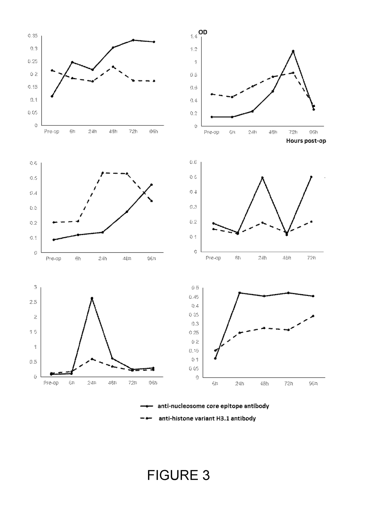
[0103]Serum samples taken from 6 patients with CRC pre-surgery and at 6, 24, 48, 72 and 96 hours post-surgery were assayed for circulating cell-free nucleosome levels using a commercial (total) nucleosome ELISA produced by Roche employing a common anti-nucleosome core epitope and a labelled anti-dsDNA antibody. The samples were then re-assayed but using an anti-histone variant H3.1, H3.2 or H3t capture antibody. The measured (total) nucleosome levels increased following the trauma of surgery using the commercial ELISA but the response to surgery was altered and muted for nucleosomes containing histone H3 variant measured using the assay employing anti-histone H3.1, H3.2 or H3t antibody. The results are shown in FIG. 3.


PUM

PropertyMeasurementUnit
volumesaaaaaaaaaa
mass spectrometryaaaaaaaaaa
Mass Spectrometryaaaaaaaaaa

Abstract

The invention relates to the use of histone binding agents for detecting, isolating and / or purifying cell free nucleosomes of tumor originor circulating tumor DNA from a biological sample. The invention also relates to methods and kits using said histone binding agents.

Description

FIELD OF THE INVENTION[0001]The invention relates to a method for the purification or enrichment of circulating cell free nucleosomes of tumor origin and associated circulating tumor DNA from blood, serum or plasma.BACKGROUND OF THE INVENTION[0002]Cellular DNA exists as a protein-nucleic acid complex called chromatin. The nucleosome is the basic unit of chromatin structure and consists of double stranded DNA (dsDNA) wound around a protein complex. The DNA is wound around consecutive nucleosomes in a structure often said to resemble “beads on a string” and this forms the basic structure of open or euchromatin. In compacted or heterochromatin this string is coiled and super coiled in a closed and complex structure.[0003]Each nucleosome in chromatin consists of a protein complex of eight highly conserved core histones (comprising of a pair of each of the histones H2A, H2B, H3, and H4). Around this complex are wrapped approximately 146 base pairs (bp) of DNA. Cell free nucleosomes are r...

Claims


Application Information

Patent Timeline
Patent Type & Authority Applications(United States)
IPC IPC(8): G01N33/68G01N27/62C12N15/10
CPCG01N33/6875C12N15/1003G01N27/622G01N2560/00G01N2800/7028
Inventor MICALLEF, JACOB VINCENTHERZOG, MARIELLEECCLESTON, MARK EDWARD
Owner BELGIAN VOLITION SPRL
Why Eureka
Try Eureka