
Treatment of eye disease
a technology for eye diseases and gene therapy, applied in the field of gene therapy of eye diseases, can solve problems such as difficulty in predicting how vision would be useful
- Summary
- Abstract
- Description
- Claims
- Application Information
AI Technical Summary
Benefits of technology
Problems solved by technology
Method used
Image

Examples
example 1
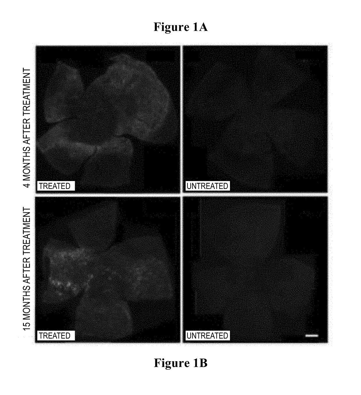
[0180]The present inventors targeted the bipolar cells and horizontal cells of the retina, which are next in line after photoreceptor degeneration, for the introduction of exogenous melanopsin. To achieve this, the inventors injected an AAV vector under the retina (i.e. by subretinal injection) in retinal degeneration (rd) mice, which have the human equivalent of end-stage RP (FIGS. 1A-1D) The treated mice developed significantly increased pupil responses to light (FIGS. 2A-2E) and the location of invoked electrical recordings correlated to the regions where bipolar and horizontal cells had been transduced (FIGS. 2A-2E).
[0181]One of the advantages of targeting the bipolar and horizontal cells is that they more closely represent the map that would previously have been formed by the photo-receptors. Although ganglion cells could be made light-sensitive, the axons of the ganglion cells run horizontally across the retina to the optic nerve head and it is difficult to predict how vision ...
example 2
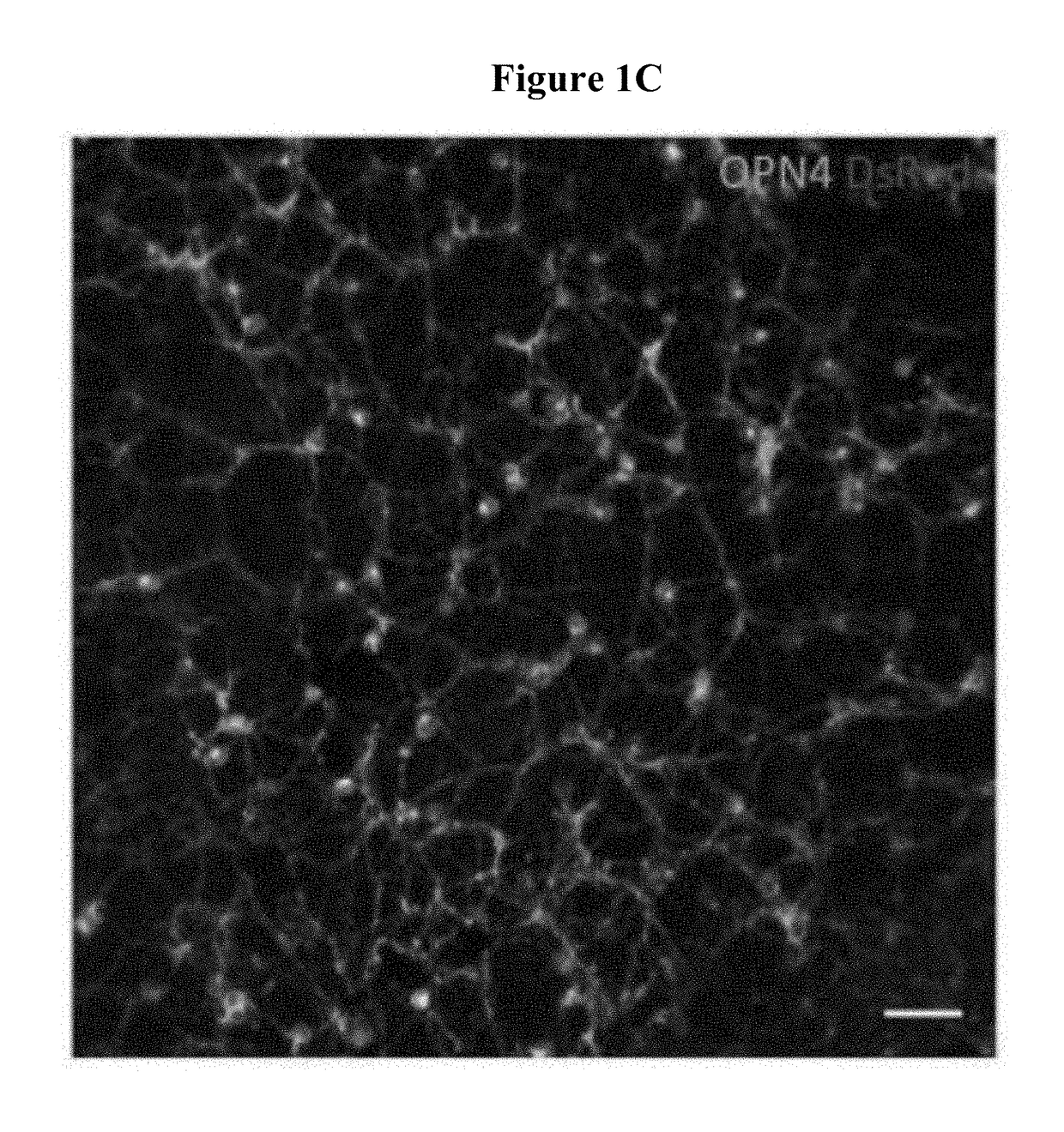
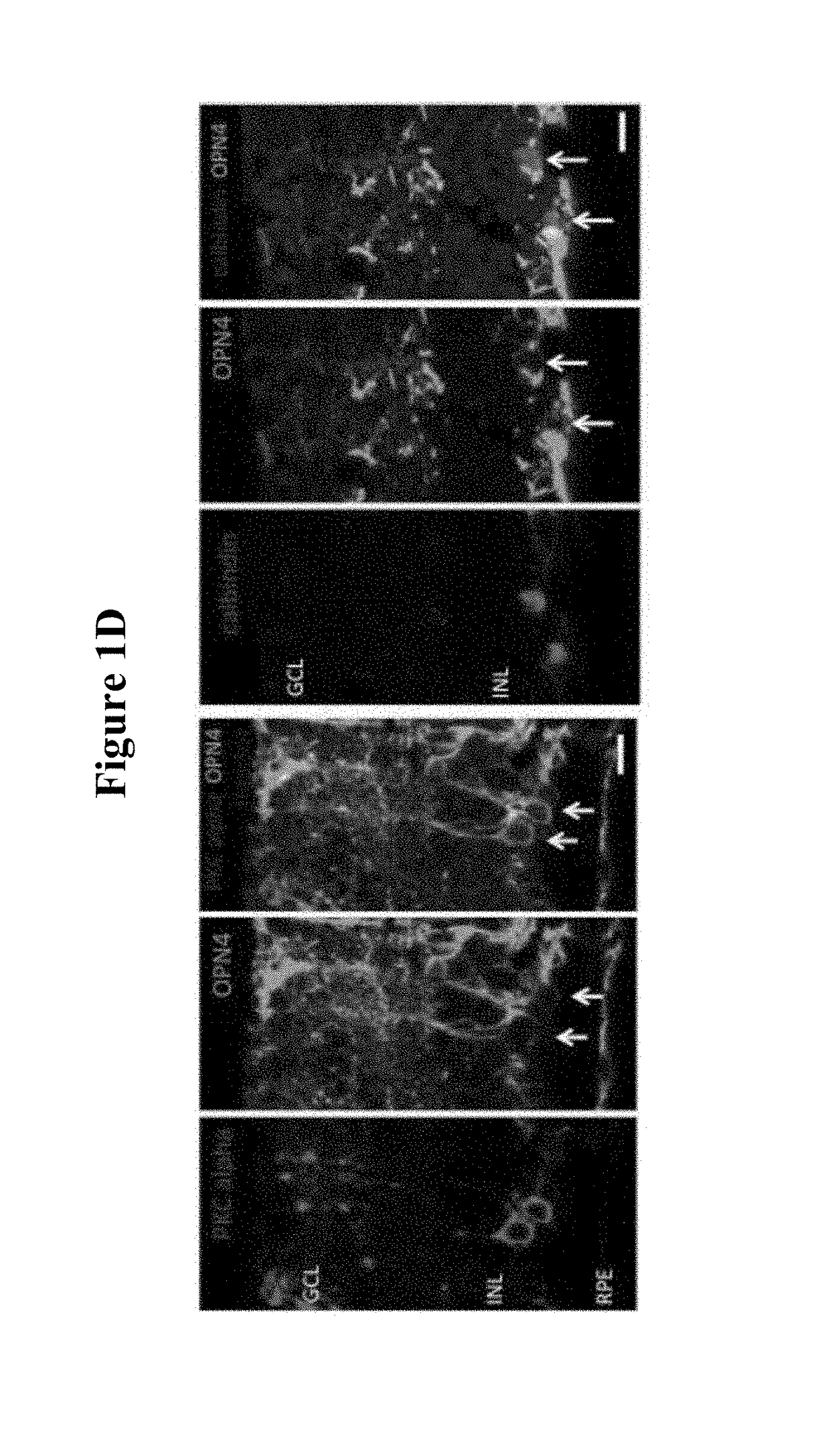
[0186]Histology confirmed the transduction of the outer retinal cells (bipolar and horizontal) that do not normally express melanopsin (FIGS. 1A-1D).
[0187]Widespread transduction of the retina of the rd1 mouse (which has a rapid photoreceptor degeneration) was observed by immune-staining for human melanopsin (FIGS. 1A-1B 4 months (FIG. 1A left panel a i) and 15 months (FIG. 1B left panel b i) after gene therapy; images of control retinae are shown for comparison showing absence of human melanopsin at 4 months (FIG. 1A right panel iii) and 15 months (FIG. 1B right panel iii)).
[0188]The red label (DsRed) in the top right panel of FIG. 1C identifies transduced bipolar and horizontal cells on the subretinal surf ace. Appropriate membrane localisation of human melanopsin was also observed, demonstrating the network of transduced cells.
[0189]Further immunostaining for PKC alpha (identifying bipolar cells) and calbindin (identifying horizontal cells) illustrated transduction of these cell ...
example 3
Methods
[0190]Capsid mutant AAV (rAA V2 / 8.Y733F) expressing human melanopsin was delivered via subretinal injection to the left eye of 15 Pde6brd1 / rd1 mice. 9 mice were injected with phosphate-buffered saline (sham group) and 13 untreated mice were also included. 12 months after treatment, pupil responses were recorded from the right eye following stimulation of the left eye with a 2 second white light stimulus of varying intensity.
Results
[0191]12 months after subretinal injection, a large area of retina in each treated eye expressed human melanopsin, as confirmed by immunohistochemistry. At a light intensity of 2×10¹⁵ photons / cm2 / s, which was in the mid-range of the light intensities tested, there was significantly more pupil constriction in the treated group (mean constriction 36.0%±7.06) versus the sham treated group (3.1%±1.45) and the untreated group (15.6%±6.61); treated versus sham p=0.0026, treated versus untreated p=0.038 2 way ANOVA, Tukey's post hoc test.
Conclusions
[0192]H...
PUM
| Property | Measurement | Unit |
|---|---|---|
| volume | aaaaa | aaaaa |
| volume | aaaaa | aaaaa |
| volume | aaaaa | aaaaa |
Abstract
Description
Claims
Application Information