Combining modified antibodies with expansion microscopy for in-situ, spatially-resolved proteomics

Inactive Publication Date: 2018-02-22
EXPANSION TECH
View PDF0 Cites 70 Cited by
  • Summary
  • Abstract
  • Description
  • Claims
  • Application Information

AI Technical Summary

Benefits of technology

[0006]In one aspect, provided herein are methods of labeling a biological sample, the methods comprising: contacting the sample with at least one binding composition under conditions where it selectively recognizes a target biomolecule, wherein the binding composition comprises a modified antibody comprising a first antibody having an antigen-binding site having an affinity for the target biomolecule, wherein the first antibody is operably linked to (i) a detectable label, and (ii) a polyelectrolyte gel binding moiety; contacting the sample with a solution comprising monomers of a polyelectrolyte gel; by free radical polymerization, polymerizing the monomers to form the polyelectrolyte gel and covalently conjugate the polyelectrolyte gel binding moiety to the polyelectrolyt

Problems solved by technology

However, because the number of fluorescent dyes that can be used is small (typically <6), this strategy is limited to imaging only a small number of proteins per sample.
Additionally, the polymerization process dampens the fluorescence of the dyes, which are pe

Method used


Image

  • Combining modified antibodies with expansion microscopy for in-situ, spatially-resolved proteomics
  • Combining modified antibodies with expansion microscopy for in-situ, spatially-resolved proteomics
  • Combining modified antibodies with expansion microscopy for in-situ, spatially-resolved proteomics

Examples


example 1

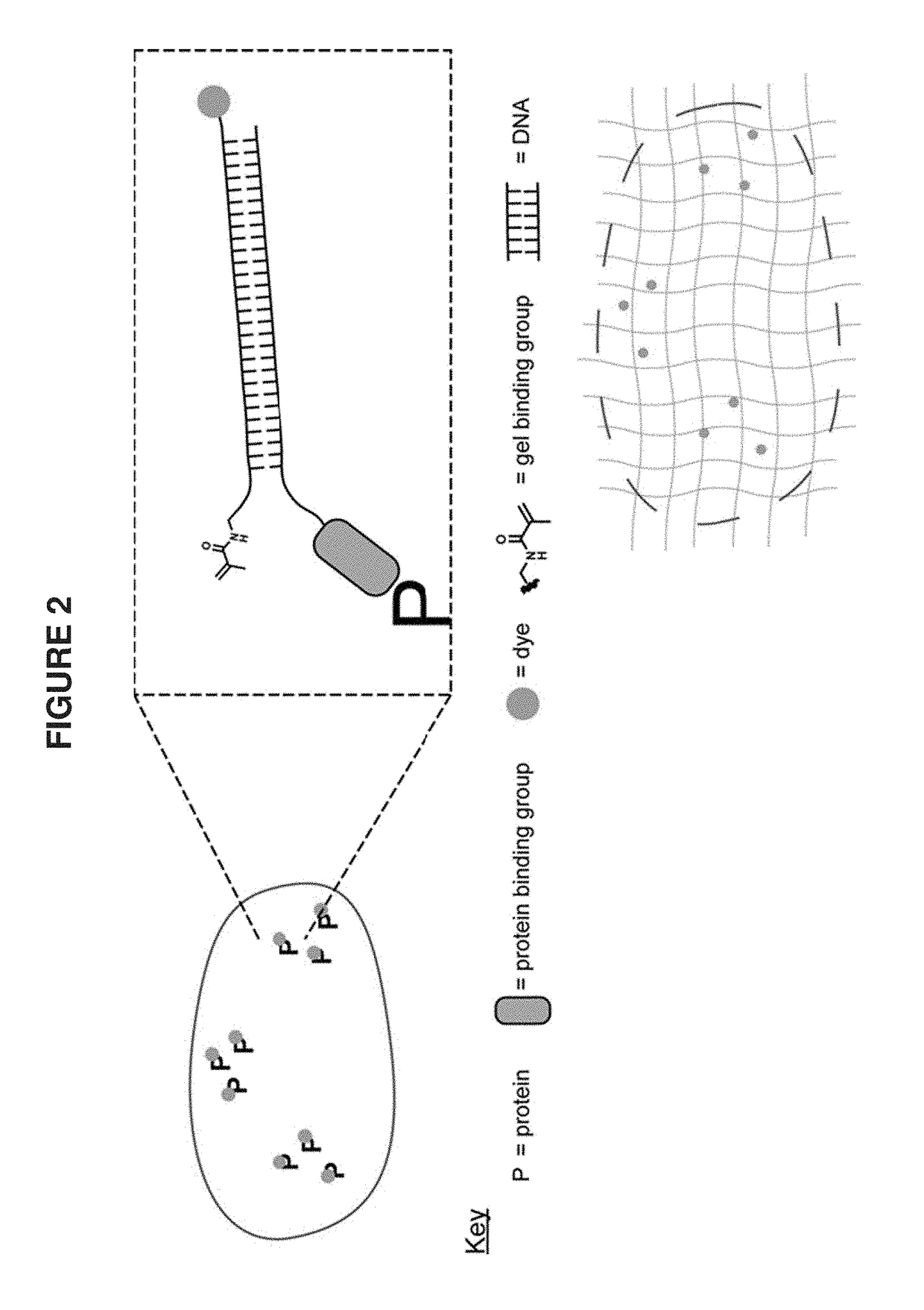
[0095]A target biomolecule of interest is identified, and an antibody having an antigen-binding site with an affinity for the target biomolecule is obtained. The antibody is modified to be operably linked to a detectable label and also operably linked to a polyelectrolyte gel binding moiety to yield a modified antibody. The modified antibody is a binding composition or a component of the binding composition.

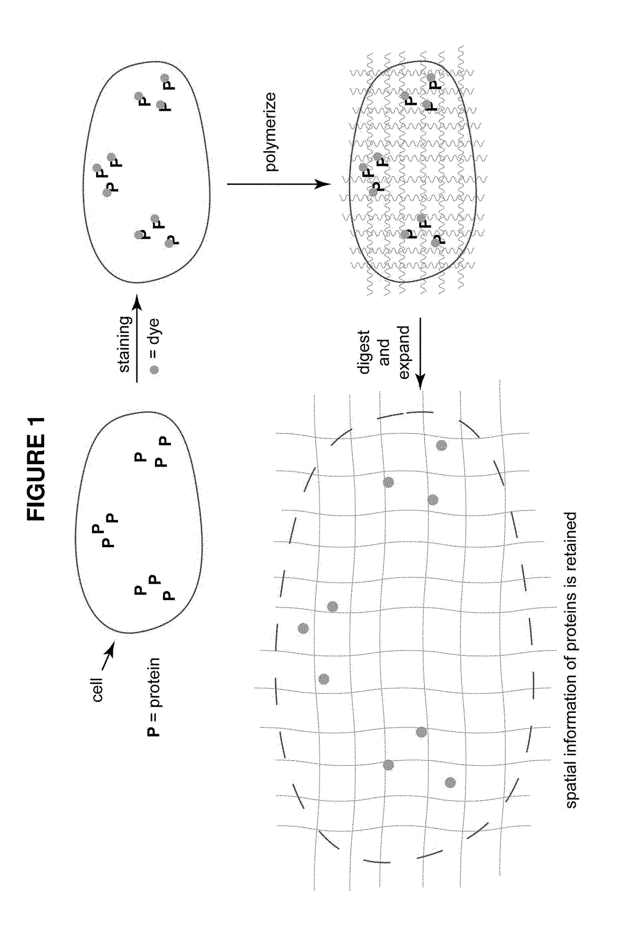
[0096]A biological sample of interest is obtained. The sample is contacted with the binding composition under conditions where it selectively recognizes the target biomolecule. The biological sample is incubated with the detection reagent, in some cases together with the labeling reagent, under conditions that allow a complex between the detection reagent (and labeling reagent) and target to form. Upon treatment with a solution of monomers and subsequent free radical polymerization, a polyelectrolyte gel is formed to which the polyelectroylte gel binding moiety (operably linked t...

example 2

[0097]A target biomolecule of interest is identified, and an antigen-binding fragment having an antigen-binding site with an affinity for the target biomolecule is obtained. The antigen-binding fragment is modified to be operably linked to a detectable label and also operably linked to a polyelectrolyte gel binding moiety to yield a modified antigen-binding fragment. The modified antigen-binding fragment is a binding composition or a component of the binding composition.

[0098]A biological sample of interest is obtained. The sample is contacted with the binding composition under conditions where it selectively recognizes the target biomolecule. The biological sample is incubated with the detection reagent, in some cases together with the labeling reagent, under conditions that allow a complex between the detection reagent (and labeling reagent) and target to form. Upon treatment with a solution of monomers and subsequent free radical polymerization, a polyelectrolyte gel is formed to...

example 3

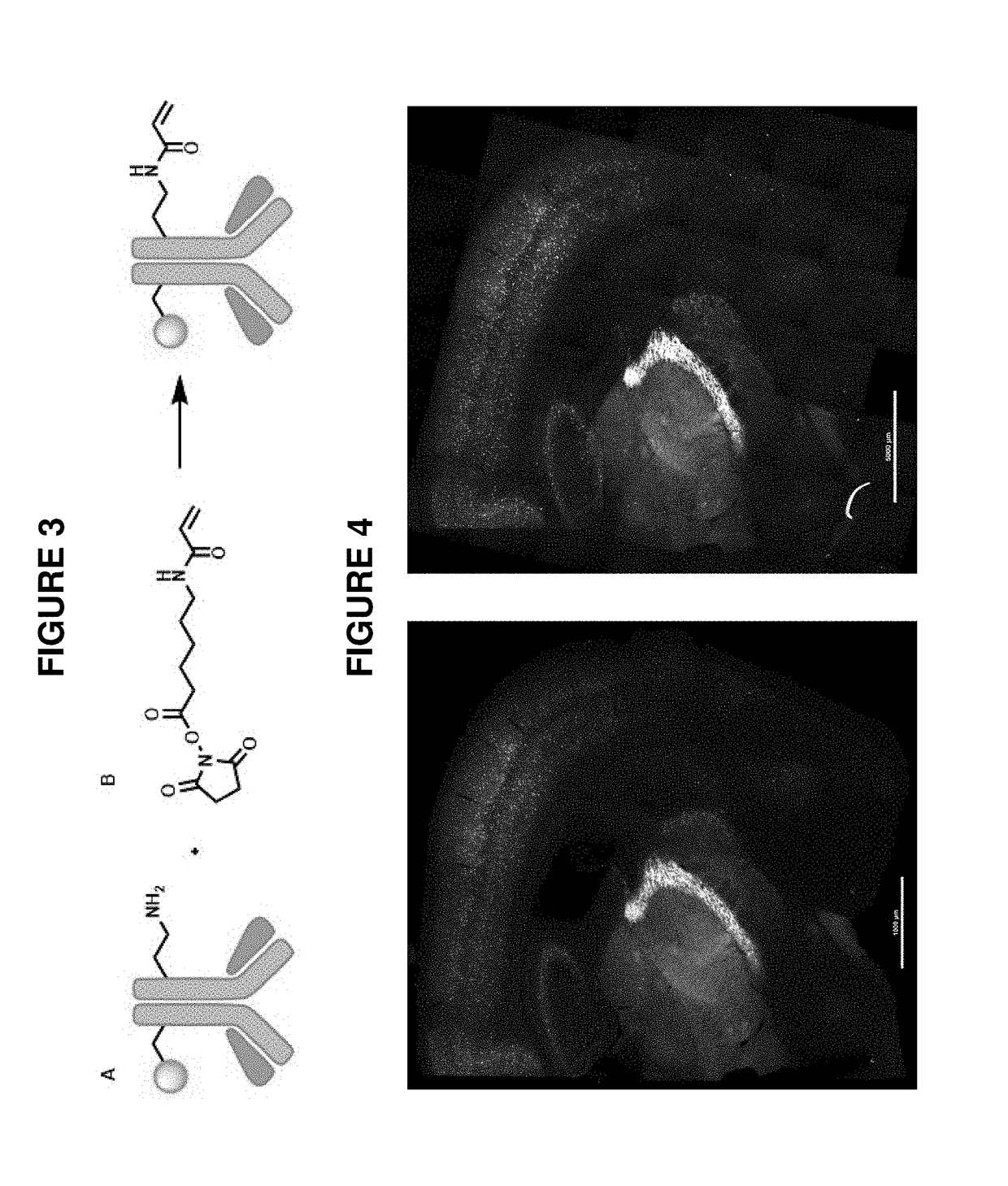
[0099]As described in Example 1(above), a target biomolecule of interest (a primary anti-PV antibody to murine parvalbumin protein) was identified, and an antibody having an antigen-binding site with an affinity for the target biomolecule was obtained (here, a secondary antibody to the primary anti-PV antibody). This secondary antibody was modified to be operably linked to a fluorophore (an atto647N dye) and also operably linked to a polyelectrolyte gel binding moiety to yield a modified antibody, as shown in FIG. 3.

[0100]A mouse brain sample was obtained. The sample was first contacted with the primary anti-PV antibody and then contacted with the binding composition comprising the modified secondary antibody under conditions where it selectively recognizes the primary anti-PV antibody target biomolecule. In each case, the sample was incubated under conditions that allow a complex between the detection reagent (and labeling reagent) and target to form. Upon treatment with a solution...


PUM


Abstract

This invention relates to imaging, such as by expansion microscopy, labelling, and analyzing biological samples, such as cells and tissues, as well as reagents and kits for doing so.

Description

CROSS REFERENCE TO RELATED APPLICATIONS[0001]This application claims the benefit of U.S. Provisional Application No. 62 / 334,628, filed on May 11, 2016, which is incorporated by refereince herein in its entirety.FIELD OF THE INVENTION[0002]The invention relates to imaging, such as by expansion microscopy, labelling, and analyzing biological samples, such as cells and tissues, as well as reagents and kits for doing so.BACKGROUND OF THE INVENTION[0003]In expansion microscopy (ExM), 3-dimensional imaging with nanoscale precision is performed on cells and tissues. This is accomplished by physically expanding the biological sample using a dense polymer matrix (FIG. 1). The first step of this process involves treating the tissue with a fluorescent protein-binding-group (typically an antibody) that selectively binds to the protein being analyzed. Next the sample is infused with a monomer solution that permeates into the tissue. Free radical polymerization of this solution creates a polymer ...

Claims


Application Information

Patent Timeline
IPC IPC(8): G01N1/30G01N33/533G01N33/543G01N33/545
CPCG01N1/30G01N33/533G01N33/54306G01N33/545G01N33/5375G01N33/58
Inventor KOHMAN, RICHIE E.
Owner EXPANSION TECH
Why Eureka
Try Eureka