Minimally Invasive Subgaleal Extra-Cranial Electroencephalography EEG Monitoring Device

Inactive Publication Date: 2018-05-03
NEW YORK UNIVERSITY
View PDF6 Cites 10 Cited by
  • Summary
  • Abstract
  • Description
  • Claims
  • Application Information

AI Technical Summary

Benefits of technology

The present invention describes a brain wave monitoring device that can be implanted in a patient for detecting brain electrical activity. This device includes a processor that analyzes the detected brain electrical activity to detect changes in brain state, such as epileptic events. The device can also generate brain state data and wirelessly transmit this data to a remote computer or other device for further analysis and treatment. The technical effects of this invention include improved accuracy and efficiency in capturing brain wave data and the ability to remotely monitor and treat brain disorders.

Problems solved by technology

Unfortunately, many patients experiencing seizures lose consciousness or are amnesic while most subtle or non-convulsive seizures may be unobserved or unrecognized.
Furthermore, unrecognized seizures increase the risk of cognitive decline, injury and death.
Video EEG studies are inpatient based and thus very expensive and only used for acute situations or for pre-surgical investigations.
Current methods for ambulatory scalp EEG recordings are not practical for long-term use and can typically be used for a maximum of 3-4 days.
These methods are also limited due to scalp EEG artifacts, battery life or the inability of patients to tolerate scalp electrodes from more than a few days.
However, not only are these highly invasive procedures expensive (generally more than $50,000), they also have a high morbidity rate.
Seizure detection systems based on body motion or motor activity, accelerometers or video detectors can only detect major convulsive events and are not implantable, limiting their value as most seizures are non-convulsive in nature and therefore not detectable with changes in motion.

Method used


Image

  • Minimally Invasive Subgaleal Extra-Cranial Electroencephalography EEG Monitoring Device
  • Minimally Invasive Subgaleal Extra-Cranial Electroencephalography EEG Monitoring Device
  • Minimally Invasive Subgaleal Extra-Cranial Electroencephalography EEG Monitoring Device

Examples


example i

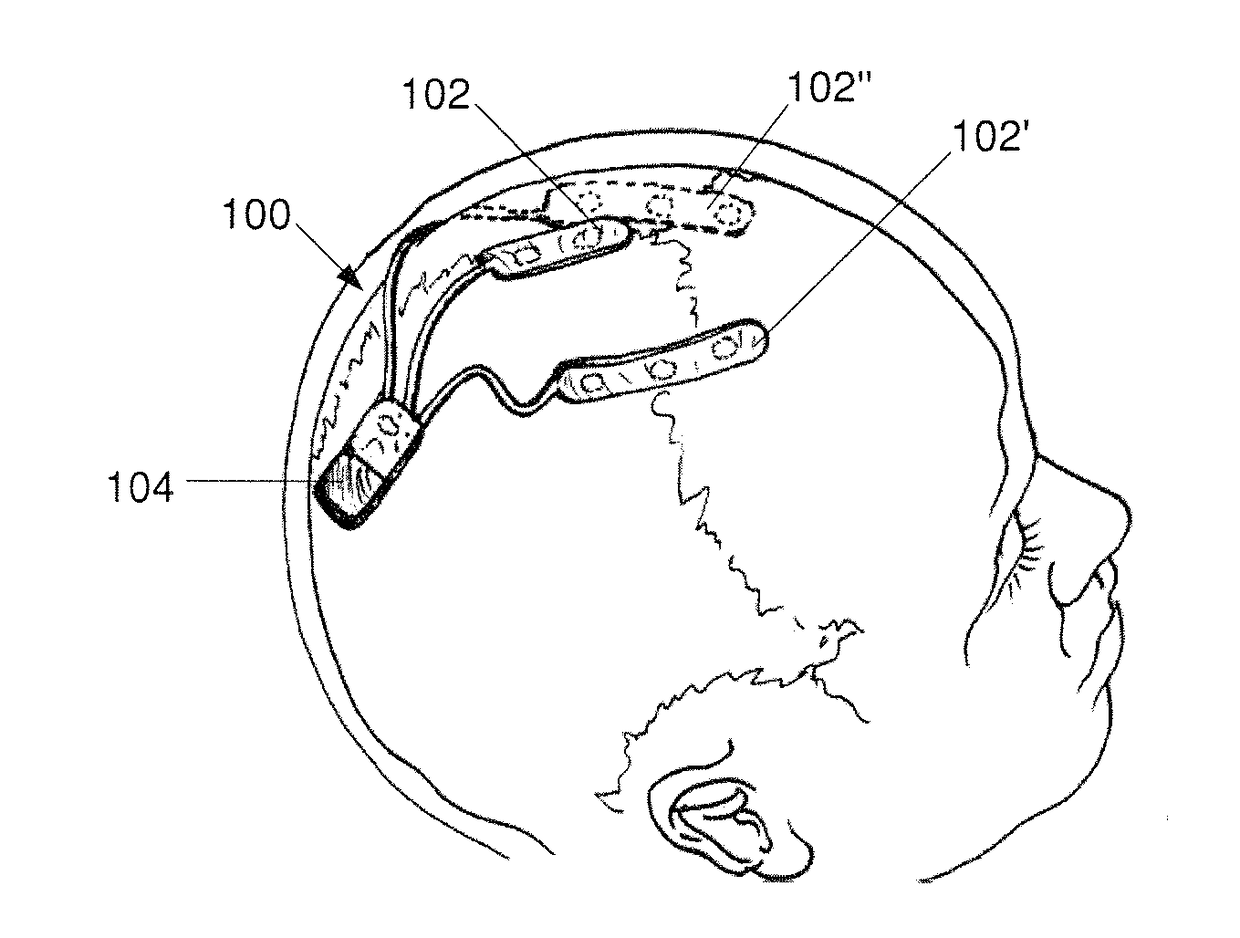
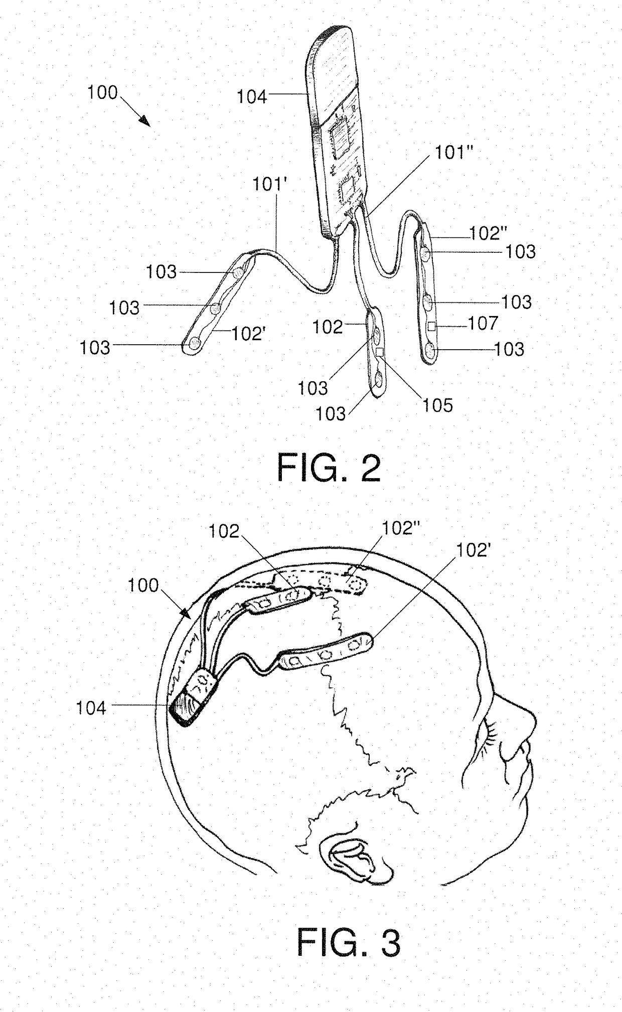
[0063]The exemplary device 100 may be implanted in a subgaleal region of a patient, near the brain, as shown in FIG. 11. As can be seen, at least one contact 103 (e.g., an electrode for recording electrophysiological data, particularly EEG data) of the device may be implanted in a subgaleal region that is between the skull 14 and the scalp 16 of the patient near the brain 12 of the patient. Specifically, the at least one contact 103 or electrode arrays 102, 102′, 102″ of the device 100 may be position perpendicular to a cranial vertex 18 of the patient. More particularly, the at least one contact 103 or electrode arrays 102, 102′, 102″ of the device 100 may lie across a mid-length of the patient's skull 14. It is contemplated that the exemplary device 200 may be alternatively implanted in the subgaleal region of the patient in the same manner described above and as shown in FIG. 11.

[0064]The device 100, 200 may be used to record EEG data from the patient extracranially without drill...

example ii

[0066]The exemplary device 100, 200, 300 may also be used to record EEG data from the patient that allows for identification of sleep stages (e.g., awake and sleep states). In particular the exemplary unitary device 300 as show in FIGS. 6-10 may detect sleep stages of a patient from a single device implanted at a single location, preferably at the cranial vertex. In Example II, the exemplary device 300 may be used to conduct a pre-surgical evaluation of a patient to record EEG data and determine electrophysiological signals for awake and sleep states of the patient. FIG. 13 shows the EEG data recorded over time in an awake patient who is being evaluated for epilepsy surgery by intracranial electrodes implanted directly onto the dura mater as well as EEG data recorded by an exemplary subgaleal device 300 of the present invention. A patient who is being considered for epilepsy surgery may be in one of two categories: (1) if their seizures are not their seizures are not adequately cont...

example iii

[0069]In Example III, the exemplary device 100, 200, 300 may be used to record EEG data from the patient and detection of epileptic activity. The exemplary device 300 may be used to conduct a pre-surgical evaluation of a patient to record EEG data and determine ictal activity of the patient. FIG. 15 shows the EEG data recorded over time in a patient who is being evaluated for epilepsy surgery by intracranial electrodes implanted directly onto the dura mater as well as EEG data recorded by an exemplary subgaleal device 300 of the present invention. In the data shown in FIG. 15, the top 17 data lines show EEG data obtained from intracranial depth and subdural electrodes, whereas the last data line, which is also labeled as “SG EEG,” shows EEG data obtained using electrodes that are implanted into the subgaleal space of the patient, such as the exemplary device 300, at the vertex on the right side. As can be seen in FIG. 15, complex partial seizure from the right temporal lobe may be c...


PUM


Abstract

A system includes an implantable body configured for implantation in a subgaleal extracranial position, the implantable body including a first electrode array including a first elongated body comprising first and second electrode contacts separated from one another by a distance selected to facilitate the detection of brain electrical activity and a unit coupled to the first electrode array. The unit includes a processor analyzing the detected brain electrical activity to determine whether an epileptic event has occurred and generating epileptic event data based on this determination and a transceiver controlled by the processor to wirelessly transmit epileptic event data to and from a remote computing device.

Description

BACKGROUND[0001]For epilepsy patients, an objective seizure detection method that is safe, accurate and does not interfere with patient activities is critical to advance patient care. Seizure frequency is the most important index for determining and monitoring seizure control. Unfortunately, many patients experiencing seizures lose consciousness or are amnesic while most subtle or non-convulsive seizures may be unobserved or unrecognized. As the majority of current determinations of seizure frequency are based on patient and / or caregiver reports, these determinations represent, at best, a crude estimate of the true frequency. Studies have shown that seizure counts reported by the patients or caregivers can have an error rate as high as 60% which has important implications for therapy. Unrecognized seizures have a major effect on epilepsy clinical trials as the error rate and placebo effect are driven largely by the subjective nature of seizure count. Furthermore, unrecognized seizur...

Claims


Application Information

Patent Timeline
IPC IPC(8): A61N1/36A61B5/0476A61B5/00
CPCA61N1/36064A61B5/0476A61N1/36135A61B5/6868A61B5/4094A61B5/4076A61B5/6814A61B5/686A61B5/6867A61N1/0529A61N1/37514A61B5/287A61B5/291A61B5/316A61B5/374A61B5/369
Inventor KUZNIECKY, RUBEN I.DOYLE, WERNER K.PACIA, STEVEFRIEDMAN, DANIEL
Owner NEW YORK UNIVERSITY
Why Eureka
Try Eureka