

Image processing apparatus, imaging apparatus, image processing method, and computer readable storage medium
- Summary
- Abstract
- Description
- Claims
- Application Information
AI Technical Summary
Benefits of technology
Problems solved by technology
Method used
Image


Examples
embodiment 1
[0031][Entire Configuration of Imaging Apparatus]
[0032]Hereafter, with reference to FIGS. 1 to 5, description will be made about an OCT apparatus 1, which is an example of the imaging apparatus that captures a tomographic image of a subject by an optical coherence tomography according to Embodiment 1. The OCT apparatus 1 is an SS-OCT apparatus using a swept source. FIG. 1 illustrates a configuration example of the OCT apparatus 1 according to the present embodiment.
[0033]The OCT apparatus 1 includes a measurement optical system 2 that is configured to measure a subject eye 100, an information processing unit 40 (image processing apparatus), and a display unit 70. The measurement optical system 2 includes a swept source 10, an OCT interference unit 20, a measurement arm 50, a reference arm 60, and a detecting unit 30 that is configured to detect interfering light. The information processing unit is configured to acquire tomographic information on a retina of the subject eye 100 based...
embodiment 2
[0125]The OCT apparatus 1 according to Embodiment 1 performs the processing of removing FPN for one B scan data, for generating a tomographic image. In contrast, an OCT apparatus according to Embodiment 2 generates a background signal based on a specified B scan data in processing a plurality of B scan data, and applies the generated background signal uniformly to signal processing of the plurality of B scan data. The present embodiment uses the background signal uniformly to the signal processing of the plurality of B scan data, so as to bring efficiency to the signal processing.
[0126]Acquiring the plurality of B scan data enables an observation of multiple portions on a fundus, construction of volume data, and generation of a high-quality tomographic image with reduced speckle noise by averaging luminances of aligned multiple tomographic images. In the present embodiment, description will be made about processing for generating a plurality of tomographic images based on the plural...
embodiment 3
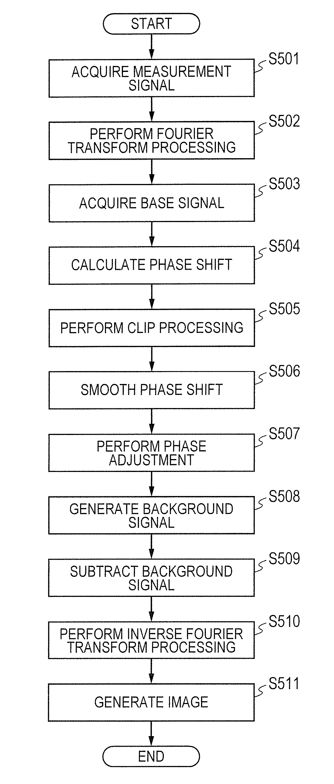
[0144]As to the signal processing according to Embodiment 1 and Embodiment 2, although dark lines illustrated in FIG. 3B are not shown in a single frame of tomographic image, when a plurality of tomographic images is averaged, an area having a slightly reduced luminance is enhanced, and dark lines appear in some cases. This appearance is a phenomenon in which a noise component is excessively subtracted from the measurement signals due to an unnaturally high accuracy of the phase adjustment. In contrast, an OCT apparatus according to Embodiment 3 clips phase shifts and performs a smoothing process, so as to adjust an accuracy of a phase adjusting amount. This process does not subtract FPN (a noise component) excessively, preventing the dark lines from occurring even when a plurality of tomographic images is averaged. The clipping in the present specification refers to a process in which an upper limit is provided for input values, so as to suppress a peak of the input values to the u...
PUM
 Abstract
Description
Claims
Application Information