
Systems, compositions, and methods for transplantation

a technology of system and composition, applied in the field of systems, compositions, and methods for transplantation, can solve the problems of inability to differentiate acute injury from acute injury, difficulty in treating spinal and joint pain, and inability of physicians to have a system or method available, etc., to achieve the reduction of the degeneration rate of tissue, cartilage and discs, synovial inflammation, or a combination thereof, and the effect of reducing the severity and occurren

Active Publication Date: 2019-02-14
CYTONICS CORP
View PDF1 Cites 6 Cited by
  • Summary
  • Abstract
  • Description
  • Claims
  • Application Information

AI Technical Summary

Benefits of technology

[0023]In one aspect, provided herein is a method of treating a subject, comprising administering to a subject in need thereof an effective amount of any composition described herein or a composition obtainable by any method described herein. In some embodiments, the composition is administered into an anatomic site relevant to a pathology of the subject. In some embodiments, protease activity is inhibited at an anatomic site of administration; thereby decreasing the degeneration rate of tissue, the degeneration rate of cartilage, the degeneration rate of discs, or synovial inflammation, or a combination thereof. In some embodiments, the subject has one or more conditions comprising: arthritis, inflammation, ligament injury, tendon injury, bone injury, cartilage degeneration, cartilage injury, an autoimmune disease, back pain, joint pain, joint degeneration, disc degeneration, spine degeneration, bone degeneration, or any combination thereof; wherein inflammation comprises joint or disc inflammation caused by surgery, joint or disc inflammation caused by a joint or disc replacement, or a combination thereof. In some embodiments, the subject has been previously diagnosed with the one or more conditions. In some embodiments, the administration is to a joint selected from the group comprising a wrist, spinal, shoulder, elbow, carpal, metacarpal, phalangeal, acromioclavicular, sternoclavicular, scapular, costal, sacroiliac, hip, knee, ankle, tarsal, and a metatarsal joint.
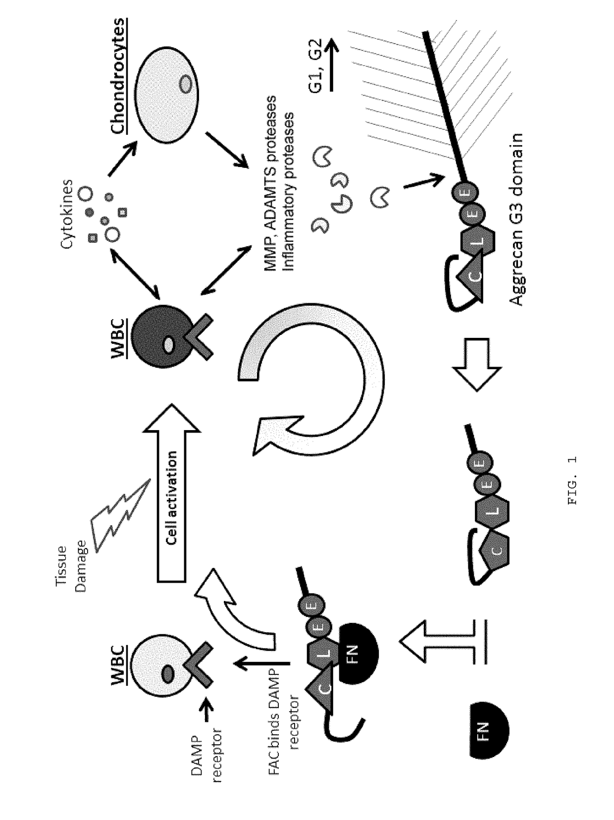
[0028]In one aspect, provided herein is a method of treating a subject with one or more conditions, comprising administering to the subject an effective amount of any composition provided herein, a wild-type A2M protein, A2M variant, or a combination thereof. In some embodiments, nonspecific inhibition of one or more proteases in the subject, inhibition Aggrecan G3 fragment formation, inhibition FAC formation, or a combination thereof, is increased. In some embodiments, the rate of degeneration of tissue, cartilage and discs, synovial inflammation, or a combination thereof, is decreased in the subject. In some embodiments, treating results in a reduction in severity, occurrence, rate of progression, or a combination thereof, of the one or more conditions. In some embodiments, any of the methods provided herein further comprise administering one or more additional carriers or drugs. In some embodiments, the one or more additional carriers or drugs comprise hydrogels, hyaluronic acid preparations, polymer microspheres, corticosteroids, microparticles, chitosan, local anaesthetics, growth factors, cytokines, protease inhibitors, steroids, hyaluranic Acid (HA), or other biologically active autogenous or endogenous mediators. In some embodiments, the one or more conditions are treatable with any composition provided herein. In some embodiments, the one or more conditions comprise cancer, degenerative diseases, traumatic diseases, and / or inflammatory diseases, whose pathogenesis includes the activity of proteases. In some embodiments, the cancer, degenerative diseases, traumatic diseases, and / or inflammatory diseases whose pathogenesis includes the activity of proteases comprises osteoarthritis, inflammatory arthritides, chondrosis, chondral injuries, enthesopathies, tendinopathies, ligamentous injuries, degenerative diseases of the bone, cartilage, tendons, and ligaments, post-operative conditions and wound healing, and other musculoskeletal diseases. In some embodiments, the one or more conditions comprise cancer, arthritis, inflammation, ligament injury, tendon injury, bone injury, cartilage degeneration, cartilage injury, an autoimmune disease, back pain, joint pain, joint degeneration, disc degeneration, spine degeneration, bone degeneration, or any combination thereof. In some embodiments, inflammation comprises joint or disc inflammation caused by surgery, joint or disc inflammation caused by a joint or disc replacement, or a combination thereof. In some embodiments, the subject is a human, pig, mouse, rat, rabbit, cat, dog, monkey, frog, horse or goat. In some embodiments, the subject has been previously diagnosed with the one or more conditions. In some embodiments, the composition is administered into an anatomic site relevant to the host pathology. In some embodiments, the administration comprises injection with a hollow-lumen device or flexible catheter combinations. In some embodiments, the hollow-lumen device comprises a needle, syringe, or combination thereof. In some embodiments, the administration occurs during a surgical procedure.

Problems solved by technology

Inflammation causing spinal and joint pain can be difficult to treat.
Increasing degrees of inflammation and force applied to joints result in joint injury.
Physicians currently do not have a system or method available to differentiate between acute injury due to trauma and age related joint deteriorations.
Presently, it can be difficult to determine the appropriate course of treatment for a given patient since it can be frequently unclear whether the particular condition the patient suffers from may be acute or chronic or if pathology in the joint is the cause of the pain.
However, no clear causal pathway leading from injury or degeneration to the painful state has been confirmed.
Although some studies have provided evidence that the epidural space can be affected by an intervertebral disc herniation, none has measured concentrations of biomolecules in the epidural space in an attempt to detect the differences between affected and non-affected persons.
Injury to tendons and ligaments causes damage not only to the connective tissue, but to the extracellular matrix as well.
Damage to the ECM can interrupt cell behavior in the connective tissue and decrease and / or limit healing.
After injury, continuing damage can be caused by production of matrix metalloproteinases (MMPs) by the body.
This can lead to an imbalance between the synthesis and degradation of the ECM, as the body tries to heal itself while the enzymes remodel the ECM.
An overabundance of remodeling by MMPs cause damage to previously connected tissue which results in the formation of scar tissue.
In addition, scar tissue adhesion to surrounding tissue can cause further pulling and / or stretching of the tendons or ligaments and resultant pain.
However, these simple remedies do not always cure the injury and often more advanced treatments are needed.

Method used


Image

  • Systems, compositions, and methods for transplantation
  • Systems, compositions, and methods for transplantation
  • Systems, compositions, and methods for transplantation

Examples


example 1

n and Selection of HEK293 Clones Expressing Recombinant A2M

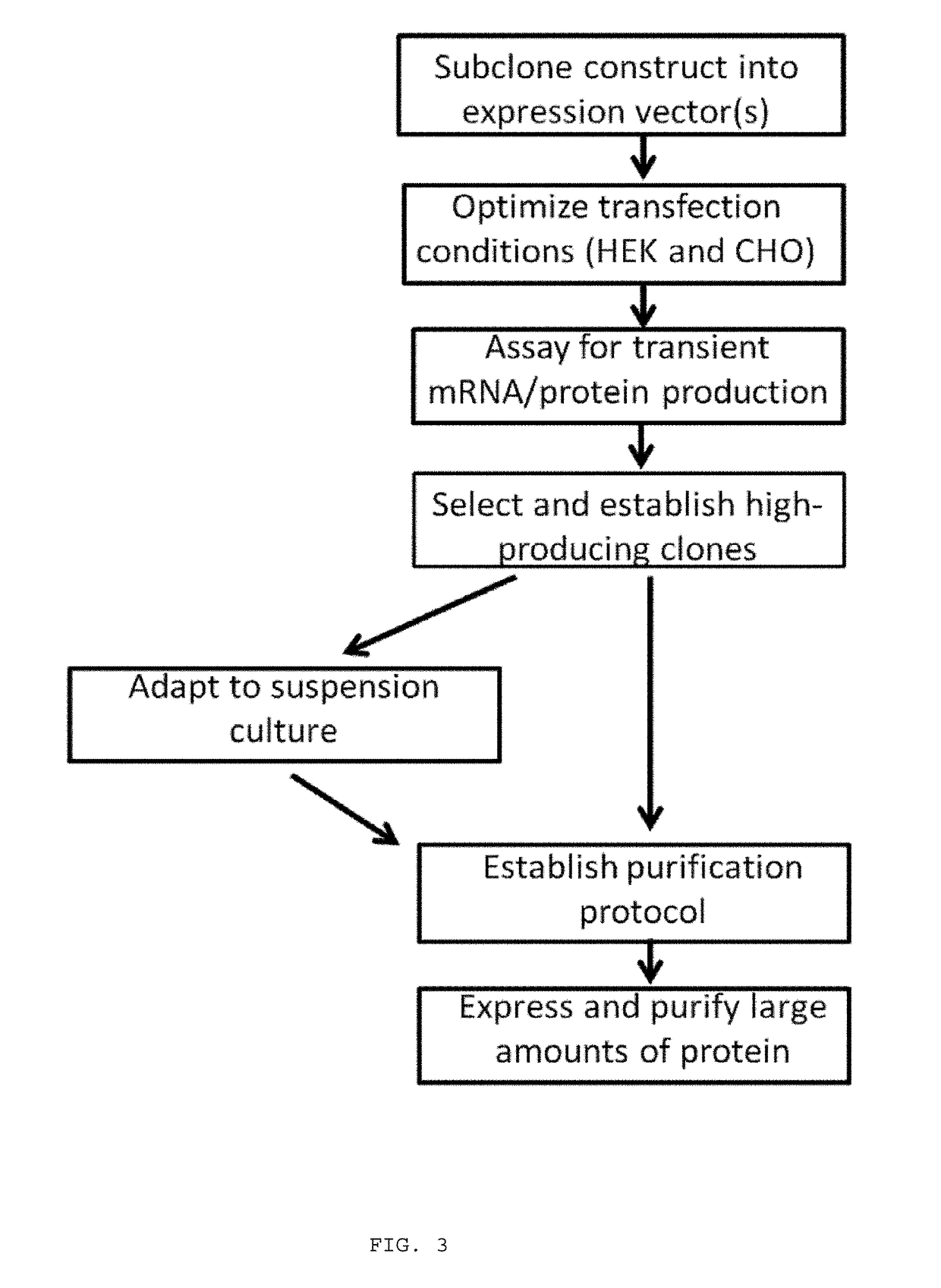
[0338]Recombinant A2M wild type sequence was expressed in HEK293F cells. Hek293F cells are plated adherently and allowed to attach overnight. Cells are transfected with XTreme Gene HP (Roche) and DNA in a 6 uL reagent: 2 ug DNA ratio. Cells are grown for 48 hours at 5% CO2 and 37 degrees Celsius. Forty-eight hours after transfection media samples are taken to confirm success of the transfection via an ELISA assay that quantifies A2M protein. Cells are split so as to be in logarithmic growth phase and selection antibiotic (blasticidin) is added at 10 μg / mL (selection concentration determined experimentally). Cells are selected in antibiotic until all of the negative control cells are dead (usually about 4 to 5 days). Another media sample is taken at this point to confirm that this newly established pool is still producing protein. Upon confirmation of protein production cells are plated at a density of −100 cells / 10 cm dish w...

example 2

n of ADAMTS-5- and ADAMTS-4-Induced Damage of Cartilage with A2M

[0339]Bovine Cartilage Explants (BCEs) were treated with 500 ng / ml ADAMTS-5 or ADAMTS-4 for 2 days, with a 3-fold serial dilution of purified A2M (FIG. 7A, B). Concentration of A2M tested were 100, 33.3, 11.1, 3.7, 1.2, 0.4 μg / mL. The A2M inhibited cartilage catabolism in a concentration dependent manner. The IC50 for inhibiting 500 ng / ml of ADAMTS-5 was calculated to be ˜7 μg / ml A2M (a 1:1 molar ratio). Maximum inhibition was observed in ˜90% with 100 μg / ml A2M (a 14:1 molar ratio). The A2M was shown to block formation of Aggrecan G3 fragments (FIG. 7A, B) and FAC formation (FIG. 9).

example 3

n of APIC Retentate and Filtrate

[0340]Fresh cartilage was treated with APIC containing ˜7 mg / ml A2M. Cartilage catabolism was efficiently blocked by 1% v / v of the Retentate of the APIC production process (concentration of proteins >500 kDa in size), but not by the Filtrate (contains proteins 99% of the protective factors of autologous blood.


PUM

PropertyMeasurementUnit
molecular weightaaaaaaaaaa
concentrationaaaaaaaaaa
pore sizeaaaaaaaaaa

Abstract

Systems and methods for purification and concentration of autologous alpha-2-macroglobulin (A2M) from whole blood are provided. Also provided are diagnostic methods for identifying sites in the synovial joints, spine, tendons or ligaments for treatment of pain, degeneration, or inflammation with autologous A2M. Methods for utilizing autologous A2M in combination with other autologous treatments (e.g. platelets and other growth factors) are provided in addition to combinations with exogenous drugs or carriers. Also provided is a method of producing recombinant A2M wild type or variants thereof where the bait region was modified to enhance the inhibition characteristics of A2M and / or to prolong the half life of the protein in joints and spine disc or epidural space.

Description

CROSS-REFERENCE[0001]This application is a continuation of U.S. application Ser. No. 14 / 380,234, filed on Aug. 21, 2014, pursuant to 35 U.S.C. § 371 as a United States National Phase Application of International Application No. PCT / US2013 / 027159, filed Feb. 21, 2013, which claims the benefit of U.S. Provisional Application No. 61 / 601,434, filed on Feb. 21, 2012, U.S. Provisional Application No. 61 / 726,815, filed on Nov. 15, 2012, U.S. Provisional Application No. 61 / 726,840, filed on Nov. 15, 2012, U.S. Provisional Application No. 61 / 727,433, filed on Nov. 16, 2012, and U.S. Provisional Application No. 61 / 740,218, filed on Dec. 20, 2012, which applications are incorporated herein by reference in their entirety.[0002]All publications and patent applications mentioned in this specification are herein incorporated by reference to the same extent as if each individual publication or patent application was specifically and individually indicated to be incorporated by reference.BACKGROUND ...

Claims


Application Information

Patent Timeline
Patent Type & Authority Applications(United States)
IPC IPC(8): A61K38/57A61M1/34A61K45/06A61K38/17A61K35/16C07K14/81A61K35/19A61K35/28
CPCA61M1/34A61K45/06A61K38/1722A61K38/57A61K35/16C07K14/8107A61K35/19A61K35/28A61K2300/00A61K38/55C07K1/34C07K14/81
Inventor HANNA, LEWISLAUGHLIN, JOHN DAVIDBROWNING, SHAWN ROBERT
Owner CYTONICS CORP
Learn More