Monitoring Treatment of Peripheral Artery Disease (PAD) Using Diffuse Optical Imaging

a peripheral artery disease and imaging technology, applied in the field of peripheral artery disease monitoring and imaging treatment using diffuse optical imaging, can solve the problems of gangrene, infection of wounds and tissue loss, and progressively shorter distances

Active Publication Date: 2019-05-02
THE TRUSTEES OF COLUMBIA UNIV IN THE CITY OF NEW YORK
View PDF0 Cites 7 Cited by
  • Summary
  • Abstract
  • Description
  • Claims
  • Application Information

AI Technical Summary

Benefits of technology

[0015]Some embodiments of the first apparatus further comprise at least one processor configured to (a) control generation of the electrical signals that drive the first plurality of light sources, accept data representative of the light detected by the first plurality of light detectors, and determine a first level of perfusion in the first angiosome based on the data representative of the light detected by the first plurality of light detectors; (b) control generation of the electrical signals that drive the second plurality of light sources, accept data representative of the light detected by the second plurality of light detectors, and determine a second level of perfusion in the second angiosome based on the data representative of the light detected by the second plurality of light detectors; and (c) control generation of the electrical signals that drive the third plurality of light sources, accept data representat

Problems solved by technology

As the disease progresses, this distance becomes progressively shorter, and in later stages the pain becomes severe and present even at rest.
Untreated, the disease eventually leads to formation of foot wounds or ulcerations, which can subsequently lead to infection of the wound and tissue loss (gangrene), and ultimately amputation.
PAD affects approximately 12 million individuals in the United States and is associated with significant morbidity and mortality.
Furthermore, patients with PAD have an increased risk of stro

Method used


Image

  • Monitoring Treatment of Peripheral Artery Disease (PAD) Using Diffuse Optical Imaging
  • Monitoring Treatment of Peripheral Artery Disease (PAD) Using Diffuse Optical Imaging
  • Monitoring Treatment of Peripheral Artery Disease (PAD) Using Diffuse Optical Imaging

Examples


Embodiment Construction

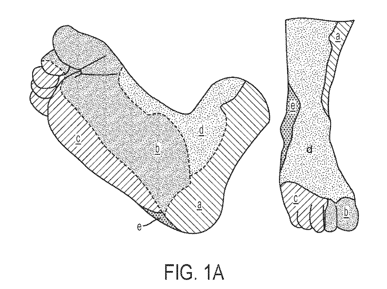
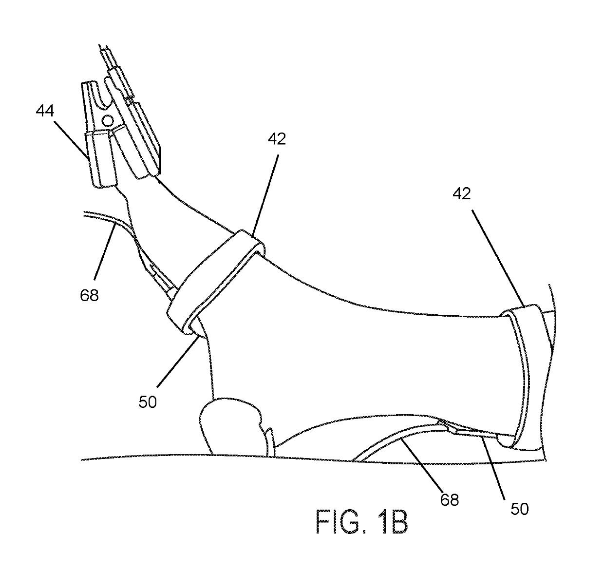
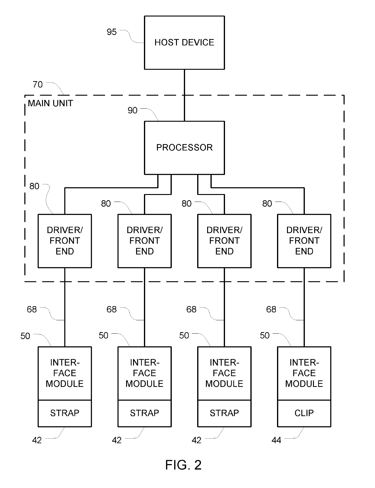
[0068]SECTION 1: Monitoring Treatment of Peripheral Artery Disease (PAD) Using Diffuse Optical Imaging.

[0069]Vascular optical tomographic imaging (VOTI) is an emerging imaging modality capable of detecting hemoglobin concentrations in tissue. VOTI is non-invasive, non-ionizing and does not require contrast injection. In a clinical pilot study involving 40 subject that it has been shown that this technology promises to diagnose peripheral arterial disease (PAD) within lower extremities of diabetic patients with calcified arteries with high sensitivity and specificity.

[0070]The VOTI system is capable of quantifying the blood volume changes within the foot during the thigh cuff occlusion and outputting diagnostic parameters, such as change in hemoglobin concentration, enabling the assessment of foot perfusion. VOTI is also capable of providing the locations of under-perfused regions within the foot and evaluating the severity of arterial disease, even within diabetic patients with calc...


PUM


Abstract

A plurality of modules are simultaneously positioned at locations that correspond to different angiosomes. Each of these modules has a front surface shaped and dimensioned for contacting a person's skin, a plurality of different-wavelength light sources aimed in a forward direction, and a plurality of light detectors aimed to detect light arriving from in front of the front surface. Each module is supported by a support structure (e.g., a strap or a clip) that is shaped and dimensioned to hold the front surface adjacent to the person's skin at a respective position. Perfusion in each of the angiosomes is monitored using these modules, and the surgeon can rely on this information to guide his or her intervention.

Description

CROSS REFERENCE TO RELATED APPLICATIONS[0001]This Application claims the benefit of U.S. Provisional Application 62 / 326,822 filed Apr. 24, 2016, and U.S. provisional application 62 / 417,741 filed Nov. 4, 2016. Each of the above identified applications is incorporated herein by reference in its entirety.STATEMENT REGARDING FEDERALLY-SPONSORED RESEARCH[0002]This invention was made with government support under HL115336 awarded by the National Institutes of Health. The government has certain rights in the invention.BACKGROUND[0003]Peripheral arterial disease (PAD) is the narrowing of arteries due to plaque accumulation and in the vascular walls (atherosclerosis). PAD typically manifests itself as claudication, which is defined as a cramping leg pain with ambulation to a set distance. As the disease progresses, this distance becomes progressively shorter, and in later stages the pain becomes severe and present even at rest. Untreated, the disease eventually leads to formation of foot wou...

Claims


Application Information

Patent Timeline
IPC IPC(8): A61B5/026A61B5/00A61B5/02A61B5/0225A61B5/1455
CPCA61B5/0261A61B5/0075A61B5/02007A61B5/6828A61B5/6829A61B5/6831A61B5/7425A61B5/02255A61B5/0073A61B5/6884A61B5/1455A61B2562/04A61B5/00A61B5/02A61B5/021A61B5/022A61B5/0245A61B2560/0437A61B5/24
Inventor HIELSCHER, ANDREAS H.FONG, CHRISTOPHER J.HOI, JENNIFERKIM, HYUN K.KHALIL, MICHAEL
Owner THE TRUSTEES OF COLUMBIA UNIV IN THE CITY OF NEW YORK
Why Eureka
Try Eureka