Microscope lens with integrated wide-field camera and beam scanning device

a wide-field camera and microscope technology, applied in the field of microscope lenses with integrated wide-field cameras and beam scanning devices, can solve the problems of limited accuracy and repeatability, inability to perform sub-surface microscopic imaging with a handheld device, and millions of biopsy of benign lesions, so as to reduce the need for biopsy, improve specificity, and reduce the effect of benign-to-malignant detection ra

Active Publication Date: 2019-05-02
MEMORIAL SLOAN KETTERING CANCER CENT +1
View PDF0 Cites 2 Cited by
  • Summary
  • Abstract
  • Description
  • Claims
  • Application Information

AI Technical Summary

Benefits of technology

[0004]Wide-field clinical examination followed by dermoscopy (magnification 4×-10×) is the current standard approach for guiding biopsy and pathology and diagnosis of skin lesions. However, this approach varies in specificity, depending on setting, and lesion and patient characteristics, resulting in highly variable benign-to-malignant detection rates, ranging from as low as 2-to-1 to as high as 600-to-1, which, consequently, results in millions of biopsies of benign lesions. Reflectance confocal microscopy (RCM), which noninvasively images beneath the skin surface and shows nuclear and cellular details in vivo, has been proven to increase specificity and lower the benign-to-malignant detection rate by about two times relative to that with dermoscopy alone. Wide-field clinical examination and dermoscopy combined with RCM imaging is now being implemented in clinical settings to triage lesions into those that appear benign (these patients are being spared biopsy) versus those that appear suspicious (these patients proceed to standard biopsy and pathology). The initial impact is promising in terms of reducing the need for biopsy of benign lesions.
[0009]A person skilled in the art will recognize that the present inventions of an integrated scanner or an integrated camera, together or separately, apply not only to reflection confocal microscopy, but any scanned-laser based imaging modality including but not limited to optical coherence tomography and optical coherence microscopy, multiphoton microscopy, second harmonic generation microscopy, photoacoustic microscopy, etc. Furthermore, approaches that do not have a scanning laser beam, such as structured light microscopy or white light optical coherence microscopy, will benefit from the integrated camera for contextualizing the microscopic field of view within the surface morphology.

Problems solved by technology

However, this approach varies in specificity, depending on setting, and lesion and patient characteristics, resulting in highly variable benign-to-malignant detection rates, ranging from as low as 2-to-1 to as high as 600-to-1, which, consequently, results in millions of biopsies of benign lesions.
What is currently lacking, therefore, is the ability to accurately and repeatably locate the RCM field of view at small areas of suspicion within the larger wide-field image.
In other words, sub-surface microscopic imaging with a handheld device is being performed with limited accuracy and repeatability within the context of clinically-observed gross surface morphology, and with only subjective and thus limited means to register the RCM image to a wide-field image.
This leads to increased variability and uncertainty in interpreting RCM images and in making a determination whether to biopsy.
The challenge in providing simultaneous confocal microscopic imaging with adequately high numerical aperture (NA) and wide field imaging at lower NA in a single device is the limited field of view supported by any high-NA objective lens.

Method used


Image

  • Microscope lens with integrated wide-field camera and beam scanning device
  • Microscope lens with integrated wide-field camera and beam scanning device
  • Microscope lens with integrated wide-field camera and beam scanning device

Examples


Embodiment Construction

[0035]Before any embodiments of the invention are explained in detail, it is to be understood that the invention is not limited in its application to the details of construction and the arrangement of components set forth in the following description or illustrated in the following drawings. The invention is capable of other embodiments and of being practiced or of being carried out in various ways.

Microscope Objective Lens with Integrated Wide-Field Camera

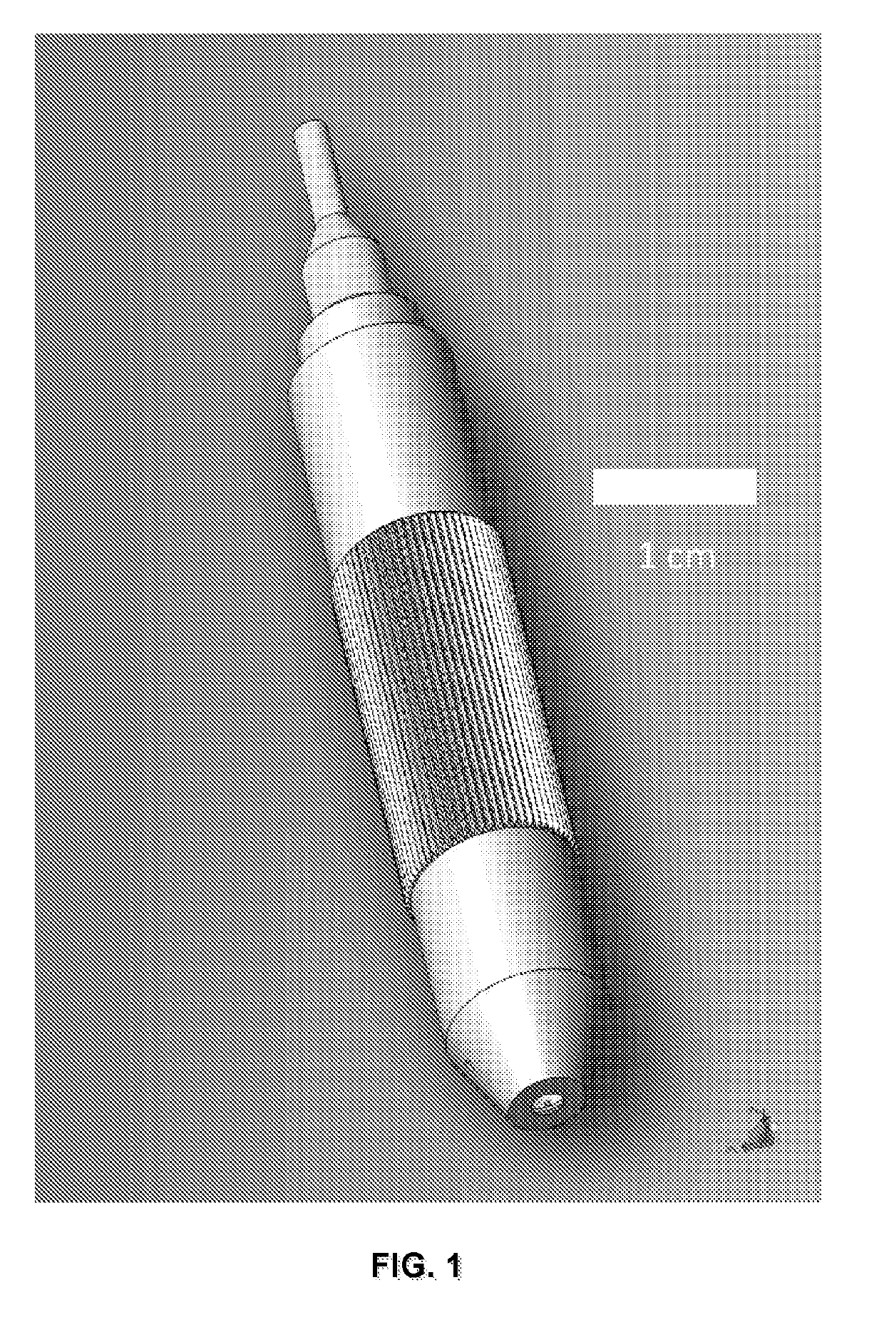
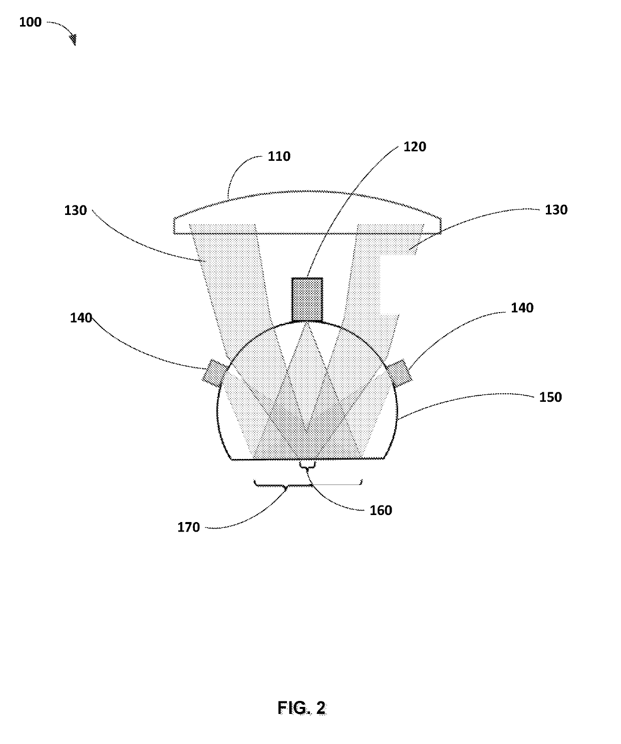
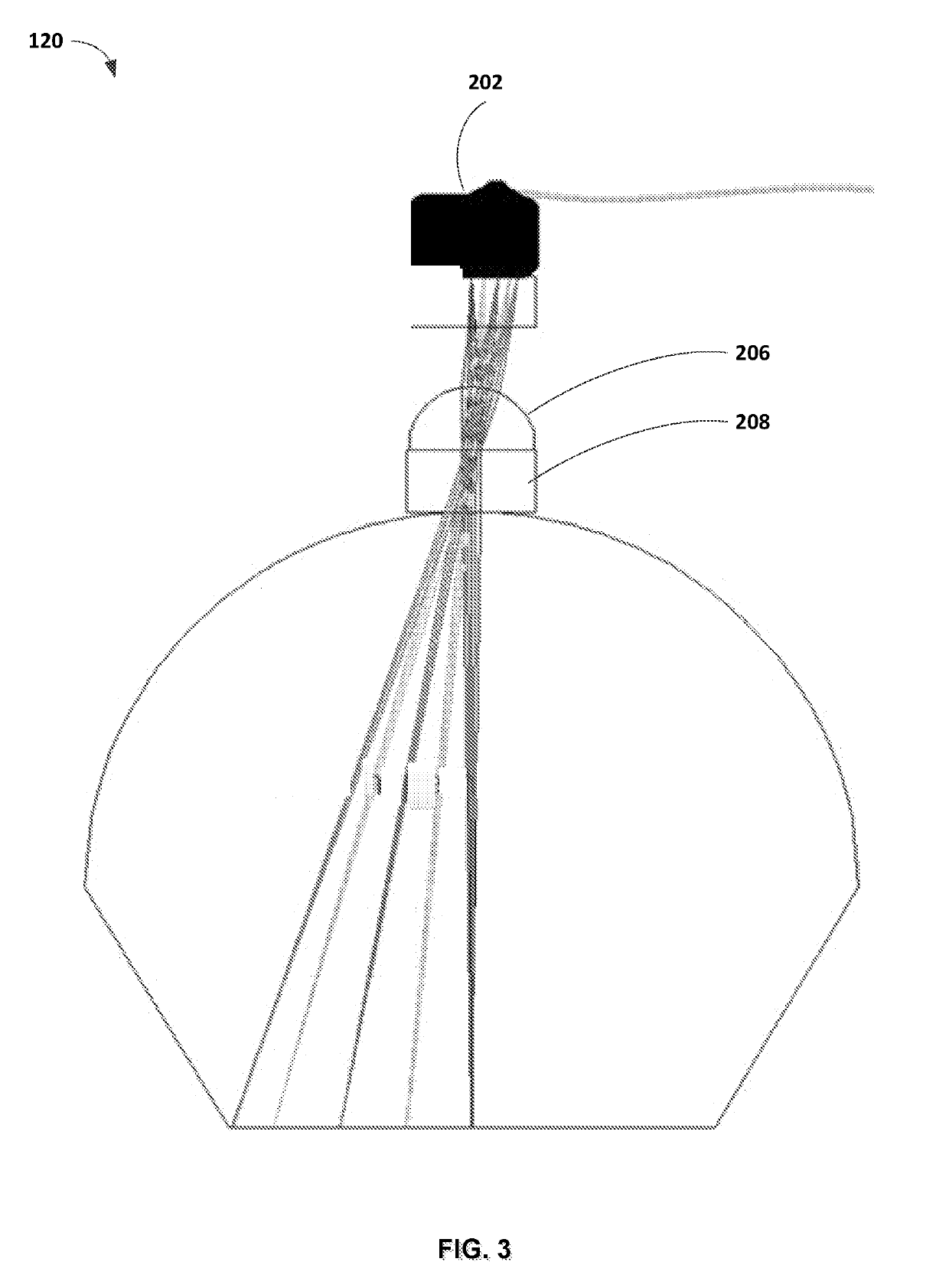
[0036]The promise of in vivo microscopy for diagnosis of diseases such as cancer is huge, and significant progress has been made to create in vivo microscopy systems. Two aspects of current state-of-the-art microscope optics are: 1) the available field of view laterally is limited to a few hundred microns, less than 1 mm, and 2) the optics are bulky, and the size of the objective lens where it is in contact with the tissue being examined is typically several mm, even in clinical microscopes designed for in vivo examination. Theref...


PUM


Abstract

Handheld devices and methods that integrate wide-field dermoscopy with reflectance confocal microscopy for non-invasive simultaneous capture of wide-field color images of the skin surface and images of the sub-surface cellular structure.

Description

CROSS-REFERENCE TO RELATED APPLICATIONS[0001]This application is a non-provisional of and claims priority to U.S. Provisional Application No. 62 / 294,149, filed on Feb. 11, 2016 and U.S. Provisional Application No. 62 / 294,705, filed on Feb. 12, 2016. The contents of both applications are incorporated herein by reference.STATEMENT OF FEDERALLY-SPONSORED RESEARCH[0002]This invention was made with government support under 1R21EB018507 and P30CA008748 awarded by the National Institutes of Health and under 1542210 awarded by the National Science Foundation. The government has certain rights in the invention.BACKGROUND OF THE INVENTION[0003]Wide-field imaging paired with reflectance confocal microscopy is advancing in clinical settings to guide noninvasive diagnosis of skin cancer. However, two separate devices are required to perform each imaging procedure.SUMMARY OF THE INVENTION[0004]Wide-field clinical examination followed by dermoscopy (magnification 4×-10×) is the current standard ap...

Claims


Application Information

Patent Timeline
Patent Type & Authority Applications(United States)
IPC IPC(8): G02B21/36G02B21/06G02B21/00A61B5/00
CPCG02B21/361G02B21/06G02B21/0028G02B21/0008A61B5/0068A61B5/0077A61B5/444G02B21/0036G02B21/02A61B5/0075
Inventor DICKENSHEETS, DAVIDRAJADHYAKSHA, MILIND
Owner MEMORIAL SLOAN KETTERING CANCER CENT
Why Eureka
Try Eureka