An injection and extraction ophthalmic device

a technology of ophthalmic devices and injections, which is applied in the field of bodily fluid extraction, can solve the problems of increased risk of blindness infection in patients, unclear safety of repeated injections over long periods of time, damage to ocular structures, etc., and achieves safe and reliable drug delivery, safe and easy, and the effect of volum

Inactive Publication Date: 2019-05-30
SINGAPORE HEALTH SERVICES PTE
View PDF1 Cites 5 Cited by
  • Summary
  • Abstract
  • Description
  • Claims
  • Application Information

AI Technical Summary

Benefits of technology

[0012]The injector device may have the potential to standardise the injection technique in a more safe and reproducible way. The volume of drug delivery may also be more reproducible if reflux is reduced. The extraction of vitreous fluid from the eye for rapid biomarker analysis, may allow a tailored, individualised treatment based on biomarker levels and give additional outcome measures for treatment.
[0013]The “diagnostic plus therapeutic” device-vitreous extractor-plus-injector may simultaneously perform ocular fluid biopsy (vitreous sampling) and injection of therapeutic drugs safely and easily. Biomarker analysis may be performed on the vitreous fluid extracted to determine specific dosage and injection frequency, enabling individualised and patient-tailored therapy.
[0014]The invention may provide a safe and reliable way to extract vitreous and inject anti-VEGF agents into the eye. Injections are currently performed using improvised syringes meant for venepuncture and blood sampling. It is unstable and exposes the patient to risk of infection, damage to ocular structures, cataract and even blindness, requiring a skilled doctor to perform. As patients will require regular injections over their lifetime, there may be a need for nurses or technicians to perform this routine procedure to reduce burden on hospitals and doctors.
[0015]The removal of a small amount of vitreous humor before the injection of the therapeutic may prevent a spike in intraocular pressure and so reduce complications in the eye. To this end, the device may include a shearing device for cutting through extracted vitreous humor, so as to separate the sample from the remaining material. The shearing device may be a dedicated cutting tool, or may be included as part of the sample extracting mechanism.
[0020]To prevent retinal detachment when the needle is removed from the eye after biopsy due to the viscous nature of the vitreous humor.

Problems solved by technology

However, a standardised method of delivery, the frequency of the injections necessary for optimum results without over-treatment, and the ocular and systemic safety of these repeated injections over long periods of time are unclear.
However, multiple injections expose the patient to an increased risk of blinding infection, damage to ocular structures (e.g., retinal detachment, cataract), a higher risk of glaucoma and even systemic complications (e.g. stroke and ischaemic heart disease.
This procedure is not commonly performed and not performed routinely in the treatment of AMD and diabetic eye disease due to the risk involved in the procedure.
Another disadvantage of the current method of intra-vitreal injection of medication into the eye is that when a fixed volume of drug is injected into an already pressurised eye, there is an increased risk of a temporary spike in intra-ocular pressure (IOP) and reflux of drug from the injection site.
Previous studies have shown that the risk of glaucoma surgery is higher in eyes receiving multiple intra-vitreal injections.
Extraction of vitreous thought a needle is challenging as the vitreous may be in a gel-like state making it impossible for suction.
In addition, the gel-like state of the vitreous and adhesions to the retina may pose additional risk from traction when the device is retracted from the eye.
Currently there is no standardised method of intra-vitreal injections.
This non-standardised technique and excessive manipulation exposes the patient to an unnecessary risk of infection.
In users, who are not used to the injection technique, there may be also a higher risk of damage to ocular structures, cataract and other complications.
The increased injection load is both a treatment burden for the patients and a challenge to any busy retinal practice.

Method used


Image

  • An injection and extraction ophthalmic device
  • An injection and extraction ophthalmic device
  • An injection and extraction ophthalmic device

Examples


Embodiment Construction

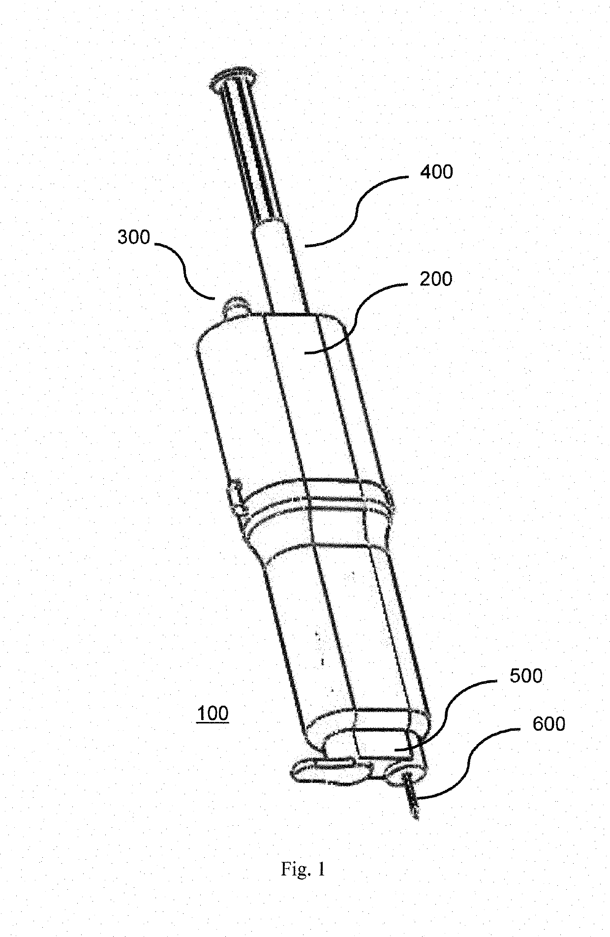
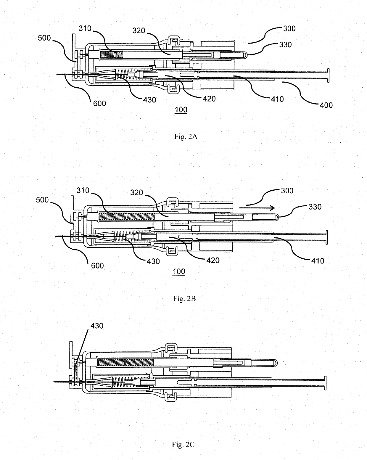
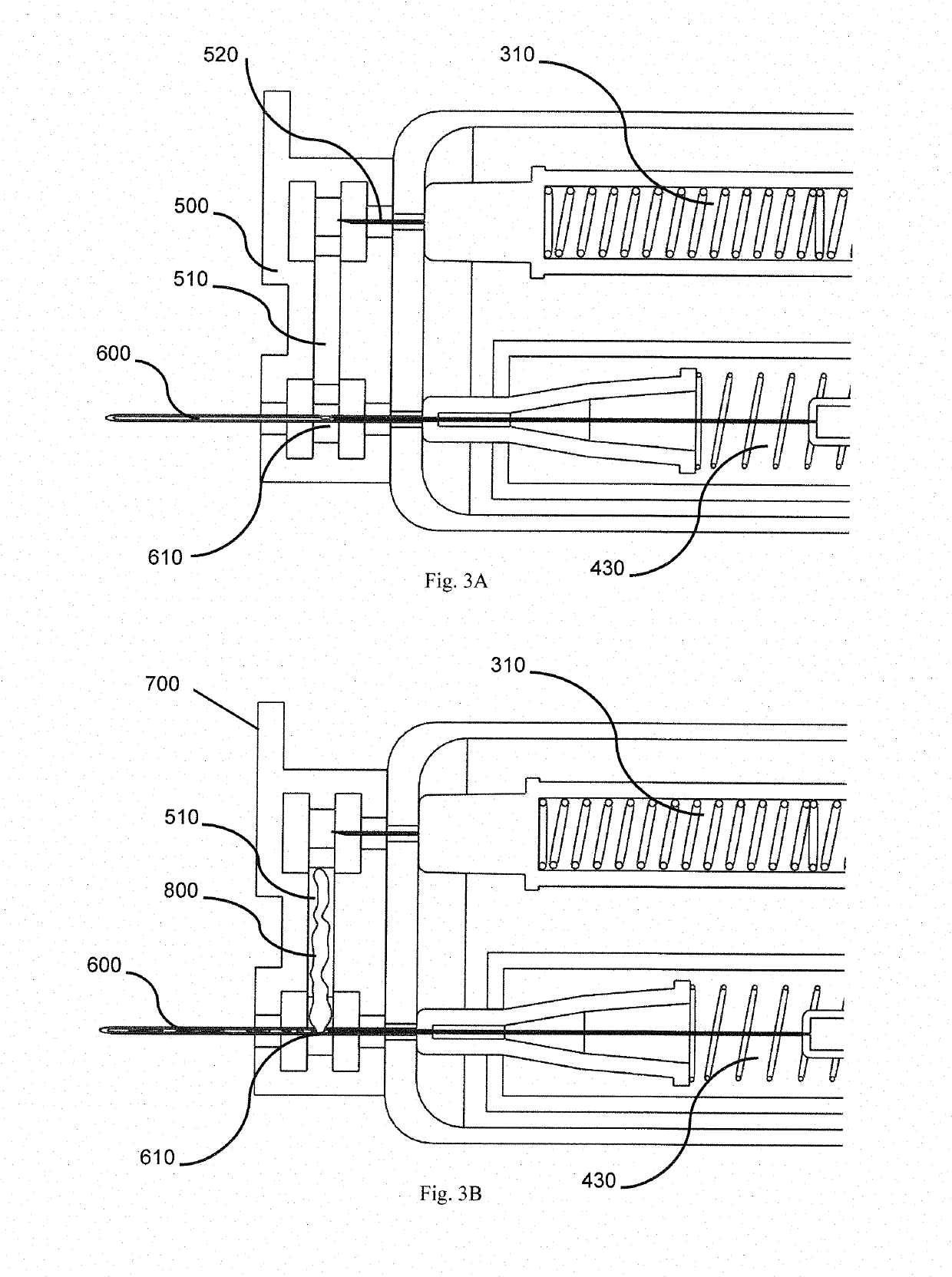
[0037]Referring to the drawings, in order to provide swift extraction of a vitreous sample, cutting of gel-like state of the vitreous sample and delivery of a therapeutic drug, in one embodiment, the present invention provides a disposable handheld extraction and delivery device 100 to facilitate extraction of the vitreous sample from an eye of a patient, the device comprising a casing 200 extending from a proximal end to a distal end and at least one opening, at the distal end, a needle portion 600 having a through hole, protruding out from the at least one opening, the needle portion 600 includes at least one side hole along a predetermined length of the needle portion 600. The at least one side hole 610 of the needle portion 600 may be sized and shaped preferably to increase suction area. The at least one side hole 610 shaped in any manner preferably rectangular to maximise streamlined flow exchange. In the alternative, the at least one side hole 610 may be shaped circularly or s...


PUM


Abstract

An extraction and injection ophthalmic device comprising; a housing having;an injection port arranged to receive an injection source; a vacuum port arranged to receive an vacuum source; an eye engagement portion, and; at least one needle for selective fluid communication with one or both of the vacuum port or injection port; wherein on activation of the vacuum source the device is arranged to extract a sample through the needle, and on activation of the injection source the device is arranged to inject an agent through the needle.

Description

FIELD OF THE INVENTION[0001]The present invention relates to retrieval of a bodily fluid for diagnosis thereafter injection of a therapeutic drug used for ophthalmology and the eye, and for similar purposes in other parts of the human body.BACKGROUND[0002]Age-related macular degeneration (AMD) and diabetic eye disease are the leading causes of blindness in elderly patients in the world, including Singapore and other Asian countries. Anti-vascular endothelial growth factor (anti-VEGF) agents, injected directly into the eye (vitreous humor), have emerged as the standard of care for these diseases. This has been shown to be superior to the other treatment modalities: photodynamic therapy for AMD and laser therapy for DR. However, a standardised method of delivery, the frequency of the injections necessary for optimum results without over-treatment, and the ocular and systemic safety of these repeated injections over long periods of time are unclear.[0003]While intra-vitreal injections ...

Claims


Application Information

Patent Timeline
Patent Type & Authority Applications(United States)
IPC IPC(8): A61F9/00A61B10/00A61M5/31A61M5/32
CPCA61F9/0017A61B10/0045A61M5/31A61M5/3202A61M5/178A61B5/154
Inventor ANG, MARCUS HAN NIANTEO, KELVIN YI CHONGTAN, ANNA CHENG SIM
Owner SINGAPORE HEALTH SERVICES PTE
Why Eureka
Try Eureka