
Method and associated device for rapid detection of target biomolecules with enhanced sensitivity

a biomolecule and target technology, applied in the field of enhanced sensitivity target biomolecule detection methods and associated devices, can solve the problems of inefficient detection methods, lack of accurate rapid point-of-care diagnostic tests, different tuberculosis, etc., and achieve the effect of rapid diagnostic testing and rapid detection of a biomolecul

Active Publication Date: 2019-06-20
GENERAL ELECTRIC CO
View PDF2 Cites 4 Cited by
  • Summary
  • Abstract
  • Description
  • Claims
  • Application Information

AI Technical Summary

Benefits of technology

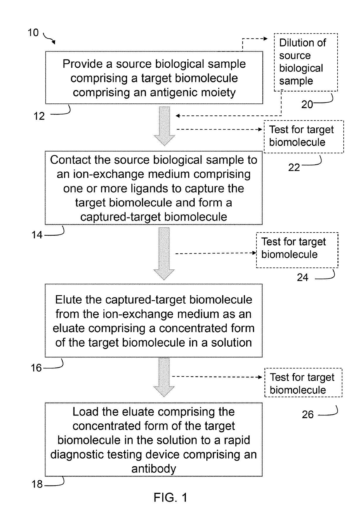
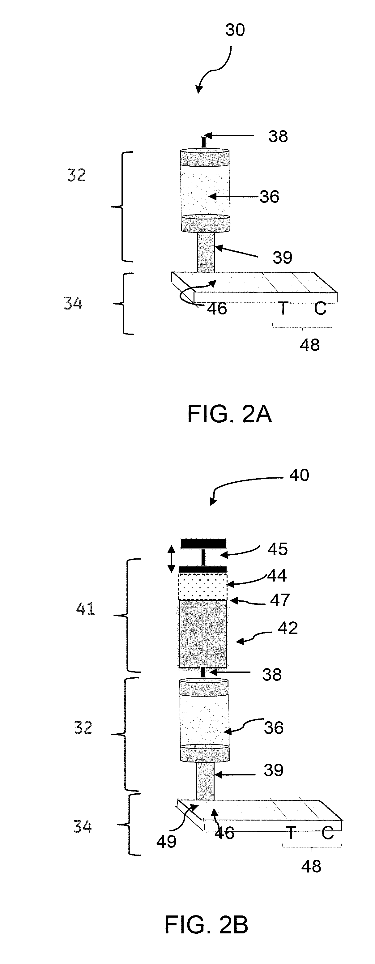
The patent text describes a rapid detection method of infectious diseases using a rapid diagnostic testing device. The method involves contacting a biological sample with an ion-exchange medium to capture the infectious disease biomolecule and form a captured-biomolecule. The captured-biomolecule is then eluted and loaded onto an antibody for detection. The method is efficient, cost-effective, and can be performed in resource-limited settings with minimal training. The patent also describes a method for detecting tuberculosis in urine samples. The device includes a concentrator and an immune-based assay unit for detecting the antigen. Overall, the patent provides a solution for early detection and control of infectious diseases.

Problems solved by technology

Among major infectious diseases, tuberculosis is different in that it lacks accurate rapid point-of-care diagnostic tests.
Inefficient detection methods and lack of timely treatment of tuberculosis are major causes of failure to control the spread of tuberculosis.
Prior methods for detecting surface polysaccharides (LAM) using different body fluids, such as serum, urine or sputum, have been investigated, but have proven ineffective.
For example, prior studies with urine sample required extensive sample processing and manipulation, rendering such methodologies complex and cumbersome, specifically in the field.

Method used


Image

  • Method and associated device for rapid detection of target biomolecules with enhanced sensitivity
  • Method and associated device for rapid detection of target biomolecules with enhanced sensitivity
  • Method and associated device for rapid detection of target biomolecules with enhanced sensitivity

Examples


example 1

Dilution of a Source Urine Sample in Concentrating TB-LAM Present in the Source Urine Sample Using an Ion Exchange Medium

[0061]A urine sample was spiked with TB-LAM to have a final concentration of 24 ng / ml for TB-LAM in the urine sample. The urine sample was diluted to 4× to reduce the conductivity to a range that is optimal for an anion exchange membrane to bind the TB-LAM antigen. 6.2 ml of the diluted urine sample containing 6 ng / ml TB-LAM was applied to a Q+ membrane column, wherein the TB-LAM was captured to the Q+ membrane column (i.e., membrane-bound TB-LAM or captured TB-LAM). The membrane-bound TB-LAM was eluted from the Q+ membrane column using 1.35 M NaCl in 20 mM Tris buffer. 0.1 mL eluate was loaded to an RDT device, wherein the eluate contained TB-LAM at a concentration of 315.1 ng / ml in an elution buffer containing 1.35 M NaCl in 20 mM Tris buffer. The TB-LAM was concentrated to 51.5-fold using the Q+ membrane column. The result yields about 13× increase in concentra...

example 2

ve Analysis of Urine Sample Containing TB-LAM Before and after Concentrating the Urine Sample

[0062]Preparation of Q+ Membrane Columns:

[0063]Predictor® ReadyToProcess Adsorber Q+96-well plate columns (or Q+ membrane columns) were prepared for binding of TB-LAM antigen from human urine samples by washing each well with 2 mL of acidified buffer solution. After washing the Q+ membrane column, the column was equilibrated with a neutral pH buffer solution (e.g., phosphate buffered saline (PBS)).

[0064]Preparation of Urine Sample Containing TB-LAM:

[0065]TB-LAM antigen was added to a urine sample to attain a TB-LAM concentration of 24 ng / mL. The urine sample was diluted (4× dilution) to generate a diluted urine sample having desired salinity and enable efficient binding to the anion exchange membrane (e.g., ˜10 mS / cm). The resulting concentration of TB-LAM antigen was 6 nanogram / mL (ng / mL), which is a level in the typical range for a human TB-infected patient and in the working range of comm...

example 3

ive Analysis of Urine Sample Containing TB-LAM Before and after Concentrating the Urine Sample

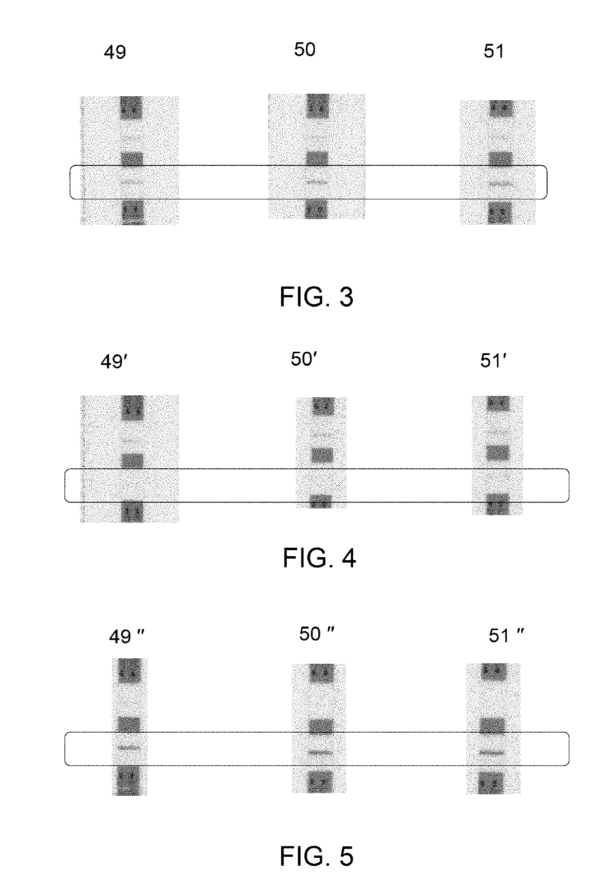
[0070]The source urine sample before applying to the column, and the eluate for different samples (N=2) were quantified by ELISA assay against a standard dilution series of TB-LAM in the solution used at each point of the assay. A quantitative estimation included determination of concentration of TB-LAM in the source urine sample and in eluate by standard ELISA bio-quantification methodology as described below. The eluate containing TB-LAM was directly applied to an ELISA reaction, wherein the eluate contained TB-LAM in an elution buffer of 1.35 M NaCl in 20 mM Tris.

[0071]Preparation of Reagents for ELISA:

[0072]Carbonate Coating Buffer was prepared by using contents of 1 capsule material into 100 mL double distilled water (ddH2O). Capture antibody (IV-03) was used as 5 μg / mL concentration. 10% BSA solution was diluted to 1:10 to 1% in 1×PBS to make a Blocking Buffer. Biotinylated antibody w...


PUM

PropertyMeasurementUnit
volumeaaaaaaaaaa
anglesaaaaaaaaaa
concentrationsaaaaaaaaaa

Abstract

A rapid detection method of a target biomolecule comprising an antigenic moiety is provided. The method includes providing a source biological sample comprising the target biomolecule; contacting the source biological sample to an ion-exchange medium; eluting the captured-target biomolecule from the ion-exchange medium as an eluate, and loading the eluate to a rapid diagnostic testing device comprising an antibody. The eluate comprises a concentrated form of the biomolecule in a solution having a salt concentration greater than 150 mM. A concentration of the target biomolecule in the eluate is in a range from about 2× to 25× compared to a concentration of the biomolecule in the source biological sample. The target biomolecule binds to the antibody under the salt concentration of greater than 150 mM. A device for rapid detection of target biomolecule is also provided.

Description

[0001]This application relates generally to a rapid detection method of infectious disease. In a particular aspect, the application relates to a rapid detection method and an associated device for rapid diagnostic testing of a target biomolecule.BACKGROUND[0002]A rapid detection method of analytes or target biomolecules performed by employing a rapid diagnostic test (RDT) is relatively less time consuming and less labor intensive as compared to conventional methods. Rapid diagnostic tests (RDTs) have been used for detection of various infectious diseases. The RDTs are suitable for preliminary and / or emergency medical screening, for example, for use in medical facilities with limited resources, and offer a useful alternative to microscopy in situations where reliable microscopic diagnosis facility is not available or is not immediately available. RDTs also allow point of care (POC) testing in primary care. RDTs can be performed independent of laboratory equipment by minimally trained...

Claims


Application Information

Patent Timeline
Patent Type & Authority Applications(United States)
IPC IPC(8): G01N33/569G01N1/40G01N33/543B01L3/00B01D15/36
CPCG01N33/5695G01N1/4005G01N33/54366B01L3/5023B01D15/363G01N2333/35G01N2469/10G01N2001/4011B01L2200/0631B01L2200/10G01N2405/06B01L2300/0825
Inventor MISNER, MATTHEW JEREMIAHGROSSMANN, GREGORY ANDREWOLSEN, CATHRYN ELLENNELSON, JOHN RICHARDBALES, BRIAN CHRISTOPHERMOORE, DAVID ROGERSMIGELSKI, JR., PAUL MICHAEL
Owner GENERAL ELECTRIC CO
Learn More